

Eye Adult Changes in Thought (Eye ACT) Study: Design and Report on the Inaugural Cohort
- PMID: 38875039
- PMCID: PMC11556780
- DOI: 10.3233/JAD-240203
Eye Adult Changes in Thought (Eye ACT) Study: Design and Report on the Inaugural Cohort
Abstract
Background: Conflicting research on retinal biomarkers of Alzheimer's disease and related dementias (AD/ADRD) is likely related to limited sample sizes, study design, and protocol differences.
Objective: The prospective Eye Adult Changes in Thought (Eye ACT) seeks to address these gaps.
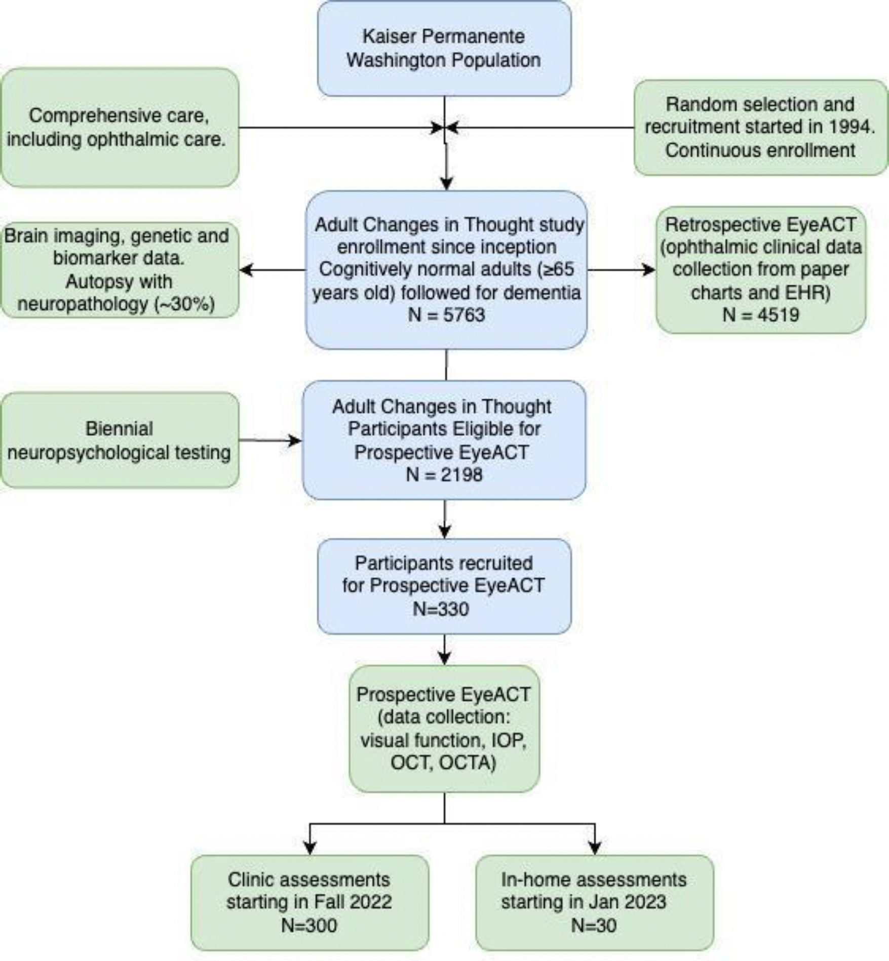
Methods: Eye ACT participants are recruited from ACT, an ongoing cohort of dementia-free, older adults followed biennially until AD/ADRD, and undergo visual function and retinal imaging assessment either in clinic or at home.
Results: 330 participants were recruited as of 03/2023. Compared to ACT participants not in Eye ACT (N = 1868), Eye ACT participants (N = 330) are younger (mean age: 70.3 versus 71.2, p = 0.014), newer to ACT (median ACT visits since baseline: 3 versus 4, p < 0.001), have more years of education (17.7 versus 16.2, p < 0.001) and had lower rates of visual impairment (12% versus 22%, p < 0.001). Compared to those seen in clinic (N = 300), Eye ACT participants seen at home (N = 30) are older (77.2 versus 74.9, p = 0.015), more frequently female (60% versus 49%, p = 0.026), and have significantly worse visual acuity (71.1 versus 78.9 Early Treatment Diabetic Retinopathy Study letters, p < 0.001) and contrast sensitivity (-1.9 versus -2.1 mean log units at 3 cycles per degree, p = 0.002). Cognitive scores and retinal imaging measurements are similar between the two groups.
Conclusions: Participants assessed at home had significantly worse visual function than those seen in clinic. By including these participants, Eye ACT provides a unique longitudinal cohort for evaluating potential retinal biomarkers of dementia.
Keywords: Adult Changes in Thought (ACT); Alzheimer’s disease; Cognitive Abilities Screening Instrument (CASI) score; Eye Adult Changes in Thought (Eye ACT); age-related macular degeneration; contrast sensitivity; ophthalmology; optical coherence measurement; prospective study; visual acuity.
Conflict of interest statement
CONFLICT OF INTEREST
Dr. A. Lee reports support from the US Food and Drug Administration, grants from Amazon, Carl Zeiss Meditec, iCareWorld, Meta, Microsoft, Novartis, NVIDIA, Regeneron, Santen Pharmaceutical, and Topcon, personal fees from Alcon, Boehringer Ingelheim, Genentech/Roche, Gyroscope, Janssen, Johnson & Johnson, and Verana Health outside of the submitted work, and nonfinancial support from Microsoft outside of the submitted work. This article does not reflect the opinions of the Food and Drug Administration. All other authors have no conflict of interest to report. Dr. Cecilia Lee is an Associate Editor of this journal but was not involved in the peer-review process of this article nor had access to any information regarding its peer-review.
Figures
References
-
- Hinton DR, Sadun AA, Blanks JC, Miller CA (1986) Optic-nerve degeneration in Alzheimer’s disease. N Engl J Med 315, 485–487. - PubMed
-
- Iseri PK, Altinaş O, Tokay T, Yüksel N (2006) Relationship between cognitive impairment and retinal morphological and visual functional abnormalities in Alzheimer disease. J Neuroophthalmol 26, 18–24. - PubMed
-
- Garcia-Martin E, Bambo MP, Marques ML, Satue M, Otin S, Larrosa JM, Polo V, Pablo LE (2016) Ganglion cell layer measurements correlate with disease severity in patients with Alzheimer’s disease. Acta Ophthalmol 94, e454–9. - PubMed
-
- Bambo MP, Garcia-Martin E, Pinilla J, Herrero R, Satue M, Otin S, Fuertes I, Marques ML, Pablo LE (2014) Detection of retinal nerve fiber layer degeneration in patients with Alzheimer’s disease using optical coherence tomography: searching new biomarkers. Acta Ophthalmol 92, e581–2. - PubMed
-
- Kirbas S, Turkyilmaz K, Anlar O, Tufekci A, Durmus M (2013) Retinal nerve fiber layer thickness in patients with Alzheimer disease. J Neuroophthalmol 33, 58–61. - PubMed
MeSH terms
Grants and funding
LinkOut - more resources
Full Text Sources
Medical
