. 2024 Jun 14;20(6):e1011642.
doi: 10.1371/journal.ppat.1011642. eCollection 2024 Jun.

Antiviral activity of intracellular nanobodies targeting the influenza virus RNA-polymerase core

Affiliations

Antiviral activity of intracellular nanobodies targeting the influenza virus RNA-polymerase core

Mélissa Bessonne et al. PLoS Pathog. .

Abstract

Influenza viruses transcribe and replicate their genome in the nucleus of the infected cells, two functions that are supported by the viral RNA-dependent RNA-polymerase (FluPol). FluPol displays structural flexibility related to distinct functional states, from an inactive form to conformations competent for replication and transcription. FluPol machinery is constituted by a structurally-invariant core comprising the PB1 subunit stabilized with PA and PB2 domains, whereas the PA endonuclease and PB2 C-domains can pack in different configurations around the core. To get insights into the functioning of FluPol, we selected single-domain nanobodies (VHHs) specific of the influenza A FluPol core. When expressed intracellularly, some of them exhibited inhibitory activity on type A FluPol, but not on the type B one. The most potent VHH (VHH16) binds PA and the PA-PB1 dimer with an affinity below the nanomolar range. Ectopic intracellular expression of VHH16 in virus permissive cells blocks multiplication of different influenza A subtypes, even when induced at late times post-infection. VHH16 was found to interfere with the transport of the PA-PB1 dimer to the nucleus, without affecting its handling by the importin β RanBP5 and subsequent steps in FluPol assembly. Using FluPol mutants selected after passaging in VHH16-expressing cells, we identified the VHH16 binding site at the interface formed by PA residues with the N-terminus of PB1, overlapping or close to binding sites of two host proteins, ANP32A and RNA-polymerase II RPB1 subunit which are critical for virus replication and transcription, respectively. These data suggest that the VHH16 neutralization is likely due to several activities, altering the import of the PA-PB1 dimer into the nucleus as well as inhibiting specifically virus transcription and replication. Thus, the VHH16 binding site represents a new Achilles' heel for FluPol and as such, a potential target for antiviral development.


Conflict of interest statement

The authors have declared that no competing interests exist.

Figures

Fig 1
Fig 1. Selection mode of intracellular neutralizing FluPol-specific nanobodies/VHHs.
Fig 2
Fig 2. FluPol polypeptides and FluPol-specific VHH sequences.
A. Schematic representation of FluPol and its structural domains, the core (colored in brown) constituted by PB1 and domains of the PA and PB2 subunits and flexibly-linked domains, PAN and PB2C (in blue and green colors) that display different packing arrangements onto the core. To select core-specific VHHs, the FluPol core was expressed as a fusion protein with a His-tag and linkers (black colors) cleavable by the TEV protease expressed in frame [20]. The core was produced and purified for alpaca immunization to generate a VHH library screening. B. ClustalW-based amino acids alignment of the anti-FluPol core VHHs with their CDR1, CDR2 and CDR3 domains represented in red characters.
Fig 3
Fig 3. Effect of VHH expression on the FluPol replication and/or transcription activities.
(A) Effect of VHHs on FluPol activity using a luciferase-reporter minireplicon assay. Plasmids expressing NP, PA, PB1, PB2 of a H3N2 strain were co-transfected in HEK 293T cells together with the WSN-NA-firefly-luciferase reporter plasmid and with a plasmid encoding a VHH or with an empty plasmid (indicated as No VHH). A plasmid encoding the nano-luciferase was co-transfected to control DNA uptake and normalize minireplicon activity. Luciferase activities were measured in cell lysates 48 hours post-transfection. (B) Same procedure than in (A), except that plasmids encoding the replicative complex of the WSN H1N1 strain and an influenza B virus (strain B/Memphis/13/2003) with its replicon were included in the assay. In (A and B), data are mean ± s.e.m. n = 2 independent transfections with n = 3 or 4 technical replicates. Kruskal-Wallis test was used to compare luminescence in the presence or absence of nanobodies. p<0.05 is considered significant. In (C), the FluPol activity was quantified in transfections containing 0 to 50 ng of the VHH16 plasmid per P96 well. An additional empty plasmid was included in each transfection to transfect the same amount of DNA. Data are mean ± s.e.m. with n = 4 technical replicates. Kruskal-Wallis test was used to compare luminescence in the presence or absence of VHHs. p<0.05 is considered significant.
Fig 4
Fig 4. MDCK cells constitutively expressing a GFP-tagged version of VHH16 (or GFP as control cells) were seeded 24 h before infection with an influenza A virus encoding a reporter nanoluciferase (WSN-Luc).
(A) Infections were carried out at different multiplicities of infection (m.o.i.) and virus replication was quantified in cell lysates 24 hours and 48 hours post-infection (n ≥ 2 replicates) (B) Light microscopy views of MDCK-VHH16-GFP and MDCK-GFP cells infected at a m.o.i. of 0.05 24 hours post-infection.
Fig 5
Fig 5. Virus permissivity of RK13 cells expressing VHH16 in an inducible manner.
(A) Scheme of the DNA construct to promote VHH16-2A-GFP gene expression in a doxycycline (Dox)-inducible manner. (B) Two different RK13 cell clones selected for VHH16-2A-GFP gene expression were incubated (or not) with doxycycline and infected with the reporter influenza virus WSN-Luc. Twenty-four hours post-infection, virus replication was quantified by measurement of the luciferase activity. Data are mean ± s.e.m. n = 2 independent transfections with n = 3 technical replicates. 2-way ANOVA test was used to compare luminescence in the presence or absence of doxycycline. p<0.05 is considered significant. (C) WSN-Luc virus replication quantification as a function of Dox concentration. (n ≥ 2 biological replicates) (D) Quantification of WSN-Luc virus replication when Dox (1 μg/mL) was added at different times post-infection (n ≥ 2 biological replicates). (E) Replication quantification of influenza virus types (H1N1, H7N1 and H3N2) encoding a reporter nanoluciferase in RK13 and RK13-VHH16 cells incubated with doxycycline. VSV-dsRed virus [36] replication was quantified by measuring mCherry fluorescence in fixed cells using a TECAN spectrophotometer Infinite 200 PRO (excitation wave length 580 nm for an emission wave length lecture at 620 nm). VHH16 expression did not inhibit VSV-dsRed replication (n ≥ 2 biological replicates).
Fig 6
Fig 6. The interaction between the VHH16 and FluPol.
(A) Complementation split-luciferase interaction assay between VHH16 and FluPol subunits. The normalized luminescence ratio (NLR) is calculated as described in the Materials and Methods section to quantify the interaction between VHH16 fused to Luc1 and FluPol subunits fused to Luc2. Luc1- and Luc-2 tagged polypeptides were expressed with or without untagged FluPol subunits. Twenty-four hours post-transfection, cells were lysed and luminescence was measured. Data are mean ± s.e.m. n = 4 technical replicates. Mann-Withney test was used to compare the NLR values, p<0.05 is considered significant. (B) BLI binding kinetics measurements between VHH16 and the PA-PB1 dimer. Equilibrium dissociation constants (KD) was determined on the basis of fits, applying a 1:1 interaction model.
Fig 7
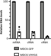
Fig 7. Quantification of viral RNAs in WSN-infected MDCK-VHH16 cells.
Specific primers (Table 1) were used for strand-specific real-time RT-PCR using tagged primers for quantification of the vRNA, cRNA, and mRNA of segment 5.
Fig 8
Fig 8. FluPol trafficking and assembly in the presence of VHHs.
(A) Complementation split-luciferase interaction assay between PA-Luc1 and PB2-Luc2 fusion proteins in the presence of PB1 and VHHs as indicated. The normalized luminescence ratio (NLR) is calculated as described in the Materials and Methods section. Twenty-four hours post-transfection, cells were lysed and luminescence was measured. Data are mean ± s.e.m. n = 4 technical replicates and are representative of several experiments. (B) Subcellular localization of the PB1-GFP fusion protein co-expressed with PA and VHHs. (C) Percentage of PB1-GFP-expressing cells with nuclear versus cytoplasmic localization of PB1-GFP. Fifty GFP-positive cells were scored for each condition.
Fig 9
Fig 9. VHH16-escape mutants selection and characterization.
(A) VHH16-escape mutants screening protocol carried out in MDCK-VHH16 cells. Plaque-forming units selected after 2 or 3 passages in MDCK-VHH16-GFP cells were amplified for PA and PB1 genomic segments sequencing. (B) Mutations selected on PA and PB1 subunits in VHH16-escape mutants (C) Lateral view of the PA structure organization (in blue) around the N-terminus of PB1 (in green). The lateral chains of the amino acids that were substituted in the mutants were shown. (D) Effect of VHH16 expression on FluPol mutants replication/transcription activities in a luciferase-reporter minireplicon assay. Plasmids expressing the subunits of the FluPol replicative complex mutants harboring the following mutations (PA-Q408R, PB1-D2N or PB1-D2A) were co-transfected in HEK 293T cells together with the WSN-NA-firefly-luciferase reporter plasmid and with a plasmid encoding VHH16, VHH7 or with an empty plasmid (indicated as No VHH). A plasmid encoding the nano-luciferase was co-transfected to control DNA uptake and normalize minireplicon activity. Luciferase activities were measured in cell lysates 48 hours post-transfection.
Fig 10
Fig 10. Amino acid sequences alignment of 25 PA and PB1 subunits representative of the influenza A virus diversity in different hosts.
Positions in which substitutions were identified are indicated by arrowheads. Strictly conserved residues are white on a red background, and partially conserved residues are red.
Fig 11
Fig 11. 3D-model of the VHH16-FluPol interaction.
(A) Pairwise alignment of VHH16 sequence with the VHH Nb8191 previously described as specific of FluPol [26]. Note the full sequence identity in their CDR2 that constitutes the VHH paratope (PDB number 7NIR). (B) Surface representations of FluPol (with the N-terminus of PB1 in yellow) in complex with the VHH Nb8191 (PDB number 7NIR), RNA-Polymerase II CTD (Pol2CTD) (PDB number 6FHH) and with ANP32A (PDB number 6XZQ). Note that the VHH binding generate a steric clash with ANP32A and RNA-polymerase II CTD.

References

    1. EFSA (European Food Safety Authority), ECDC (European Centre for Disease Prevention and Control), EURL (European Reference Laboratory for Avian Influenza), Adlhoch C, Fusaro A, Gonzales JL, Kuiken T, Marangon S, Niqueux É, Staubach C, Terregino C, Aznar I,Chuzhakina K, Muñoz Guajardo I and Baldinelli F, 2022. Scientific report: Avian influenza overview June–September2022.EFSA Journal 2022;20(10):7597,58pp. 10.2903/j.efsa.2022.7597 - DOI
    1. Shaw M, Palese P. 2013. Orthomyxoviridae, p 1151–1185. In Knipe DM, Howley P (ed), Fields virology, 6th ed, vol 1. Lippincott Williams & Wilkins, Philadelphia, PA.
    1. Pflug A, Lukarska M, Resa-Infante P, Reich S, Cusack S. 2017. Structural insights into RNA synthesis by the influenza virus transcription-replication machine. Virus Res. 234:103–117. doi: 10.1016/j.virusres.2017.01.013 - DOI - PubMed
    1. Boulo S, Akarsu H, Ruigrok RW, Baudin F. 2007. Nuclear traffic of influenza virus proteins and ribonucleoprotein complexes. Virus Res 124: 12–21. doi: 10.1016/j.virusres.2006.09.013 - DOI - PubMed
    1. Deng T, Sharps J, Fodor E, Brownlee GG. 2005. In vitro assembly of PB2 with a PB1-PA dimer supports a new model of assembly of influenza A virus polymerase subunits into a functional trimeric complex. J Virol 79:8669–8674. doi: 10.1128/JVI.79.13.8669-8674.2005 - DOI - PMC - PubMed