

Multiphase enhanced CT-based transformer for differential diagnosis and predicting surgical risk events of solid abdominal tumors in children
- PMID: 38875936
- PMCID: PMC11225889
- DOI: 10.1016/j.tranon.2024.102034
Multiphase enhanced CT-based transformer for differential diagnosis and predicting surgical risk events of solid abdominal tumors in children
Abstract
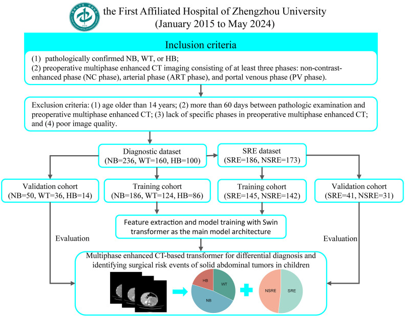
Background: For pediatric patients with solid abdominal tumors, early diagnosis can guide clinical treatment decisions, and comprehensive preoperative evaluation is essential to reduce surgical risk. The aim of this study was to explore the feasibility of multiphase enhanced CT-based transformer in the early diagnosis of tumors and prediction of surgical risk events (SRE).
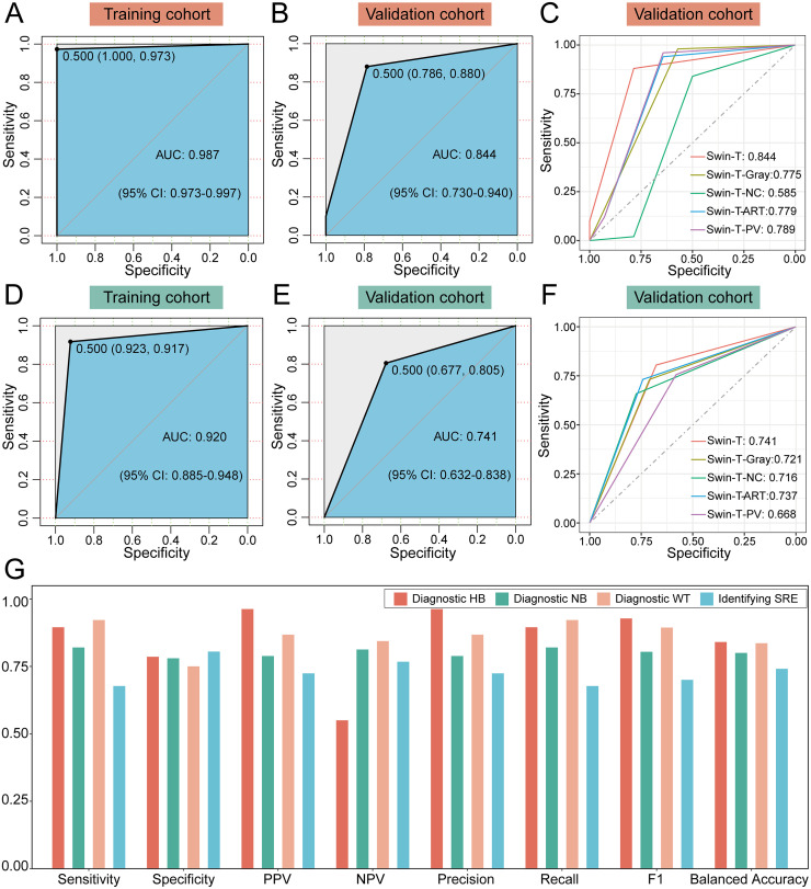
Methods: A total of 496 pediatric patients with solid abdominal tumors were enrolled in the study. With Swin transformer, we constructed and trained two Swin-T models based on preoperative multiphase enhanced CT for personalized prediction of tumor type and SRE status. Subsequently, we comprehensively evaluated the performance of each model and constructed four benchmark models for performance comparison.
Results: There was no significant difference in SRE status between tumor types. In the diagnostic task, areas under the receiver operating characteristic curves (AUC) of the Swin-T model were 0.987 (95 % CI, 0.973-0.997) and 0.844 (95 % CI, 0.730-0.940) in the training and validation cohorts, respectively. In predicting SRE, AUCs of the Swin-T model were 0.920 (95 % CI, 0.885-0.948) and 0.741 (95 % CI, 0.632-0.838) in the training and test cohorts, respectively. The Swin-T model achieved the best performance in both classification tasks compared to benchmark models.
Conclusion: The Swin-T model is a promising tool to assist pediatricians in the differential diagnosis of abdominal tumors and in comprehensive preoperative evaluation.
Keywords: Classifier; Diagnosis; Multiphase enhanced CT; Swin transformer.
Copyright © 2024. Published by Elsevier Inc.
Conflict of interest statement
Declaration of competing interest The authors declare no potential conflicts of interest to disclose.
Figures

Similar articles
-
Swin Transformer Improves the IDH Mutation Status Prediction of Gliomas Free of MRI-Based Tumor Segmentation.J Clin Med. 2022 Aug 8;11(15):4625. doi: 10.3390/jcm11154625. J Clin Med. 2022. PMID: 35956236 Free PMC article.
-
Predicting microsatellite instability and key biomarkers in colorectal cancer from H&E-stained images: achieving state-of-the-art predictive performance with fewer data using Swin Transformer.J Pathol Clin Res. 2023 May;9(3):223-235. doi: 10.1002/cjp2.312. Epub 2023 Feb 1. J Pathol Clin Res. 2023. PMID: 36723384 Free PMC article.
-
Deep learning-assisted LI-RADS grading and distinguishing hepatocellular carcinoma (HCC) from non-HCC based on multiphase CT: a two-center study.Eur Radiol. 2023 Dec;33(12):8879-8888. doi: 10.1007/s00330-023-09857-w. Epub 2023 Jul 1. Eur Radiol. 2023. PMID: 37392233
-
Classification of Mobile-Based Oral Cancer Images Using the Vision Transformer and the Swin Transformer.Cancers (Basel). 2024 Feb 29;16(5):987. doi: 10.3390/cancers16050987. Cancers (Basel). 2024. PMID: 38473348 Free PMC article.
-
Predicting lung cancer bone metastasis using CT and pathological imaging with a Swin Transformer model.J Bone Oncol. 2025 Apr 17;52:100681. doi: 10.1016/j.jbo.2025.100681. eCollection 2025 Jun. J Bone Oncol. 2025. PMID: 40342492 Free PMC article.
References
-
- Gatta G., Botta L., Rossi S., Aareleid T., Bielska-Lasota M., Clavel J., Dimitrova N., Jakab Z., Kaatsch P., Lacour B., Mallone S., Marcos-Gragera R., Minicozzi P., Sánchez-Pérez M.J., Sant M., Santaquilani M., Stiller C., Tavilla A., Trama A., Visser O., Peris-Bonet R. Childhood cancer survival in Europe 1999-2007: results of EUROCARE-5–a population-based study. Lancet Oncol. 2014;15(1):35–47. doi: 10.1016/s1470-2045(13)70548-5. - DOI - PubMed
-
- Lambin P., Leijenaar R.T.H., Deist T.M., Peerlings J., de Jong E.E.C., van Timmeren J., Sanduleanu S., Larue R., Even A.J.G., Jochems A., van Wijk Y., Woodruff H., van Soest J., Lustberg T., Roelofs E., van Elmpt W., Dekker A., Mottaghy F.M., Wildberger J.E., Walsh S. Radiomics: the bridge between medical imaging and personalized medicine. Nat. Rev. Clin. Oncol. 2017;14(12):749–762. doi: 10.1038/nrclinonc.2017.141. - DOI - PubMed
LinkOut - more resources
Full Text Sources
