. 2024 Jul 9;19(7):946-956.
doi: 10.1016/j.stemcr.2024.05.006. Epub 2024 Jun 13.

Self-assembling 3D vessel-on-chip model with hiPSC-derived astrocytes

Affiliations

Self-assembling 3D vessel-on-chip model with hiPSC-derived astrocytes

Dennis M Nahon et al. Stem Cell Reports. .

Abstract

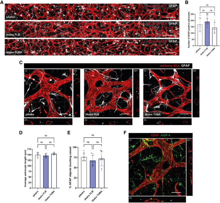
Functionality of the blood-brain barrier (BBB) relies on the interaction between endothelial cells (ECs), pericytes, and astrocytes to regulate molecule transport within the central nervous system. Most experimental models for the BBB rely on freshly isolated primary brain cells. Here, we explored human induced pluripotent stem cells (hiPSCs) as a cellular source for astrocytes in a 3D vessel-on-chip (VoC) model. Self-organized microvascular networks were formed by combining hiPSC-derived ECs, human brain vascular pericytes, and hiPSC-derived astrocytes within a fibrin hydrogel. The hiPSC-ECs and pericytes showed close interactions, but, somewhat unexpectedly, addition of astrocytes disrupted microvascular network formation. However, continuous fluid perfusion or activation of cyclic AMP (cAMP) signaling rescued the vascular organization and decreased vascular permeability. Nevertheless, astrocytes did not affect the expression of proteins related to junction formation, transport, or extracellular matrix, indicating that, despite other claims, hiPSC-derived ECs do not entirely acquire a BBB-like identity in the 3D VoC model.

Keywords: BBB; blood-brain barrier; hiPSC-Astro; hiPSC-ECs; hiPSC-derived astrocytes; hiPSC-derived endothelial cells; human induced pluripotent stem cells; microfluidics; organ-on-chip; vessel-on-chip.


Conflict of interest statement

Declaration of interests The authors declare no competing interests.

Figures

None
Graphical abstract
Figure 1
Figure 1
iAstros incorporated into 3D VoC model (A) Schematic of VoC protocol using hiPSC-ECs with HBVPs and pAstros or iAstros. (B) Representative immunofluorescence images of a 3D VoC triple culture containing hiPSC-ECs, HBVPs, and iAstros at day 1, 2, 4, and 6 showing hiPSC-mCherry ECs (red) and iAstros differentiated from the TUBA hiPSC line (green, TUBA-GFP). Scale bars: 250 μm. (C) Representative immunofluorescence images of microvascular networks in microfluidic chips on day 7 showing hiPSC-mCherry ECs (red). Images showing microvascular networks from a VoC double culture (hiPSC-ECs and HBVPs) or 3D VoC triple cultures including either pAstros or iAstros from two independent hiPSC lines (FLB or TUBA). Scale bars: 250 μm. (D–F) Quantification of full channel images of microvascular networks showing vascular density at endpoint day 7 showing (D), average vessel diameter (E), and average vessel length (F). Data are shown as mean ± SD. All conditions are N = 3, n = 12–18; three independent experiments with minimum of 3 microfluidic channels per experiment. One-way ANOVA with Sidak’s multiple comparison test. p < 0.05, ∗∗p < 0.01, ∗∗∗p < 0.001, ∗∗∗∗p < 0.0001; ns, non-significant. See also Figures S1 and S2.
Figure 2
Figure 2
Comparable structural properties of primary and hiPSC-Astros in a 3D VoC model (A) Representative immunofluorescence images of microvascular networks in microfluidic chips on day 7 showing hiPSC-mCherry ECs (red) and astrocytes (silver; GFAP). Images showing VoC triple cultures of hiPSC-ECs with HBVPs and pAstros or iAstros from FLB or TUBA hiPSC line. Scale bars: 500 μm. (B) Quantification of astrocytes in VoC triple cultures showing number of GFAP-positive astrocytes in ±80% of full microfluidic channel. ns, non-significant. (C) Representative immunofluorescence confocal images of microvascular networks in microfluidic chips showing hiPSC-mCherry ECs (red) and astrocytes (silver; GFAP). Images displaying xyz (i), xy (ii), and yz cross-sectional perspectives (iii). Images showing VoC triple cultures of hiPSC-ECs with HBVPs and pAstros or iAstros from the FLB or TUBA hiPSC lines. Scale bars: 100 μm. (D and E) Quantification of astrocytes in VoC model showing average astrocyte length (D) and % of GFAP objects touching the microvascular network (E). Data are shown as mean ± SD. For (B) and (D) N = 3, n = 6–8; three independent experiments with a minimum of two microfluidic channels per experiment. For (E) N = 3, n = 3; three independent experiments, one microfluidic channel per experiment with three regions of interest (ROIs) per channel. One-way ANOVA with Tukey’s multiple comparison. ns, non-significant. (F) Representative immunofluorescence confocal image of microvascular network in microfluidic chips showing ECs (red; CD31) and astrocytes (green; Aqp4) in a VoC triple culture of hiPSC-ECs with HBVPs and iAstros from the FLB hiPSC line. Images displaying xyz (i), xy (ii), and yz cross-sectional perspectives (iii). Scale bar: 100 μm. See also Video S1.
Figure 3
Figure 3
Comparable structural properties of HBVPs in a 3D VoC model including astrocytes (A) Representative immunofluorescence confocal image of microvascular network in microfluidic chips showing ECs (red; CD31) and HBVPs (silver; NG2, green; SM22) in a VoC double culture of hiPSC-ECs with HBVPs. Images displaying xyz (i), xy (ii), and yz cross-sectional perspectives (iii). Scale bar: 100 μm. (B) Representative immunofluorescence confocal images of microvascular networks in microfluidic chips on day 7 showing hiPSC-mCherry ECs (red), HBVPs (green; SM22), and pAstros (silver, FABP7) or iAstros from the TUBA hiPSC line (silver, TUBA-GFP), surface-rendered images and color-coded images of HBVPs distance to the vessel surface. Images showing VoC double cultures (hiPSC-ECs with HBVPs) and VoC triple cultures including either pAstros or iAstros from the TUBA hiPSC line. Scale bars: 100 μm. (C–F) Quantification of HBVPs in VoC double and triple cultures containing iAstros from the TUBA hiPSC line showing number of SM22-positive objects (C), average SM22 object volume (D), normalized average SM22 intensity per object (E), and percentage of SM22 objects touching the microvascular network (F). Data are shown as mean ± SD. For (C–F) N = 3, n = 9–10; three independent experiments, one microfluidic channel per experiment with three or four ROIs per channel. Scale bars: 100 μm. One-way ANOVA with Sidak’s multiple comparison test. p < 0.05, ∗∗∗∗p < 0.0001; ns, non-significant. See also Video S1.
Figure 4
Figure 4
Improved microvascular network of 3D VoC triple culture including hiPSC-Astros upon activation of cAMP signaling or application of continuous perfusion (A) Schematic of experimental setup for improving microvascular network formation of VoC triple cultures containing hiPSC-ECs, HBVPs, and iAstros from the FLB iPSC line. In the iAstro cAMP condition, the medium was daily supplemented with 250 μM dbcAMP to activate cAMP signaling. In the iAstro continuous flow (cFlow) condition, the VoC triple culture was subjected to continuous flow from day 3 onwards. (B) Quantification of vessel density over time from daily images of hiPSC-mCherry ECs for the four VoC culture conditions. (C) Representative immunofluorescence images of microvascular networks from the four VoC culture conditions at day 7 showing hiPSC-mCherry ECs (red). iAstro conditions are triple cultures containing iAstros from the FLB hiPSC line. Scale bars: 200 μm. (D–F) Quantification of microvascular networks showing vascular density at endpoint day 7 (D), average vessel diameter (E), and average vessel length (F). Data are shown as mean ± SD. Data shown are N = 3–4, n = 14–22; three or four independent experiments with a minimum of 3 microfluidic channels per experiment. iAstro conditions are triple cultures containing iAstros from the FLB hiPSC line. (G) Representative immunofluorescence images of microvascular networks (red; hiPSC-mCherry ECs) perfused with 70 kDa FITC-dextran (green) on day 7, 30 s after start of perfusion. Images show VoC double cultures (hiPSC-ECs with HBVPs) and VoC triple cultures also containing iAstros from the FLB hiPSC line. Scale bars: 200 μm. (H) Quantification of permeability coefficient for the four VoC culture conditions at endpoint day 7 from N = 3–4, n = 4–11; three or four independent experiments with one to six microfluidic channels per experiment. In the iAstro conditions, data are pulled from triple cultures containing both iAstros from the FLB and the TUBA hiPSC lines. One-way ANOVA with Sidak’s multiple comparison test. p < 0.05, ∗∗p < 0.01 ∗∗∗p < 0.001, ∗∗∗∗p < 0.0001; ns, non-significant. See also Figures S3 and S4.

References

    1. Boyer-Di Ponio J., El-Ayoubi F., Glacial F., Ganeshamoorthy K., Driancourt C., Godet M., Perrière N., Guillevic O., Olivier Couraud P., Uzan G. Instruction of circulating endothelial progenitors in vitro towards specialized blood-brain barrier and arterial phenotypes. PLoS One. 2014;9 doi: 10.1371/journal.pone.0084179. - DOI - PMC - PubMed
    1. Campinho P., Vilfan A., Vermot J. Blood Flow Forces in Shaping the Vascular System: A Focus on Endothelial Cell Behavior. Front. Physiol. 2020;11:552. doi: 10.3389/fphys.2020.00552. - DOI - PMC - PubMed
    1. Campisi M., Shin Y., Osaki T., Hajal C., Chiono V., Kamm R.D. 3D self-organized microvascular model of the human blood-brain barrier with endothelial cells, pericytes and astrocytes. Biomaterials. 2018;180:117–129. doi: 10.1016/j.biomaterials.2018.07.014. - DOI - PMC - PubMed
    1. Cecchelli R., Aday S., Sevin E., Almeida C., Culot M., Dehouck L., Coisne C., Engelhardt B., Dehouck M.P., Ferreira L. A stable and reproducible human blood-brain barrier model derived from hematopoietic stem cells. PLoS One. 2014;9:e99733. doi: 10.1371/journal.pone.0099733. - DOI - PMC - PubMed
    1. Gastfriend B.D., Nishihara H., Canfield S.G., Foreman K.L., Engelhardt B., Palecek S.P., Shusta E.V. Wnt signaling mediates acquisition of blood–brain barrier properties in naïve endothelium derived from human pluripotent stem cells. Elife. 2021;10:1–33. doi: 10.7554/eLife.70992. - DOI - PMC - PubMed

Publication types

LinkOut - more resources