Review
. 2025 May:71:227-262.
doi: 10.1016/j.jare.2024.06.010. Epub 2024 Jun 13.

Pyroptosis: A spoiler of peaceful coexistence between cells in degenerative bone and joint diseases

Affiliations
Review

Pyroptosis: A spoiler of peaceful coexistence between cells in degenerative bone and joint diseases

Zhichao Li et al. J Adv Res. 2025 May.

Abstract

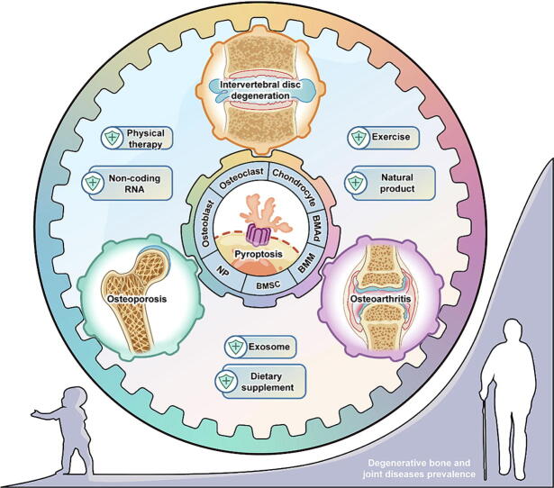
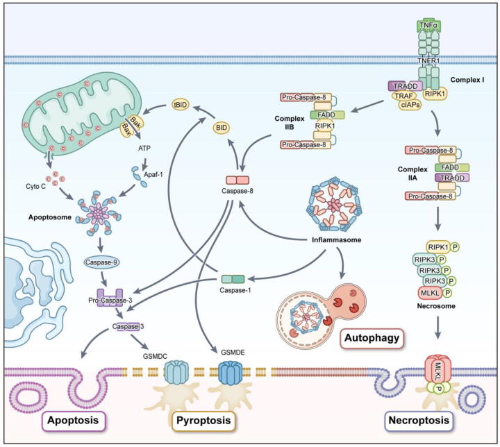
Background: As people age, degenerative bone and joint diseases (DBJDs) become more prevalent. When middle-aged and elderly people are diagnosed with one or more disorders such as osteoporosis (OP), osteoarthritis (OA), and intervertebral disc degeneration (IVDD), it often signals the onset of prolonged pain and reduced functionality. Chronic inflammation has been identified as the underlying cause of various degenerative diseases, including DBJDs. Recently, excessive activation of pyroptosis, a form of programed cell death (PCD) mediated by inflammasomes, has emerged as a primary driver of harmful chronic inflammation. Consequently, pyroptosis has become a potential target for preventing and treating DBJDs.

Aim of review: This review explored the physiological and pathological roles of the pyroptosis pathway in bone and joint development and its relation to DBJDs. Meanwhile, it elaborated the molecular mechanisms of pyroptosis within individual cell types in the bone marrow and joints, as well as the interplay among different cell types in the context of DBJDs. Furthermore, this review presented the latest compelling evidence supporting the idea of regulating the pyroptosis pathway for DBJDs treatment, and discussed the potential, limitations, and challenges of various therapeutic strategies involving pyroptosis regulation.

Key scientific concepts of review: In summary, an interesting identity for the unregulated pyroptosis pathway in the context of DBJDs was proposed in this review, which was undertaken as a spoiler of peaceful coexistence between cells in a degenerative environment. Over the extended course of DBJDs, pyroptosis pathway perpetuated its activity through crosstalk among pyroptosis cascades in different cell types, thus exacerbating the inflammatory environment throughout the entire bone marrow and joint degeneration environment. Correspondingly, pyroptosis regulation therapy emerged as a promising option for clinical treatment of DBJDs.

Keywords: Bone and joint diseases; Intervertebral disc degeneration; Osteoarthritis; Osteoporosis; Pyroptosis.


Conflict of interest statement

Declaration of competing interest The authors declare that they have no known competing financial interests or personal relationships that could have appeared to influence the work reported in this paper.

Figures

None
Graphical abstract
Fig. 1
Fig. 1
A milestone in the research of pyroptosis pathway.
Fig. 2
Fig. 2
Three pathways of pyroptosis: classical pathway, non-classical pathway, and other pathways. In the classical pathway, the inflammasome sensor activated by a variety of signals can aggregate ASC and recruit pro-caspase-1. The activated caspase-1 cleaves GSDMD and pro-IL-1β/18, and the resulting GSDMD-NT forms a plasma membrane pore and activates IL-1β/18. In the non-classical pathway, caspase-4/5/11 can be directly activated by intracellular LPS and cleave GSDMD to mediate pore formation and pyroptosis. In addition, there are other pyroptosis pathways mediated by caspase-3/8, granzyme A/B, and SpeB.
Fig. 3
Fig. 3
Excessive activation of the pyroptosis pathway impairs bone formation activities involving BMSCs, OBs, and osteocytes. Pyroptosis induced by various stress environments (nutrient deprivation, LPS plus ATP stimulation and ATRA accumulation) impairs the activity of BMSCs and induces imbalanced osteogenesis/adipogenesis, which will lead to a decrease in the number of OBs. Meanwhile, bacterial infection, high glucose, and oxidative stress environment induce pyroptosis of OBs and damage matrix mineralization. Long-lived osteocytes undergo pyroptosis due to long-term effects of environmental pollutants or implant wear particles, increasing bone brittleness. In addition, pyroptosis of OBs and osteocytes upregulate RANKL expression and downregulate OPG, enhancing bone resorption activity.
Fig. 4
Fig. 4
The pyroptosis pathway drives varying cells in the bone marrow to participate in bone resorption. Under the influence of increased RANKL activity induced by OB pyroptosis, osteocyte pyroptosis, and other factors (such as estrogen deficiency), the NLRP3 inflammasome is activated in the OC precursors to mediate PARylation of ARTD1, thus losing its inhibitory effect on classical NF-κB. The increase in NF-κB-dependent IL-1β promotes NFATc1 autocrine to promote osteoclastogenesis in a cell-autonomous manner. The increase of active IL-1β/18 mediated by pyroptosis pathway accelerates bone resorption by regulating the migration of OCs and promoting the release of bone matrix degrading enzymes. In addition to the direct effect on the bone resorption function of OCs, IL-1β/18 can also induce dendritic cells differentiate into OCs, enhance the function of IFN-γ in promoting osteoclastogenesis, drive the naive T cells differentiate into CD4+ T cells, and elevate the expression of RANKL in T cells, OBs, and osteocytes, thus indirectly participating in bone resorption.
Fig. 5
Fig. 5
The pathological mechanisms of different types of OP and the involvement of pyroptosis pathway. A. In the early PMOP period, due to the rapid loss of estrogen, the absorption of cortical bone is significantly stimulated, leading to the rapid thinning of cortical bone and enhancing the loss of trabecular bone. The rapid loss of estrogen induces pyroptosis to limit the increase in the number of OBs, and the up-regulated RANKL activity promotes osteoclastogenesis by activating the NLRP3 inflammasome in OC precursors. The increased number of OCs leads to a simultaneous increase of degraded matrix fragments, which are perceived by the NLRP3 inflammasome in OC precursors, forming a positive feedback loop of osteoclastogenesis-bone resorption activity. B. The second phase of PMOP is similar to SOP. In this phase, both cortical and trabecular bones are slowly lost while BMAT gradually deposits. BMAds and macrophages form a positive feedback loop of the pyroptosis pathway to promote a chronic inflammatory environment, which enhances osteoclastogenesis and bone resorption. Osteocytes undergo pyroptosis under the influence of chronic factors such as aging, ROS accumulation, environmental pollutants, and implant particles, resulting in the destruction of the lacunar-canalicular network. C. Secondary OP is mediated by a variety of factors, with DOP being a very common example. High glucose environment and AGEs directly damage the bone formation by inducing pyroptosis of BMSCs and OBs while increasing macrophage M1 polarization and osteoclastogenesis to promote inflammatory environment and accelerate bone resorption, resulting in bone loss.
Fig. 6
Fig. 6
The development model of OA centered on the synovial cell pyroptosis. A. Aging, mechanical damage, and enhanced catabolic activity cause microcrystals deposition of articular cartilage, which activates synovial macrophage pyroptosis to release IL-1β/18. This process leads to chondrocyte hypertrophy and differentiation, accelerated aging, and upregulated proteolytic enzymes and chemokines, thus forming a positive feedback loop. B. Obesity leads to the accumulation of extra-articular adipocytes. Meanwhile, lipid metabolites oxidize low-density lipoprotein, cholesterol, and ceramide, which enter the synovium through blood circulation, contributing to macrophage pyroptosis. C. Macrophages can induce the activation of FLSs. Activated FLSs enhance macrophage infiltration by transmitting mechanical signals and affect macrophage phenotype and function. Pyroptosis macrophages can elevate the expressions of fibrosis markers and matrix degradation enzymes of FLSs. D. Both activated macrophages and FLSs express more VEGF to promote angiogenesis. Adhesion molecules in neovascularization promote immunocyte infiltration, forming a positive feedback loop. IL-1β/18 induces CD4+ T cells differentiate to promote inflammatory environment and enhance osteoclastogenesis to mediate subchondral bone remodeling.
Fig. 7
Fig. 7
NP immune exposure mediates a strong pyroptosis pathway and promotes the development of IVDD. As individual age, the cell dialogue between NCs and NPCs gradually diminishes, and the matrix anabolism/catabolism is imbalanced under the influence of genetics and systemic metabolic disorders. The NPC pyroptosis induced by endogenous metabolites and matrix degradation products may be one of the initial factors involving in catabolic activities. Weakening NP hydration leads to biomechanical changes in IVD. Mechanical load-induced pyroptosis of AFCs and CEPCs also accelerates the formation of microcracks. The breakdown of the immune barrier formed by the AF and CEP leads to the loss of NP immune privilege. The exposed NP quickly attracts significant infiltration from immunocytes. The pyroptosis pathway induced by macrophages is strongly activated as an executioner during this process, promoting the self-absorption of NP. Additionally, macrophages will infiltrate into deep healthy NP, continuously induce pyroptosis pathway to enhance the production of cytokines in IVD, and accelerate the transformation of catabolism. The changes in permeability of calcified CEP lead to degradation and metabolite accumulation, forming a positive feedback loop of DAMPs-NLRP3. IL-1β/18 induces IVD cells and immunocytes to express more VEGF and neurogenic factors, thereby promoting angiogenesis and innervation.
Fig. 8
Fig. 8
Pyroptosis, apoptosis, and necroptosis act as alternative pathways to ensure cell death, with autophagy involving in the regulation of pyroptosis pathway. Intrinsic apoptosis pathway is regulated by the Bcl-2 protein family and modulates the release of Cyto-C by controlling the permeability of mitochondrial membrane through interaction. Cyto-C binds to Apaf-1 to induce the assembly of apoptosomes and recruit pro-caspase-9. The activated caspase-9, in turn, activates caspase-3/6/7 for apoptosis. In the extrinsic apoptosis pathway, the binding of TNF ligands to TNF receptors recruits downstream components, creating a complex I. Several components of complex I are arranged to form complex II under the influence of various factors, and the type of complex II (A or B) and the state of caspase-8 (activation or inhibition) determine whether the cells will undergo apoptosis or necroptosis. Caspase-8 triggers intrinsic apoptosis pathway by truncation of Bid. When activity of caspase-8 is inhibited, complex IIB recruits RIPK3 to form necrosomes and phosphorylates MLKL to translocate and permeabilize the plasma membrane, thus inducing necroptosis. In addition, caspase-3/8 can promote pore formation and pyroptosis by cleaving GSDME/C. MLKL can also participate in the pyroptosis pathway by activating the NLRP3 inflammasome. In addition, NLRP3 can interact with IRGM to recruit p62, Beclin-1, and ATG16L1 to complete selective autophagy degradation to prevent excessive inflammation.

Similar articles

Cited by

References

    1. Hadjidakis D.J., Androulakis I.I. Bone remodeling. Ann N Y Acad Sci. 2006;1092:385–396. - PubMed
    1. Sims N.A., Gooi J.H. Bone remodeling: Multiple cellular interactions required for coupling of bone formation and resorption. Semin Cell Dev Biol. 2008;19(5):444–451. - PubMed
    1. Song S., Guo Y., Yang Y., Fu D. Advances in pathogenesis and therapeutic strategies for osteoporosis. Pharmacol Ther. 2022;237 - PubMed
    1. Xiong Y., Mi B.B., Lin Z., Hu Y.Q., Yu L., Zha K.K., et al. The role of the immune microenvironment in bone, cartilage, and soft tissue regeneration: from mechanism to therapeutic opportunity. Mil Med Res. 2022;9(1):65. - PMC - PubMed
    1. Sanchez-Lopez E., Coras R., Torres A., Lane N.E., Guma M. Synovial inflammation in osteoarthritis progression. Nat Rev Rheumatol. 2022;18(5):258–275. - PMC - PubMed