Review
. 2025 Apr:70:423-444.
doi: 10.1016/j.jare.2024.06.009. Epub 2024 Jun 13.

Targeting colorectal cancer at the level of nuclear pore complex

Affiliations
Review

Targeting colorectal cancer at the level of nuclear pore complex

Muhammad Mahtab Aslam Khan Khakwani et al. J Adv Res. 2025 Apr.

Abstract

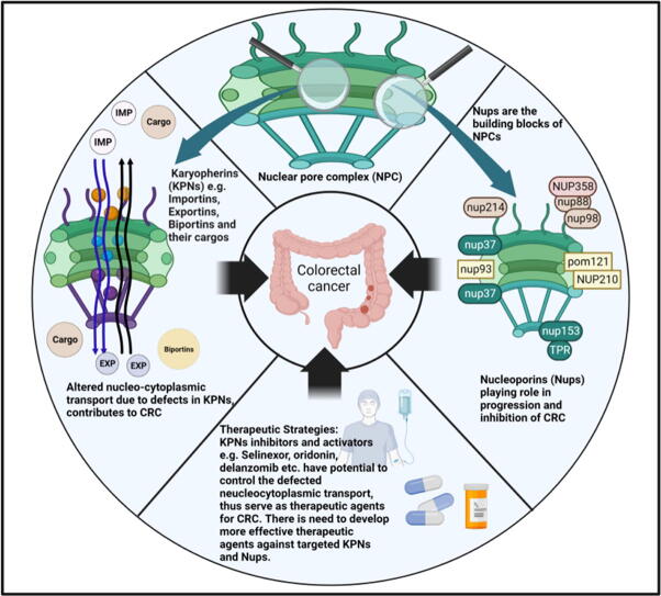
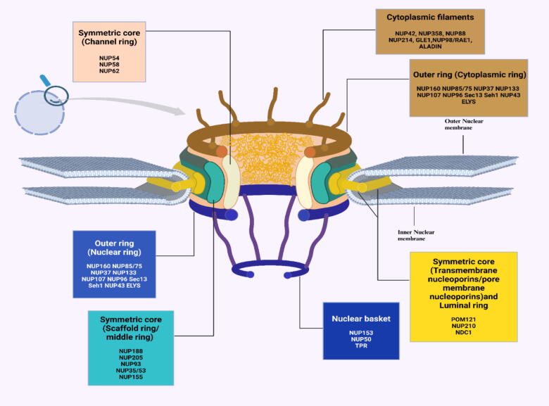
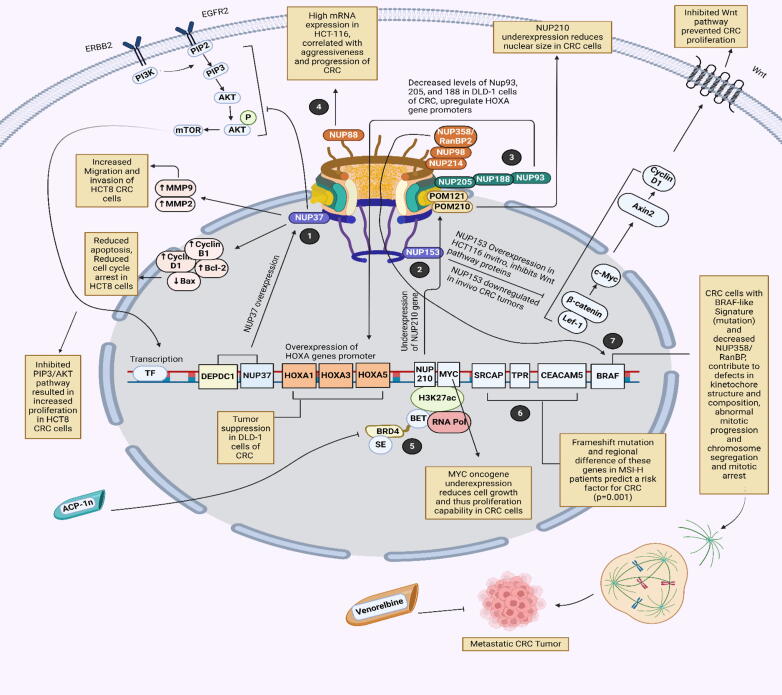
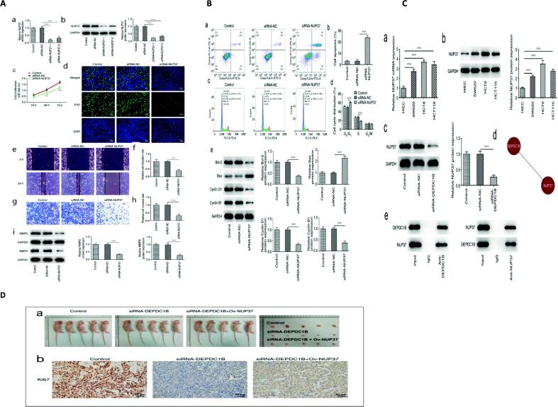
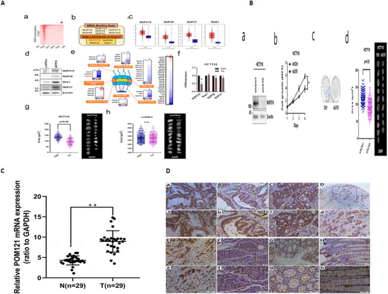
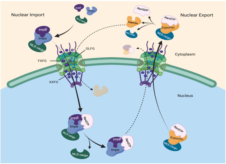
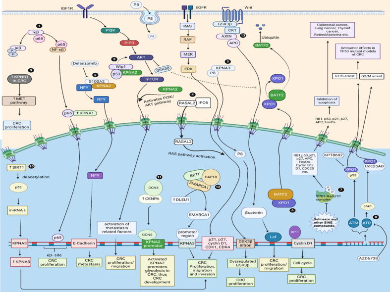
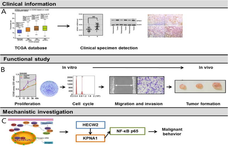
Background: Nuclear pore complexes (NPCs) are the architectures entrenched in nuclear envelop of a cell that regulate the nucleo-cytoplasmic transportation of materials, such as proteins and RNAs for proper functioning of a cell. The appropriate localization of proteins and RNAs within the cell is essential for its normal functionality. For such a complex transportation of materials across the NPC, around 60 proteins are involved comprising nucleoporins, karyopherins and RAN system proteins that play a vital role in NPC's structure formation, cargo translocation across NPC, and cargoes' rapid directed transportation respectively. In various cancers, the structure and function of NPC is often exaggerated, following altered expressions of its nucleoporins and karyopherins, affecting other proteins of associated signaling pathways. Some inhibitors of karyopherins at present, have potential to regulate the altered level/expression of these karyopherin molecules.

Aim of review: This review summarizes the data from 1990 to 2023, mainly focusing on recent studies that illustrate the structure and function of NPC, the relationship and mechanisms of nucleoporins and karyopherins with colorectal cancer, as well as therapeutic values, in order to understand the pathology and underlying basis of colorectal cancer associated with NPC. This is the first review to our knowledge elucidating the detailed updated studies targeting colorectal cancer at NPC. The review also aims to target certain karyopherins, Nups and their possible inhibitors and activators molecules as a therapeutic strategy.

Key scientific concepts of review: NPC structure provides understanding, how nucleoporins and karyopherins as key molecules are responsible for appropriate nucleocytoplasmic transportation. Many studies provide evidences, describing the role of disrupted nucleoporins and karyopherins not only in CRC but also in other non-hematological and hematological malignancies. At present, some inhibitors of karyopherins have therapeutic potential for CRC, however development of more potent inhibitors may provide more effective therapeutic strategies for CRC in near future.

Keywords: Colorectal cancer (CRC); Karyopherins; Nuclear pore complex (NPC); Nuclear transporters; Nucleocytoplasmic transport; Nucleoporins.


Conflict of interest statement

Declaration of competing interest The authors declare that they have no known competing financial interests or personal relationships that could have appeared to influence the work reported in this paper.

Figures

None
Graphical abstract
Fig. 1
Fig. 1
Structural representation of a nuclear pore complex alongwith organization of its building blocks (nucleoporins).
Fig. 2
Fig. 2
Molecular Mechanisms of development and suppression of colorectal cancer by different nucleoporins. (1) Nup37 and DEPDC1B expression is higher in HCT8 CRC cells, which increases the levels of MMP9 and MMP2 proteins, thereby promoting the migration and invasion capability in HCT8 CRC cells. Higher expression of Nup37 elevates the levels of cyclin B1, cyclin D1and Bcl-2, while reducing the level of Bax, and results in reduced apoptosis and reduced cell cycle arrest in HCT8 CRC cells. Moreover, increased Nup37 expression inhibits PIP3/AKT pathway, thus increasing proliferation of CRC cells. (2) Nup 153 expression was downregulated in CRC tumors invivo. However, Nup153 inhibited Wnt pathway by reducing the levels of Wnt signaling proteins such as Axin-2, β catenin, Lef-1, and then inhibited Wnt signaling pathway, which prevented CRC proliferation. (3) Decreased levels of Nup93 alongwith its interacting partners Nup205 and Nup188, result in HOXA promoter overexpression, thus play a tumor suppressive role in DLD-1 cells of CRC. (4) High mRNA levels of Nup88 were observed in HCT-116 CRC cells, which were significantly correlated with tumor invasion and TNM stages. (5) BRD4-driven Nup210 increases nuclear size and promotes overexpression of oncogene MYC, hence increasing cell growth and cell proliferation capability in CRC cells. However, Aminocyclopropenon-1 (ACP1-n)-an inhibitor, blocked BRD4 functions and prevented oncogenic activity in HCT-116 CRC cells. (6) Frameshift mutations and regional variations in SRCAP, TPR, and CEACAM5 genes were found only in CRC patients with MSI-H status, and predict a risk factor for CRC development (p = 0.001). (7) BRAF-like signature in association with Nup358/RanBP, contribute to the defects in kinetochore structure and composition, abnormal mitotic progression, abnormal chromosome segregation, and mitotic arrest in CRC cells which result in metastatic tumor. However, a drug known as venorelbine, when injected in mice, reduced the growth of CRC tumor.
Fig. 3
Fig. 3
Experimental visualization of NUP37 association with progression of CRC (A) NUP37 knockdown inhibits the migration, proliferation and invasion and of CRC cells. (a) The mRNA and (b) protein levels of NUP37 were detected by RT qPCR and western blotting, after transfection with siRNA-NUP37-1/2. Cell proliferation was assessed by (c) CCK-8 and (d) EdU assays (e and f) Cell migration was assessed by wound healing assay (g and h) Cell invasion was evaluated by Transwell assay (i) Levels of MMP2 and MMP9 were measured by western blotting. (B) NUP37 silencing accelerates the apoptosis and cycle arrest of CRC cells. (a and b) Cell apoptosis was detected by flow cytometry. (c and d) Cell cycle was measured by flow cytometry analysis. (e) Western blotting was used to assess apoptosis- and cell cycle-related proteins. (C) DEPDC1B binds to NUP37 directly. The (a) mRNA and (b) protein levels of NUP37 were detected in CRC cell lines by reverse transcription-quantitative PCR and western blotting. (c) Protein level of NUP37 in cells after the transfection with siRNA-DEPDC1B was detected by western blotting. (d) DEPDC1B coexpression with NUP37, was analyzed by Coexpedia database (e) Co-immunoprecipitation assay verified the combination of DEPDC1B and NUP37. (D) NUP37 inhibits CRC growth invivo by after silencing DEPDC1B, confirming the association between NUP37 and DEPDC1B. Images are reproduced from reference Copyright: © Xiong et al.
Fig. 4
Fig. 4
Experimental visualization of NUP210, NUP58 and POM121 association with CRC (A) Nuclear size reduction of colorectal cancer cells by Aminocyclopropenone (a) Heatmap showing the highest enrichment of BRD4 ChIP-seq over the gene bodies in HCT116. (b) Venn diagram showing the overlap between NUPs that are highly expressed in colorectal cancer and those that bind BRD4. (c) The expression of NUP210, NUP37, NUP58, and RAE1 in colorectal cancer via GEPIA tool. (d) Western blotting analysis of NUP210, NUP37, NUP58, and Rae1 levels in HCT116 and ccd18co cells. (e) Heatmap demonstrating NUP transcript levels in HCT116 cells after treatment with aminocyclopropenone 1n (24 h, 10 µM). (f) qRT-PCR analysis of candidate nucleoporin gene mRNA in HCT116 cells after treatment with aminocyclopropenone 1n (24 h, 10 μM). (g) DAPI-stained nuclei montages and measurement of size of the nucleus after treatment with 1n (24 h, 10 µM) in HCT116 cells. (h) DAPI-stained nuclei montages and measurement of the size of nucleus after treatment with aminocyclopropenone 1n (24 h, 10 µM) in ccd18co cells. (B) Expression level of NUP210 adjusts the proliferative capacity and nucleus size of colorectal cancer cells. (a) NUP210 levels after western blotting analysis for HCT116 cells, expressing shRNA NUP210. (b) NUP210-silenced HCT116 proliferation, examined by MTT assay. (c) Long-term proliferation assay of HCT-116 cells, expressing shRNA NUP210. (d) DAPI-stained montages of nuclei and measurement of their size in HCT116 cells expressing shRNA NUP210. Images are reproduced from reference . Copyright © 2022 Hiroya Kondo et al. (C) POM121 mRNA expression in CRC tissues as compared to those of normal colorectal tissues. qRT-PCR demonstrated that the levels of POM121 mRNA in the 29 CRC samples. Expression of Tumor was higher than those in normal colorectal samples (N) (**p < 0.001). (D) IHC staining for POM121 expression in colorectal samples. (a), (b) IHC positive staining of POM121 in well-differentiated adenocarcinoma. (c), (d) POM121 in moderately differentiated adenocarcinoma after positive IHC staining (e), (f) POM121 positive IHC staining in poorly-differentiated adenocarcinoma.(g), (h) POM121 negative IHC staining in normal colorectal tissue. Images are reproduced from reference . © 2019 Informa UK Limited, trading as Taylor & Francis Group.
Fig. 6
Fig. 6
Nuclear import and export of materials: During nuclear import, importin-NLS cargo complex forms in the cytosol and is carried to the nucleus by channel FG Nups of NPC. The XXFG (small sized ball), GLFG (mid-sized ball), and FXFG (large sized ball) repeats in the NPC diffusion barrier are present in concentration gradients rather than being evenly distributed. When NLS cargo binds to importin in the cytoplasm, importin repeated HEAT sequences are rearranged to create big pockets that have a strong affinity for FXFG repeats which moves the importin-cargo complex towards nucleus by the help of FG gradient. Binding of RanGTP with importin-cargo complex inside the nucleus, makes the surface pockets for importin XXFG tiny and specific, thus propelling the export complex. During nuclear export, exportin attaches to NES of its cargo, the complex is then bound by RanGTP and exported from the nucleus to cytosol. This differential in FG concentration can be advantageous to exportin as well. In cytosol, RanGTP is converted into RanGDP and the complex dissociates.
Fig. 7
Fig. 7
Molecular Mechanisms involved in the progression of CRC by different Karyopherins. (1) KPNA1 expression was higher in CRC cells resulting in activated NF-κβ p65 pathway. Higher KPNA1 expression translocates P65 into the nucleus which binds on κβ-site of its gene and promotes CRC proliferation. However, depleted KPNA1 levels inhibited proliferation, migration and invasion in CRC cells. (2) KPNA2 alongwith S100A2 protein, translocates nuclear transcription factor (NFYA) to the nucleus, where NFY inhibits the transcriptional activity of E- cadherin, resulting CRC metastasis. However, a drug Delanzomib inhibits S100A2/KPNA2 complex, was found to be a therapeutic agent playing positive role in inhibition of CRC metastasis. (3) Higher expression of Wip1 and KPNA2 was observed in CRC. Wild-type p53-induced phosphatase 1 (Wip1) and KPNA2 interact with each other, potentially in a p53-dependent mechanism and activate the downstream AKT/GSK-3β pathway alongwith activation of CRC metastasis related factors, thus control CRC cell migration and proliferation. (4) IPO5 recognizes its cargo RASAL2 bearing nuclear import signal, and translocates it into the nucleus. RASAL2 inside the nucleus activates RAS signaling pathway, resulting transcription of p21, p27, cyclin D1, CDK4, CDK1 genes, thus contributing to the CRC proliferation, migration and invasion. (5) Mechanistically, first of all the endocytosed P8 in the cytoplasm was found to be transported into the nucleus by importin and KPNA3, where it binds to GSK3β introns, inhibiting its transcription. Dysregulated GSK3β inhibits Wnt signaling pathway controlling CRC proliferation. GSK3β is a crucial protein kinase in the Wnt signaling pathway and causes strong phosphorylation of β-catenin. As a result, even in CRC cells with active Wnt signaling, P8 can cause cell cycle arrest. (6) Basic Leucine Zipper ATF-Like Transcription Factor 2 (BATF2) is exported out of the nucleus and broken down by ubiquitin in the cytosol by directly attaching to XPO1 through its NES region, eventually aiding the proliferation of CRC, a process that involves the AP-1/cyclin D1/pRb signaling cascade. (7) Increased levels of export of many tumor-suppressor proteins, such as RB1, p53, APC, and others, are brought about by higher expression of XPO1 which promote the oncogenesis of colorectal cancer (CRC) and other malignancies by preventing apoptosis. However, Selinexor and other inhibitors of XPO1 serve as potential therapeutics in controlling oncogenesis. (8) TP53-mutant CRC cells showed increased sensitivity to progressive XPO1 inhibitor (KPT8602) therapy followed by ATR inhibition (AZD6738) both in vitro and in vivo, indicating a logical therapeutic strategy. The administration of KPT-8602, an XPO1 inhibitor, and AZD-6738, an ATR inhibitor, had striking antitumor effects via ATM/ATR-CHK1/2 axis and increased survival. (9) According to in vitro data, a decrease in KPNB1 significantly lowered CRC cell invasion, migration, and proliferation. This reduction was positively correlated with the expression of the proto-oncogene MET. KPNB1 and MET may interact to control CRC cell growth and metastasis. Through its interaction with MET, KPNB1 deletion further inhibited the metastasis of CRC cells, suggesting that it may be used as a possible prognostic biomarker in CRC patients. (10) Sirtuin 1 (SIRT1), an important modulator of multiple cancer types, including colorectal cancer (CRC), involves p53/miR-101/KPNA3 axis and acts as an oncogene in accelerating CRC cell metastasis and growth. (11) By attracting the HAT GCN5 to the KPNA2 promoter area, Centromere protein A (CENPA) epigenetically activates transcription of KPNA2, thereby causing glycolysis and the malignant growth of colon cancer cells. (12) Higher expressions of DELU1 and KPNA3 are associated with CRC proliferation, invasion and migration. Mechanistically, SMARCA1 is a subunit of nucleosome remodeling complex, and that increased expression of DELU1 recruits SMARCA1 to the promoter region of KPNA3, thus contribute significantly to the CRC proliferation, migration and invasion. (13) Wnt signailing pathway in CRC. GSK3β is a crucial protein kinase in the Wnt signaling pathway and causes strong phosphorylation of β-catenin. Dysregulated Dysregulated GSK3β inhibits Wnt signaling pathway controlling CRC proliferation.
Fig. 5
Fig. 5
Karyopherin alpha 1 (KPNA1) promoting colorectal cancer via NF-κB p65 axis. (A) KPNA1 expression in colon adenocarcinoma patients was significantly higher than non-cancerous samples using the GEPIA database. Moreover, Western blot results further revealed that KPNA1 expression was comparatively higher in HCT116 cells, while it was lower in SW480 cells. Moreover, invitro analysis showed that, KPNA1 in colon adenocarcinoma samples of colon cancer cell lines, including SW620, HCT116, HT29, SW948, LoVo, SW480, LS174T, and SW1116 cells was significantly elevated when compared to non-cancerous normal intestinal epithelial NCM460 cells (B) The proliferation capacity was higher in SW480 cells overexpressing KPNA1, but proliferation was clearly inhibited in HCT116 cells lacking KPNA1. Additionally, flow cytometry analysis showed that KPNA1 knockdown in HCT116 cells might reverse the accelerated cell cycle caused by the overexpression of that KPNA1 in SW480 cells. In SW480 cells, KPNA1 overexpression also increased migration and invasion, while KPNA1 inhibition in HCT116 cells produced the opposite effects. Subcutaneous injections were made into nude mice of KPNA1-depleted HCT116 cells and KPNA1-overexpressing SW480 cells, respectively. The in vivo findings showed that KPNA1 facilitated tumor development as colon cancer advanced. (C) Following a KPNA1 gain-of-function method, the nuclear distribution of NF-κB p65 was significantly elevated in SW480 cells, but reduced in HCT116 cells. Western blot study supported the immunofluorescence results by confirming that nuclear NF-κB p65 expression was elevated in SW480 cells, together with KPNA1 overexpression. This behavior was the reverse in HCT116 cells with silent KPNA1. In colon cancer cells, KPNA1 functioned as a transporter to cause the nuclear accumulation of NF-κB p65 and activate the NF-κB signaling pathway. Images are reproduced from reference . Copyright © 2023, Lianrong Zhao et al under exclusive licence to Springer Science Business Media, LLC, part of Springer Nature.

Similar articles

References

    1. Macara I.G. Transport into and out of the nucleus. Microbiol Mol Biol Rev. 2001;65(4):570–594. - PMC - PubMed
    1. Tai L., Yin G., Sun F., Zhu Y. Cryo-electron Microscopy Reveals the Structure of the Nuclear Pore Complex. J Mol Biol. 2023;435(9) - PubMed
    1. Miao L., Schulten K. Transport-related structures and processes of the nuclear pore complex studied through molecular dynamics. Structure. 2009;17(3):449–459. - PMC - PubMed
    1. Hoelz A., Debler E.W., Blobel G. The structure of the nuclear pore complex. Annu Rev Biochem. 2011;80:613–643. - PubMed
    1. Watson M.L. Further observations on the nuclear envelope of the animal cell. J Cell Biol. 1959;6(2):147–156. - PMC - PubMed

MeSH terms