. 2024 Aug:86:101967.
doi: 10.1016/j.molmet.2024.101967. Epub 2024 Jun 12.

Hepatic ketogenesis is not required for starvation adaptation in mice

Affiliations

Hepatic ketogenesis is not required for starvation adaptation in mice

Kyle Feola et al. Mol Metab. 2024 Aug.

Abstract

Objective: In response to bacterial inflammation, anorexia of acute illness is protective and is associated with the induction of fasting metabolic programs such as ketogenesis. Forced feeding during the anorectic period induced by bacterial inflammation is associated with suppressed ketogenesis and increased mortality. As ketogenesis is considered essential in fasting adaptation, we sought to determine the role of ketogenesis in illness-induced anorexia.

Methods: A mouse model of inducible hepatic specific deletion of the rate limiting enzyme for ketogenesis (HMG-CoA synthase 2, Hmgcs2) was used to investigate the role of ketogenesis in endotoxemia, a model of bacterial inflammation, and in prolonged starvation.

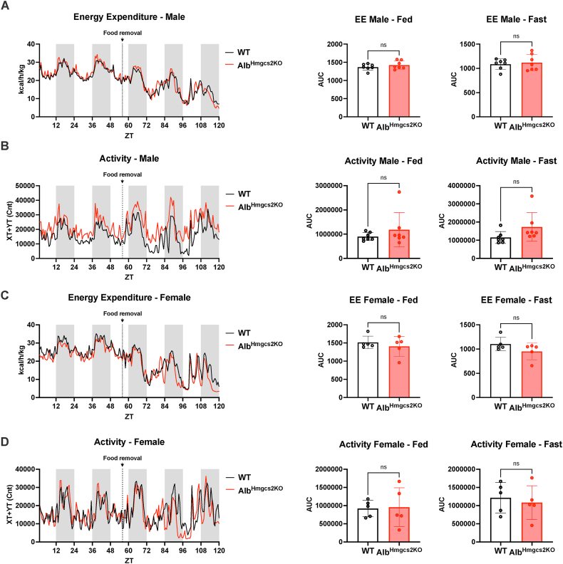
Results: Mice deficient of hepatic Hmgcs2 failed to develop ketosis during endotoxemia and during prolonged fasting. Surprisingly, hepatic HMGCS2 deficiency and the lack of ketosis did not affect survival, glycemia, or body temperature in response to endotoxemia. Mice with hepatic ketogenic deficiency also did not exhibit any defects in starvation adaptation and were able to maintain blood glucose, body temperature, and lean mass compared to littermate wild-type controls. Mice with hepatic HMGCS2 deficiency exhibited higher levels of plasma acetate levels in response to fasting.

Conclusions: Circulating hepatic-derived ketones do not provide protection against endotoxemia, suggesting that alternative mechanisms drive the increased mortality from forced feeding during illness-induced anorexia. Hepatic ketones are also dispensable for surviving prolonged starvation in the absence of inflammation. Our study challenges the notion that hepatic ketogenesis is required to maintain blood glucose and preserve lean mass during starvation, raising the possibility of extrahepatic ketogenesis and use of alternative fuels as potential means of metabolic compensation.

Keywords: Endotoxemia; Fasting metabolism; HMGCS2; Ketogenesis; Starvation adaptation.


Conflict of interest statement

Declaration of competing interest The authors declare that they have no known competing financial interests or personal relationships that could have appeared to influence the work reported in this paper.

Figures

Image 1
Graphical abstract
Figure 1
Figure 1
Lack of ketosis does not affect survival from endotoxemia. (A–B) C57BL/6J mice were challenged with 10 mg/kg i.p. LPS. (A) Food intake shown as grams per hour. n = 8. (B) Plasma total ketones after LPS challenge. n = 7. (C–D) WT and AlbHmgcs2KO (KO) mice were ad libitum fed or fasted for 24 h 2 weeks after tamoxifen administration. Whole tissue protein lysates immunoblotted for HMGCS2. (E) Liver triglyceride concentrations (mg/g tissue) in ad libitum fed mice 2 weeks after tamoxifen administration. n = 4–5/group. (F) Plasma total ketones 24 h after PBS vehicle or 10 mg/kg i.p LPS challenge. n = 4–11/group. (G–I) WT and AlbHmgcs2KO mice were challenged with 10 mg/kg i.p. n = 28/group (male), n = 14–16/group (female). (G) Kaplan–Meier survival curve. (H) Rectal body temperatures. (I) Blood glucose. Data expressed as mean ± SD, ∗∗∗∗P < 0.0001, ns (not significant); two sided unpaired t-test (E), two-way ANOVA with Sidak's multiple comparisons test (F) with mixed effects (H, I), Mantel–Cox test (G).
Figure 2
Figure 2
Mice with hepatic ketogenic deficiency do not exhibit any defects in starvation adaptation. (A–D) WT and AlbHmgcs2KO mice were fasted for 72 h n = 19–20/group (male), n = 15–16/group (female). (A) Blood ketones, beta-hydroxybutyrate (BOHB) measured by ketone meter. (B) Blood glucose. (C) Body weight. (D) Rectal body temperatures. (E) WT and AlbHmgcs2KO mice were fasted for 24 h. Plasma BOHB and Acetoacetate (AcAc, which was calculated by the difference between total ketones and BOHB) measured by colorimetric assay (Wako), and plasma non esterified fatty acids (NEFA). n = 7–13/group. (F) Survival after WT and AlbHmgcs2KO mice were fasted for 96 h and then refed. n = 19–20/group (male), n = 15–16/group (female). Data expressed as mean ± SD, ∗∗P < 0.01, ∗∗∗∗P < 0.0001, ns (not significant); two-way ANOVA with Sidak's multiple comparisons test (E) with mixed effects (A–D), Mantel–Cox test (F).
Figure 3
Figure 3
Hepatic ketogenesis does not preserve lean mass during prolonged fasting. WT and AlbHmgcs2KO mice were fasted for 72 h, two weeks after tamoxifen administration. Fat and lean mass were measured by nuclear magnetic resonance (NMR) at baseline and every 24 h n = 13–14/group (male) n = 15–16/group (female). (A–B) Lean mass by percent body weight (BW) and absolute mass. (C) Total percent lean mass loss at 72 h of fasting compared to baseline. (D) Gastrocnemius skeletal muscle mRNA expression shown relative to Rpl13a. (E–F) Fat mass by percent body weight and absolute mass. (G) Total percent fat mass loss at 72 h of fasting compared to baseline. (H) Epididymal white adipose tissue (eWAT) mRNA expression shown relative to Rpl13a. Data expressed as mean ± SD, ∗P < 0.05, ∗∗P < 0.01, ∗∗∗P < 0.001, ns (not significant); two-way ANOVA with Sidak's multiple comparisons test (A, B, D–F, H), two-sided, unpaired t test (C, G).
Figure 4
Figure 4
After matching for baseline fat mass, male mice with hepatic ketogenic deficiency have improved survival from prolonged starvation. (A–C) WT and AlbHmgcs2KO male mice were ad libitum fed or fasted for 24 h, 72 h after tamoxifen administration. (A) Overview of tamoxifen timing and start of fast for (B, C). (B) Whole liver protein lysates immunoblotted for HMGCS2. (C) Plasma total ketones measured by colorimetric assay (Wako). n = 3–4/group. (D) Liver triglyceride concentrations (mg/g tissue) in ad libitum fed WT and AlbHmgcs2KO male mice 72 h after tamoxifen administration. n = 5/group. (E–L) WT and AlbHmgcs2KO male mice were fasted 72 h after tamoxifen administration. n = 8–12/group. (E) Blood ketones (BOHB ketone meter). (F) Blood glucose. (G) Body weight. (H) Rectal body temperature. (I–L) Fat and lean mass were measured by nuclear magnetic resonance (NMR) at baseline and every 24 h. (I) Lean mass by percent body weight and absolute mass. (J) Total percent lean mass loss at 72 h of fasting compared to baseline. (K) Fat mass by percent body weight and absolute mass. (L) Total percent fat mass loss at 72 h of fasting compared to baseline. (M) Survival after WT and AlbHmgcs2KO mice were fasted for 96 h and then refed. n = 8–12/group. Data expressed as mean ± SD, ∗∗P < 0.01, ∗∗∗∗P < 0.0001, ns (not significant); two-way ANOVA with Sidak's multiple comparisons test (C, I, K) with mixed effects (E–H), two-sided, unpaired t test (D, J, L), Mantel–Cox test (M).
Figure 5
Figure 5
Hepatic ketogenic deficit mice exhibit expected changes in plasma amino acids and organic acids during fasting. (A–C) WT and AlbHmgcs2KO male mice were ad libitum fed or fasted for 24 h 2 weeks after tamoxifen administration. n = 5–6/group. (A) Plasma amino and organic acids. (B) Plasma urea. (C) Liver mRNA expression shown relative to Rpl13a. (D–F) WT and AlbHmgcs2KO male mice were fasted for 24 h 72 h after tamoxifen administration. n = 5–6/group. (D) Plasma amino and organic acids. (E) Plasma urea. (F) Liver mRNA expression shown relative to Rpl13a. (G) Glycogen content measured from livers harvested from ad libitum fed WT and AlbHmgcs2KO male mice 72 h after tamoxifen administration. n = 6–9/group. Data expressed as mean ± SD, ∗P < 0.05, ∗∗P < 0.01, ∗∗∗P < 0.001, ∗∗∗∗P < 0.0001, ns (not significant); two-way ANOVA with Sidak's multiple comparisons test (A–C), two-sided, unpaired t test (D–G).
Figure 6
Figure 6
Hepatic ketogenic deficiency does not affect activity or energy expenditure during prolonged fasting. WT and AlbHmgcs2KO mice were fasted 72 h after tamoxifen administration. (A–B) male mice n = 7/group, (C–D) female mice n = 5/group. (A, C) Energy expenditure (EE) and area under the curve (AUC) calculated during the fed and fasted periods. (B, D) Activity and area under the curve (AUC) calculated during the fed and fasted periods. Data expressed as mean ± SD, ns (not significant); two-sided, unpaired t test (A–D).
Figure 7
Figure 7
Alternative pathways of metabolic compensation. (A–F) Immunostaining of kidney sections from 24-h fasted wild-type mice using various co-staining comparisons: Lotus lectin (LTL), a proximal tubule (PT) marker; anti-HMGCS2; anti-GFP against a reporter mouse (PvAlb-MT) for the distal convoluted tubule (DCT); anti-sodium chloride co-transporter (NCC, a DCT marker), and/or anti-SCOT. (F) represents region indicated in (E). Scale bar represents 100 μm. (G) Immunostaining of brain sections (midbrain, anterior to the hypothalamus) from ad libitum fed wild-type mice using various co-staining comparisons: anti-glial fibrillary acidic protein (GFAP, an astrocyte marker), anti-microtubule-associated protein 2 (MAP-2, a neuronal marker), and anti-HMGCS2. Scale bar represents 30 μm. (H) WT and AlbHmgcs2KO mice were fasted for 24 h or fed ad libitum, 72 h after tamoxifen administration. Plasma acetate levels measured by GC/MS. n = 7–10/group. (I) WT and AlbHmgcs2KO mice were fasted for 24 h, 72 h after tamoxifen administration. Liver mRNA shown relative to Rpl13a. n = 5/group. Data expressed as mean ± SD, ∗P < 0.05, ∗∗∗P < 0.001, ns (not significant); two-way ANOVA with Sidak's multiple comparisons test (H), two-sided, unpaired t test (I). (J) Model of fasting metabolic compensation in hepatic ketogenic deficiency. Image created by BioRender.com.

Similar articles

Cited by

References

    1. Hart B.L. Biological basis of the behavior of sick animals. Neurosci Biobehav Rev. 1988;12:123–137. - PubMed
    1. Wang A., Huen S.C., Luan H.H., Yu S., Zhang C., Gallezot J.D., et al. Opposing effects of fasting metabolism on tissue tolerance in bacterial and viral inflammation. Cell. 2016;166:1512–1525.e12. - PMC - PubMed
    1. Wang A., Huen S.C., Luan H.H., Baker K., Rinder H., Booth C.J., et al. Glucose metabolism mediates disease tolerance in cerebral malaria. Proc Natl Acad Sci U S A. 2018;115:11042–11047. - PMC - PubMed
    1. Puchalska P., Crawford P.A. Multi-dimensional roles of ketone bodies in fuel metabolism, signaling, and therapeutics. Cell Metab. 2017;25:262–284. - PMC - PubMed
    1. Venable A.H., Lee L.E., Feola K., Santoyo J., Broomfield T., Huen S.C. Fasting-induced HMGCS2 expression in the kidney does not contribute to circulating ketones. Am J Physiol Ren Physiol. 2022;322:F460–F467. - PMC - PubMed

LinkOut - more resources