. 2024 Jul;300(7):107460.
doi: 10.1016/j.jbc.2024.107460. Epub 2024 Jun 12.

Loss-of-function G6PD variant moderated high-fat diet-induced obesity, adipocyte hypertrophy, and fatty liver in male rats

Affiliations

Loss-of-function G6PD variant moderated high-fat diet-induced obesity, adipocyte hypertrophy, and fatty liver in male rats

Shun Matsumura et al. J Biol Chem. 2024 Jul.

Abstract

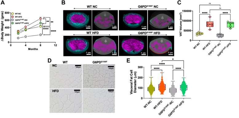
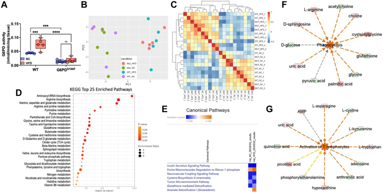
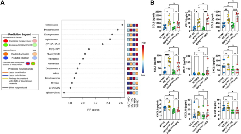
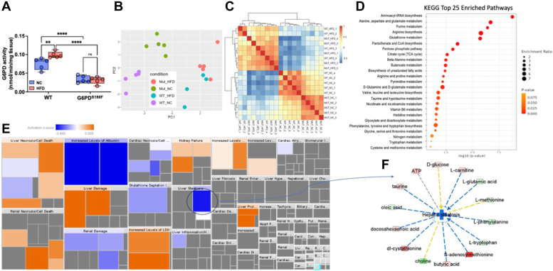
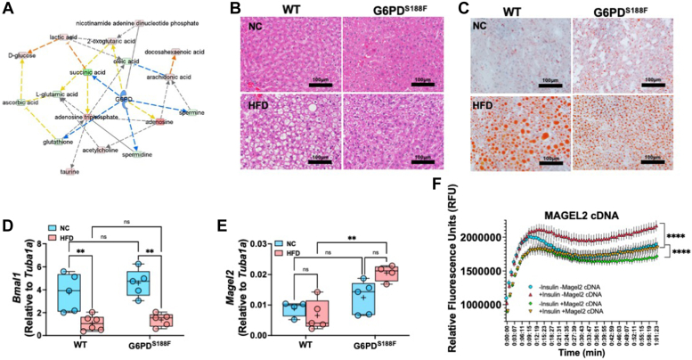
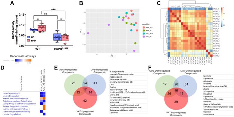
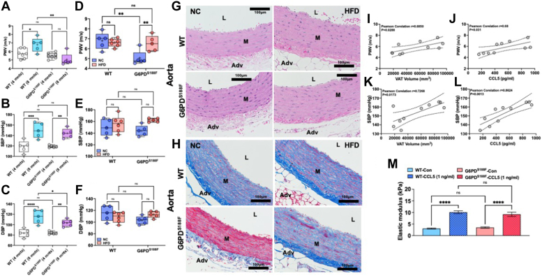
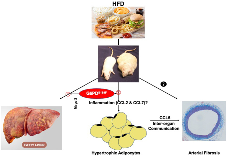
Obesity is a major risk factor for liver and cardiovascular diseases. However, obesity-driven mechanisms that contribute to the pathogenesis of multiple organ diseases are still obscure and treatment is inadequate. We hypothesized that increased , glucose-6-phosphate dehydrogenase (G6PD), the key rate-limiting enzyme in the pentose shunt, is critical in evoking metabolic reprogramming in multiple organs and is a significant contributor to the pathogenesis of liver and cardiovascular diseases. G6PD is induced by a carbohydrate-rich diet and insulin. Long-term (8 months) high-fat diet (HFD) feeding increased body weight and elicited metabolic reprogramming in visceral fat, liver, and aorta, of the wild-type rats. In addition, HFD increased inflammatory chemokines in visceral fat. Interestingly, CRISPR-edited loss-of-function Mediterranean G6PD variant (G6PDS188F) rats, which mimic human polymorphism, moderated HFD-induced weight gain and metabolic reprogramming in visceral fat, liver, and aorta. The G6PDS188F variant prevented HFD-induced CCL7 and adipocyte hypertrophy. Furthermore, the G6PDS188F variant increased Magel2 - a gene encoding circadian clock-related protein that suppresses obesity associated with Prader-Willi syndrome - and reduced HFD-induced non-alcoholic fatty liver. Additionally, the G6PDS188F variant reduced aging-induced aortic stiffening. Our findings suggest G6PD is a regulator of HFD-induced obesity, adipocyte hypertrophy, and fatty liver.

Keywords: chemokines; cytokines; fat tissue; inflammation; inter-organ communication; liver; metabolic reprogramming; vascular biology.


Conflict of interest statement

Conflict of interest The authors declare that they have no conflicts of interest with the contents of this article.

Figures

Figure 1
Figure 1
Effect of long-term high-fat diet feeding on body weight and visceral adipose tissue of wild-type and G6PDS188Frats.A, difference in body weight of wild-type (WT) and G6PDS188F rats before and after feeding high-fat diet (HFD) show body weight increased in HFD fed (WT-HFD and G6PDS188F-HFD) groups compared to their respective NC diet fed (WT-NC and G6PDS188F-NC) groups. However, G6PDS188F-HFD rats gained less weight than WT-HFD rats. B and C, two representative micro-CT scans of wild-type and G6PDS188F rats on NC and HFD showed visceral (pink) and subcutaneous (blue) adipose tissue, and summary results showing visceral adipose tissue volume increased in both the genotypes on HFD. D and E, representative images and summary results of visceral adipose cell sizes demonstrate cell size is increased in WT-HFD but not G6PDS188F-HFD rats as compared with the respective controls (NC). N = 5 in panel AD and individual cells from five different samples. Two-way ANOVA with post hoc Tukey’s multiple comparison tests was used to compare multiple groups. ∗p < 0.05; ∗∗p < 0.01; ∗∗∗∗p < 0.001.
Figure 2
Figure 2
Effect of long-term high-fat diet feeding on G6PD activity and metabolomic reprogramming, and inflammatory chemokines synthesis, in visceral adipose tissue of wild-type and G6PDS188Frats.A, G6PD activity increased in visceral adipose tissue (VAT) of wild-type (WT) rats but not G6PDS188F rats fed with HFD. B and C, PCA plot and sample correlation heat map demonstrating differential metabolism in VAT of WT and G6PDS188F fed with normal chow (NC) and high fat diet (HFD). D, KEGG enrichment pathway analysis identified the top 25 pathways in response to G6PD mutation. E, IPA core analysis predicted seven canonical pathways are significantly and differentially (Absolute z-score ≥ 2 and log10(p-value) ≥ 1.3) changed in response to G6PD mutation. F and G, IPA disease and function network analysis of metabolomic results predicted that a number of inflammatory response functions are activated in response to G6PD mutation. The majority of inflammatory responses including phagocytosis, activation of T lymphocytes, and immune response of cells changed more in HFD when compared to normal chow. Prediction legends show various symbols and arrows for interpretation of the IPA analysis. N = 5 in each group. Two-way ANOVA with post hoc Tukey’s multiple comparison test were used to compare multiple groups. ∗p < 0.05; ∗∗p < 0.01; ∗∗∗p < 0.005; and ∗∗∗∗p < 0.001.
Figure 3
Figure 3
Effect of long-term high-fat diet feeding on inflammatory chemokines synthesis in visceral adipose tissue of wild-type and G6PDS188Frats.A, VIP score plot shows metabolites including oxylipins differed in HFD from NC group. B, summary results of multiplex analysis demonstrate chemokines are increased in visceral adipose tissue of WT and G6PDS188F rats. N = 5 in each group. Two-way ANOVA with post hoc Tukey’s multiple comparison test were used to compare multiple groups. ∗p < 0.05; ∗∗p < 0.01; ∗∗∗p < 0.005; and ∗∗∗∗p < 0.001.
Figure 4
Figure 4
Effect of long-term high-fat diet feeding on liver toxicity, non-alcoholic fatty liver, and circadian rhythm gene expression in the liver of wild-type and G6PDS188Frats.A, G6PD activity decreased in the liver of G6PDS188F rats as compared to wild-type (WT) rats fed with normal chow (NC) and high-fat diet (HFD). B and C, PCA plot and sample correlation heat map demonstrating differential metabolism in the liver of WT and G6PDS188F fed with NC and HFD. D, KEGG enrichment pathway analysis identified the top 25 pathways in response to G6PD mutation. E, IPA Tox Analysis result based on the Liver HFD metabolic dataset shows that liver steatosis is significantly (activation z-score = −1.88) inhibited in response to G6PD mutation. F, G6PD can regulate eight out of 14 compounds that affect hepatic steatosis as an upstream regulator. Prediction legends showing various symbols and arrows for interpretation of the IPA analysis are from Figure 2. Two-way ANOVA with post hoc Tukey’s multiple comparison tests was used to compare multiple groups. ∗∗p < 0.01 and ∗∗∗∗p < 0.001.
Figure 5
Figure 5
Effect of long-term high-fat diet feeding non-alcoholic fatty liver and circadian rhythm gene expression in the liver of wild-type and G6PDS188Frats.A, G6PD can regulate metabolic pathways. Prediction legends showing various symbols and arrows for interpretation of the IPA analysis are from Figure 2. B, representative images of H&E staining showing lipid accumulation and fatty liver in HFD-fed wild-type (WT) rats and to a lesser extent in G6PDS188F rats. Images were reused to demonstrate rigor and reproducibility in Fig. S6. C, representative images of Oil Red staining indicate that lipid droplets are reduced in the liver of G6PDS188F rats than in wild-type (WT) rats. D and E, expression of circadian clock gene Bmal1 decreased in the liver of both genotypes fed with HFD, while circadian clock and Prader-Willi Syndrome associated Magel2 gene expression selectively increased in HFD-fed G6PDS188F rats. F, overexpression of MAGEL2 significantly reduced insulin-induced fatty acid uptake and accumulation in HepG2 cells. Two-way ANOVA with post hoc Tukey’s multiple comparison test was used to compare multiple groups. ∗∗p < 0.01 and ∗∗∗∗p < 0.001.
Figure 6
Figure 6
Effect of long-term high-fat diet feeding on G6PD activity and metabolomic reprogramming in the aorta of wild-type and G6PDS188Frats.A, G6PD activity decreased in the aorta of G6PDS188F rats as compared to wild-type (WT) rats fed with normal chow (NC) and high-fat diet (HFD). B and C, PCA plot and sample correlation heat map demonstrating differential metabolism in the liver of WT and G6PDS188F fed with NC and HFD. D, IPA core analysis showing differential changes in canonical pathway function. EandF, venn diagram shows up (17) or down (16) regulated common metabolites in visceral adipose tissue, liver, and aorta, of HFD-fed rats. N = 5 in each group. Two-way ANOVA with post hoc Tukey’s multiple comparison test was used to compare multiple groups. ∗∗p < 0.01.
Figure 7
Figure 7
Effect of aging and long-term high fat diet feeding on aortic stiffness and blood pressure of wild-type and G6PDS188Frats and VAT-derived CCL5 positively correlates with aortic stiffness and incubation of aorta with CCL5 augments elastic modulus.AC, aging increased pulse wave velocity (PWV; index of aortic stiffness) and systolic and diastolic blood pressure in both genotypes. However, diastolic pressure in older G6PDS188F was significantly lower than in age-matched wild-type (WT) rats. DF, HFD feeding for 8 months did not increase PWV and blood pressure in older wild-type rats, and blood pressure in older G6PDS188F rats, as compared with age-matched rats on a normal chow (NC) diet. However, HFD feeding increased PWV in G6PDS188F rats as compared with age-matched rats on the NC diet. G and H, representative H&E and Mason’s Trichrome staining of aorta isolated from wild-type and G6PDS188F rats fed with high-fat diet (HFD) or normal chow (NC) is shown. H&E staining images show hypertrophy of the medial layer in HFD-fed rats and Manson’s Trichrome staining shows collagen and fibrosis of the aorta is less pronounced in G6PDS188F rats as compared with wild-type rats on NC but not on long-term HFD. Images were reused to demonstrate rigor and reproducibility in Fig. S10. I and J, Pearson’s correlation shows a positive correlation between visceral adipose tissue (VAT) volume-systolic blood pressure (SBP) and -pulse wave velocity (PWV). K and L, Pearson’s correlation shows a positive correlation between VAT-derived CCL5-SBP and -PWV in G6PDS188F rats but not wild-type rats. M, isolated aorta from wild-type and G6PDS188F rats was incubated with CCL5 (1 ng/ml) ex vivo and after 72 h elastic modulus was determined by atomic force microscopy. Application of CCL5 increased elastic modulus. Two-way ANOVA with post hoc Tukey’s multiple comparison test were used to compare multiple groups. ∗p < 0.05; ∗∗p < 0.01; ∗∗∗p < 0.005; and ∗∗∗∗p < 0.001.
Figure 8
Figure 8
Schematic illustration of key findings. Long-term (8 months) high-fat diet (HFD) feeding increased body weight in wild-type rats more than G6PDS188F rats. In addition, in G6PDS188F rats, HFD-induced non-alcoholic fatty liver (fatty liver) and adipocyte hypertrophy (growth) were reduced as compared to wild-type rats, respectively, by presumably increasing Magel2 and reducing inflammation (CCL2 and CCL7). Moreover, we found HFD-elicited maladaptive inter-organ communication between adipose tissue and aorta via CCL5 that mediated aortic fibrosis and increased aortic stiffness.

References

    1. Lassale C., Tzoulaki I., Moons K.G.M., Sweeting M., Boer J., Johnson L., et al. Separate and combined associations of obesity and metabolic health with coronary heart disease: a pan-European case-cohort analysis. Eur. Heart J. 2018;39:397–406. - PMC - PubMed
    1. Wang Y., Chen X., Song Y., Caballero B., Cheskin L.J. Association between obesity and kidney disease: a systematic review and meta-analysis. Kidney Int. 2008;73:19–33. - PubMed
    1. Fabbrini E., Sullivan S., Klein S. Obesity and nonalcoholic fatty liver disease: biochemical, metabolic, and clinical implications. Hepatology. 2010;51:679–689. - PMC - PubMed
    1. Bodine S.C., Brooks H.L., Bunnett N.W., Coller H.A., Frey M.R., Joe B., et al. An American physiological Society cross-journal Call for Papers on "Inter-Organ communication in homeostasis and disease". Am. J. Physiol. Lung Cell. Mol. Physiol. 2021;321:L42–L49. - PMC - PubMed
    1. Owen M.K., Witzmann F.A., McKenney M.L., Lai X., Berwick Z.C., Moberly S.P., et al. Perivascular adipose tissue potentiates contraction of coronary vascular smooth muscle: influence of obesity. Circulation. 2013;128:9–18. - PMC - PubMed

Publication types

MeSH terms

Substances

LinkOut - more resources