. 2024 Sep;21(5):e00376.
doi: 10.1016/j.neurot.2024.e00376. Epub 2024 Jun 14.

MeCP2 gene therapy ameliorates disease phenotype in mouse model for Pitt Hopkins syndrome

Affiliations

MeCP2 gene therapy ameliorates disease phenotype in mouse model for Pitt Hopkins syndrome

Cassandra N Dennys et al. Neurotherapeutics. 2024 Sep.

Abstract

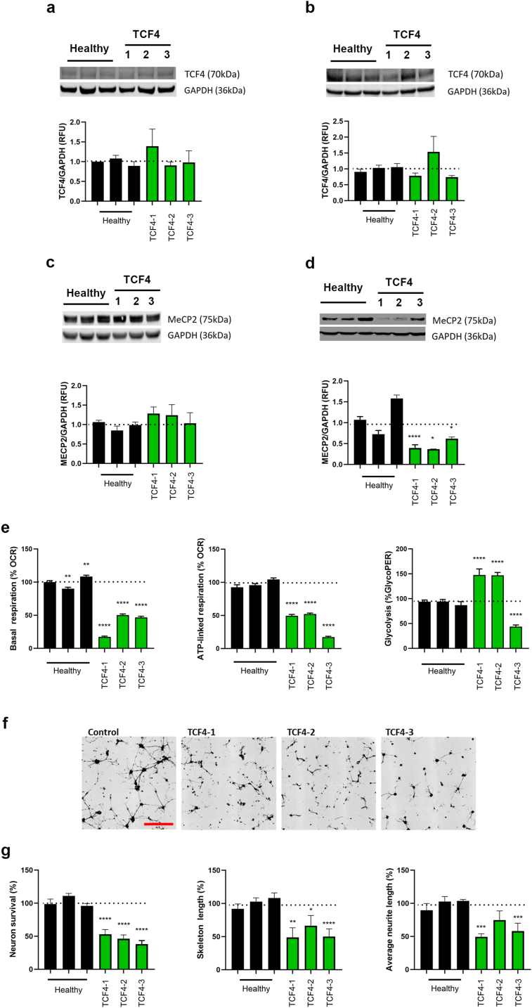
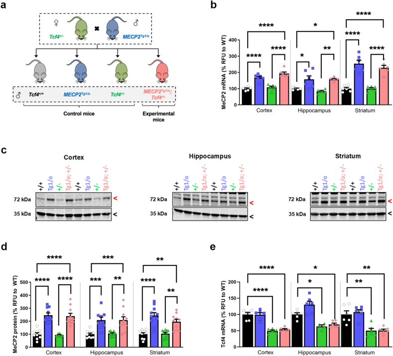
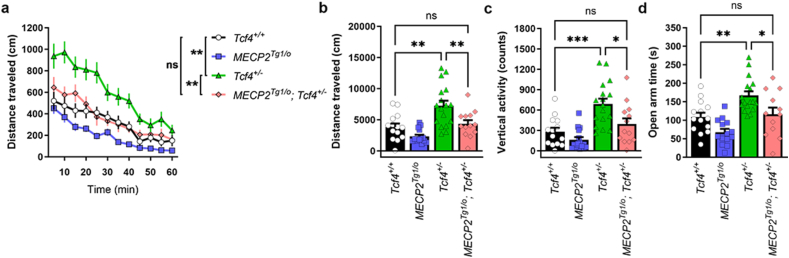
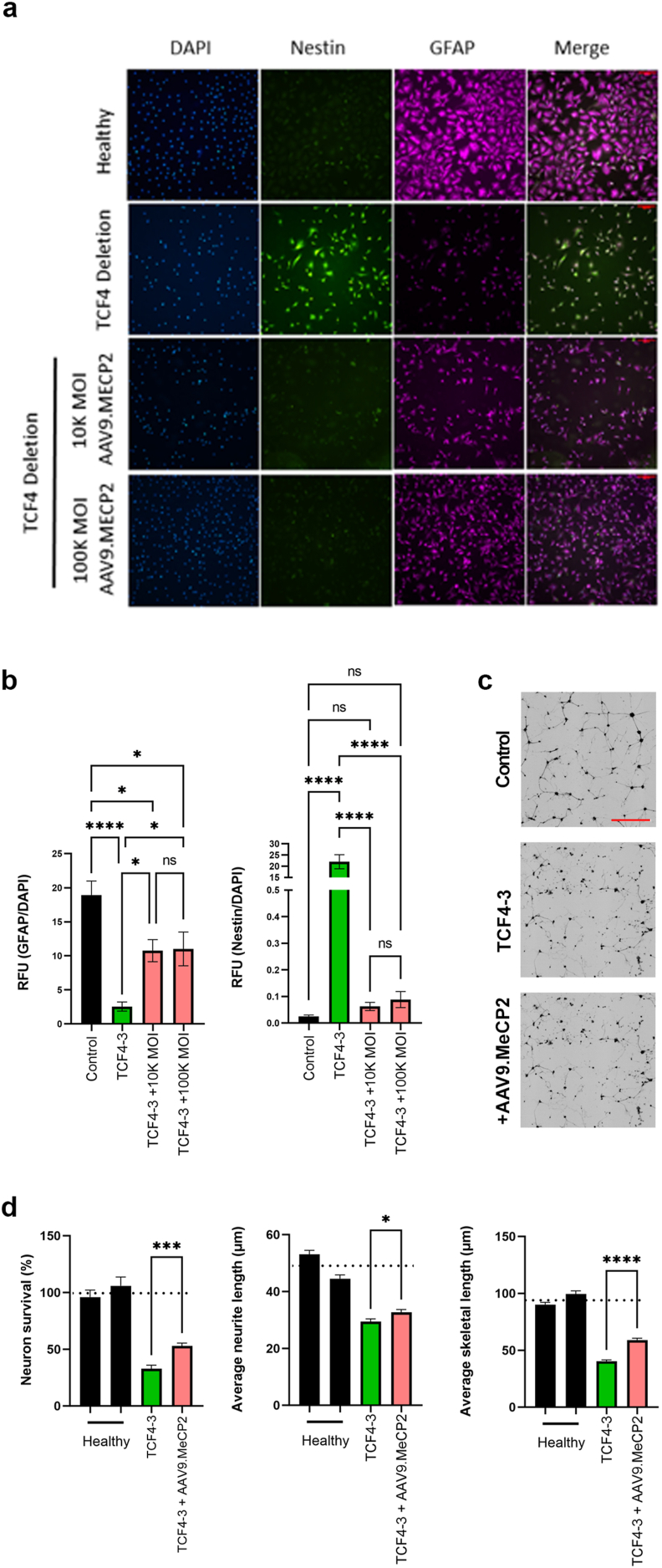
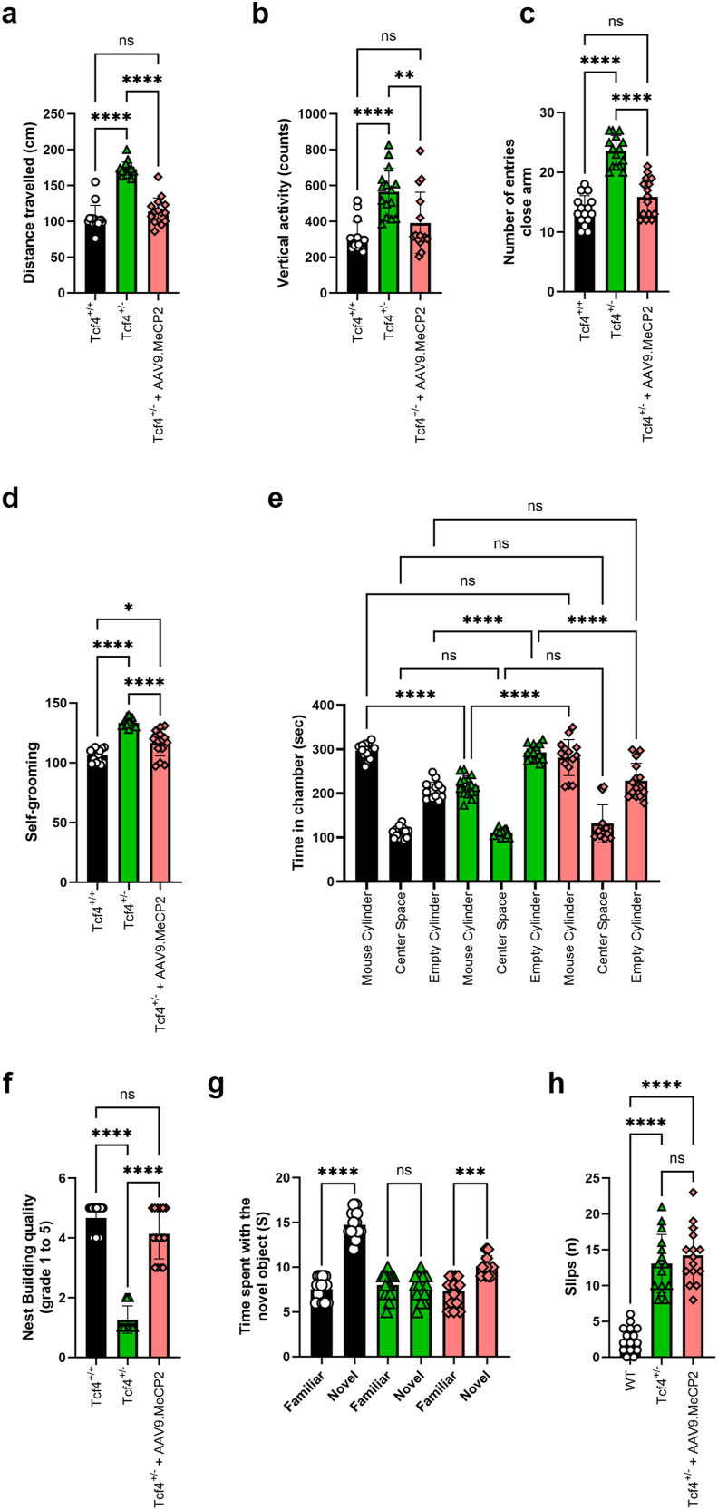
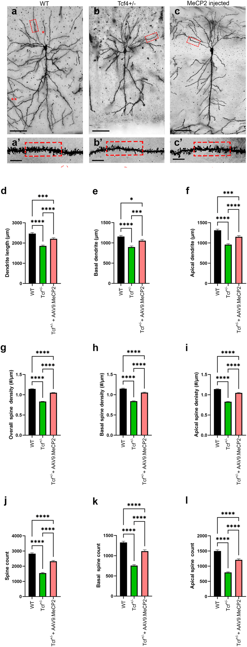
The neurodevelopmental disorder Pitt Hopkins syndrome (PTHS) causes clinical symptoms similar to Rett syndrome (RTT) patients. However, RTT is caused by MECP2 mutations whereas mutations in the TCF4 gene lead to PTHS. The mechanistic commonalities underling these two disorders are unknown, but their shared symptomology suggest that convergent pathway-level disruption likely exists. We reprogrammed patient skin derived fibroblasts into induced neuronal progenitor cells. Interestingly, we discovered that MeCP2 levels were decreased in PTHS patient iNPCs relative to healthy controls and that both iNPCs and iAstrocytes displayed defects in function and differentiation in a mutation-specific manner. When Tcf4+/- mice were genetically crossed with mice overexpressing MeCP2, molecular and phenotypic defects were significantly ameliorated, underlining and important role of MeCP2 in PTHS pathology. Importantly, post-natal intracerebroventricular gene replacement therapy with adeno-associated viral vector serotype 9 (AAV9)-expressing MeCP2 (AAV9.P546.MeCP2) significantly improved iNPC and iAstrocyte function and effectively ameliorated histological and behavioral defects in Tcf4+/- mice. Combined, our data suggest a previously unknown role of MeCP2 in PTHS pathology and common pathways that might be affected in multiple neurodevelopmental disorders. Our work highlights potential novel therapeutic targets for PTHS, including upregulation of MeCP2 expression or its downstream targets or, potentially, MeCP2-based gene therapy.

Keywords: AAV gene therapy; Mecp2; Pitt Hopkins syndrome; TCF4.


Conflict of interest statement

Declaration of competing interest The authors declare the following financial interests/personal relationships which may be considered as potential competing interests: Kathrin C Meyer, Patricia Cogram, Colleen M. Niswender reports financial support was provided by Pitt Hopkins Research Foundation. Colleen M. Niswender reports was provided by International Rett Syndrome Foundation. Rocco G. Gogliotti, Sheryl Anne D. Vermudez reports financial support was provided by National Institutes of Health. Kathrin Meyer, Cassandra Dennys has patent pending to Nationwide Children's Hospital. If there are other authors, they declare that they have no known competing financial interests or personal relationships that could have appeared to influence the work reported in this paper.

Figures

Fig. 1
Fig. 1
Patient fibroblast-derived NPCs have reduced MeCP2 levels and exhibit reduced ability to differentiate into astrocytes. Fibroblast (A and C) and NPC (B and C) lysates were analyzed by Western blot and levels of TCF4 (A and B) and MeCP2 (C and D) were quantified. NPCs were subsequently differentiated into astrocytes and their metabolic state, in addition to impact on neurons, was evaluated. E) Extracellular flux analysis was used to quantify oxygen consumption rate (OCR) and extracellular acidification rate (ECAR) to determine mitochondrial basal respiration, ATP-linked respiration and glycolytic protein efflux rate (glycoPER). Mitochondrial-based respiration was downregulated in TCF4 mutant astrocytes and a mutation-specific effect on glycoPER was identified (as measured by lactate mediated acidification). F) Representative images of neurons co-cultured with differentiated astrocytes (scale bar: 200 ​uM). G) Quantification of survival and neuronal morphology. Neurons cultured on TCF4 patient-induced astrocytes have reduced survival and shorter neurite and skeletal length. Dotted line represents average control values. Data have been generated from at least 3 independent experiments. Statistical analysis was performed using One-way ANOVA with Dunnett post-hoc analysis comparing the mean of each data set to the combined average of the controls (dotted line). ∗p ​< ​0.05, ∗∗p ​< ​0.01, ∗∗∗p ​< ​0.001, ∗∗∗∗p ​< ​0.0001.
Fig. 2
Fig. 2
Increased MeCP2 dosage does not affect Tcf4 expression in mice. (A) Breeding scheme to genetically introduce the MECP2 transgene (MECP2Tg1/o) into Tcf4+/− animals. MeCP2 mRNA (B) and protein levels (C-D) were quantified in the cortex, hippocampus and striatum of wildtype (Tcf4+/+, black), MECP2Tg1/o (blue), Tcf4+/− (green), and MECP2Tg1/o; Tcf4+/− (pink) P120 mice. (B) qPCR with values normalized to corresponding wild type controls. MeCP2 mRNA expression was significantly increased in the presence of the Tg1 transgene and was not affected by Tcf4 heterozygosity. (C) Representative images of fluorescent Western blot for MeCP2/Mecp2 (72 ​kDa, red arrow) and Gapdh (35 ​kDa, black arrow). (D) MeCP2 protein (72 ​kDa band) levels were normalized to Gapdh (35 ​kDa band) and then normalized against the corresponding wildtype controls. MeCP2 protein levels were elevated in MECP2Tg1/o and MECP2Tg1/o; Tcf4+/− mice. (E) Tcf4 mRNA expression is decreased in the brains of Tcf4+/− and MECP2Tg1/o; Tcf4+/− mice, but is unaffected by the Tg1 transgene. mRNA: n ​= ​5–6 mice per genotype. Protein: n ​= ​9–10 mice per genotype. 1-way ANOVA with Tukey's post-hoc test within each brain region. ∗p ​< ​0.05, ∗∗p ​< ​0.01, ∗∗∗p ​< ​0.001, ∗∗∗∗p ​< ​0.0001. Open circle/black bars ​= ​Tcf4+/+, blue squares/bars ​= ​MECP2Tg1/o, green triangles/bars ​= ​Tcf4+/−, pink diamonds/bars ​= ​MECP2Tg1/o; Tcf4+/−.
Fig. 3
Fig. 3
Increasing MeCP2 levels reverses abnormal phenotypes in Tcf4+/− mice. (A-B) Tcf4+/− mice (20–26 weeks of age) exhibit hyperlocomotive activity in an open field chamber, (C) enhanced vertical activity in an open field chamber, and (D) increased time spent in the open arms of an elevated zero maze (green versus black bars). All of these abnormal responses in Tcf4+/− compared animals are normalized to Tcf4+/+ levels in the presence of the MeCP2 transgene. n ​= ​8–16 mice per genotype. Statistical analysis was performed using 1-way ANOVA with Tukey's post-hoc test. ∗p ​< ​0.05, ∗∗p ​< ​0.01, ∗∗∗p ​< ​0.001, ∗∗∗∗p ​< ​0.0001, ns (not significant) for panels B, C, and D and 2-way ANOVA was used in time course locomotion data (A). Open circle/black bars ​= ​Tcf4+/+, blue squares/bars ​= ​MECP2Tg1/o, green triangles/bars ​= ​Tcf4+/−, pink diamonds/bars ​= ​MECP2Tg1/o; Tcf4+/−.
Fig. 4
Fig. 4
Treatment of patient fibroblast-derived NPCs with AAV9.MECP2 restores differentiation potential into astrocytes that are supportive of neuron morphology and survival. NPCs were transduced with AAV9.MECP2 and subsequently differentiated into astrocytes. (A) Cells were stained with the NPC marker, nestin, and an astrocyte marker, GFAP. (B) Quantification of GFAP and nestin staining intensity following differentiation. Transduction with AAV9.MeCP2 improved differentiation potential as indicated by reduced nestin and increased GFAP staining (scale bar: 100 ​μM). Astrocytes were seeded in co-culture with GFP+ ​neurons (black) and imaged 3 days later. (C) Representative images of neurons co-cultured with differentiated astrocytes. (D) Quantification of survival and neuronal morphology (scale bar: 200 ​μM). AAV9.MeCP2 significantly improved survival and neuronal morphology in the TCF4 deletion line (TCF4-3). Dotted line represents average control values. Data were generated from at least 3 independent experiments. Statistical analysis was performed using Student's T-test comparing the mean of untreated vs treated TCF4 cell lines. ∗p ​< ​0.05, ∗∗p ​< ​0.01, ∗∗∗p ​< ​0.001, ∗∗∗∗p ​< ​0.0001.
Fig. 5
Fig. 5
AAV9 delivery of MeCP2 reverses disease phenotypes in Tcf4+/− mice. Tcf4+/− pups were injected at p1 with scAAV9.P546.MeCP2. Fifteen mice per treatment group were aged to p60 prior to motor and behavioral analysis. (A) After 30 ​min, Tcf4+/− mice showed hyperactivity in the open field and treatment with scAAV9.P546.MeCP2 significantly normalized the distance travelled compared to Tcf4+/+ littermates. Anxiety levels were quantified by number of rearing events in the open field (B) and the number of times a mouse entered the closed arm of the elevated plus maze within a 5 ​min period (C). Treatment with scAAV9.P546.MeCP2 significantly reduced rearing and time spent in the closed arm in Tcf4+/− treated mice. (D) Time spent self-grooming was significantly elevated in Tcf4+/− mice and rescued by scAAV9.P546.MeCP2 injection when compared to untreated controls. (E) Three chamber social interaction. Relative to littermate controls, Tcf4+/− mice spend significantly more time with the empty cylinder, and this deficit is partially rescued by the scAAV9.P546.MeCP2 construct. (F) Nesting activities were quantified by transferring mice into a test cage containing 3 ​g of a Nestlet and the next morning nests were scored on a scale of 1–5 (5 being normal) as described in Materials and Methods. scAAV9.P546.MeCP2-injection significantly improved nesting capabilities when compared to uninjected controls. (G) Novel Object Recognition. Tcf4+/− mice do not distinguish between familiar and novel objects, and the recognition/preference for the novel object was restored by scAAV9.P546.MeCP2-injection. (H) The number of total slips of Tcf4+/− and WT littermates was evaluated using a Beam Walk test. The number of foot faults was counted in treated and untreated animals and compared to WT (Tcf4+/+) mice. scAAV9.P546.MeCP2 treatment significantly reduced the number of foot falls when compared to untreated animals. Statistical analysis was performed using one way ANOVA followed by Sidak's (A, B, C, E, H), Dunnett's (D) or Dunn's (G, F) post hoc test. ∗p ​< ​0.05, ∗∗p ​< ​0.01, ∗∗∗p ​< ​0.001, ∗∗∗∗p ​< ​0.0001.
Fig. 6
Fig. 6
Increasing Mecp2 levels leads to improvement in Tcf4+/− pyramidal cell morphology. Mice were treated as described in Fig. 5. (A–C) Representative images of dendrites from pyramidal cells from p120 Tcf4+/+, Tcf4+/− and Tcf4+/−scAAV9.P546.MeCP2-injected mice. When compared to the WT group, both treated and untreated Tcf4+/− samples showed significant reductions in dendritic length (upper panel, A to C, Scale bar: 50 ​μm), the number of the dendritic spines, and overall spine density (boxes in the lower panel, A’ to C’; Scale bar: 5 ​μm) compared to control. These observations were consistent for both basal and apical dendrites. Quantification of dendritic length (D–F), number of dendritic spines (G–I), and overall spine density (J–L) was performed using Neurolucida (MBF Bioscience, VT). Importantly, treatment with AAV9.MECP2 significantly improved total dendrite length, spine counts and spine density overall and in both basal and apical dendrites. Statistical analysis was performed using One-way ANOVA with Tukey post-hoc analysis comparing the mean of each data set to the combined average of the controls (dotted line). ∗p ​< ​0.05, ∗∗p ​< ​0.01, ∗∗∗p ​< ​0.001, ∗∗∗∗p ​< ​0.0001.

References

    1. Zweier C., Peippo M.M., Hoyer J., Sousa S., Bottani A., Clayton-Smith J., et al. Haploinsufficiency of TCF4 causes syndromal mental retardation with intermittent hyperventilation (Pitt-Hopkins syndrome) Am J Hum Genet. 2007;80(5) - PMC - PubMed
    1. Sweetser D.A., Elsharkawi I., Yonker L., Steeves M., Parkin K., Thibert R. In: GeneReviews® [Internet] Adam M.P., Feldman J., Mirzaa G.M., Pagon R.A., Wallace S.E., Bean L.J.H., editors. University of Washington, Seattle; Seattle (WA): 2012 Aug 30. Pitt-hopkins syndrome.https://www.ncbi.nlm.nih.gov/books/NBK100240/ [Updated 2018 Apr 12] 1993-2024. Available from: - PubMed
    1. Giurgea I., Missirian C., Cacciagli P., Whalen S., Fredriksen T., Gaillon T., et al. TCF4 deletions in Pitt-Hopkins syndrome. Hum Mutat. 2008 Nov;29(11) - PubMed
    1. Dong J.Y., Fan P.D., Frizzell R.A. Quantitative analysis of the packaging capacity of recombinant adeno-associated virus. Hum Gene Ther [Internet] 1996 Nov 10;7(17):2101–2112. https://pubmed.ncbi.nlm.nih.gov/8934224/ [cited 2023 Jun 21] Available from: - PubMed
    1. Amir R.E., Van Den Veyver I.B., Wan M., Tran C.Q., Francke U., Zoghbi H.Y. Rett syndrome is caused by mutations in X-linked MECP2, encoding methyl- CpG-binding protein 2. Nat Genet. 1999 Oct;23(2):185–188. - PubMed

Supplementary concepts