. 2024 Jun 14;15(1):5016.
doi: 10.1038/s41467-024-49037-y.

Single-cell and spatially resolved interactomics of tooth-associated keratinocytes in periodontitis

Affiliations

Single-cell and spatially resolved interactomics of tooth-associated keratinocytes in periodontitis

Quinn T Easter et al. Nat Commun. .

Abstract

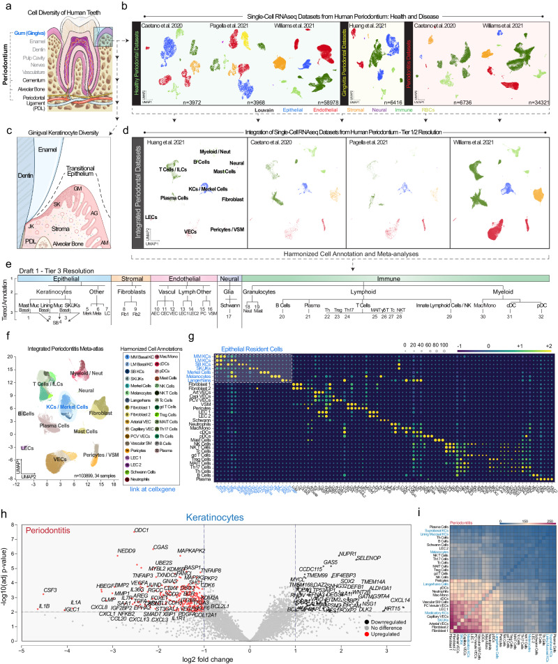
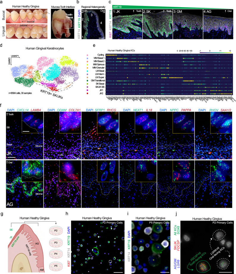
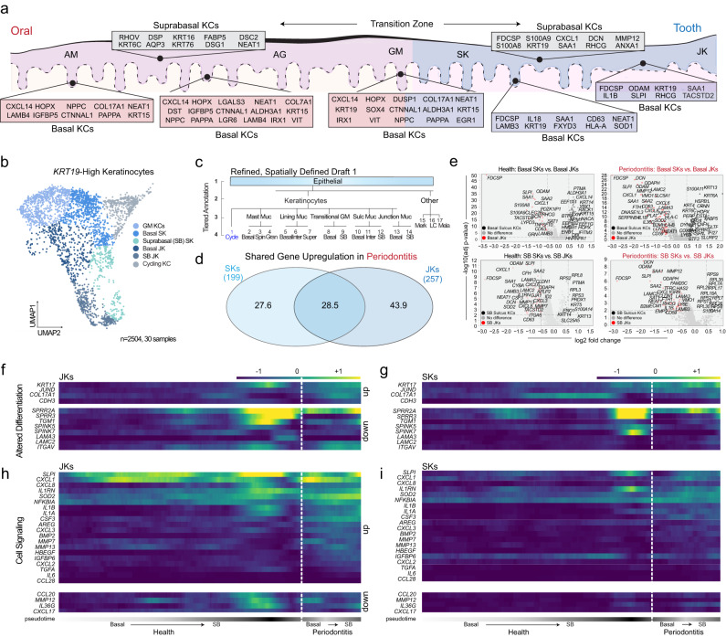
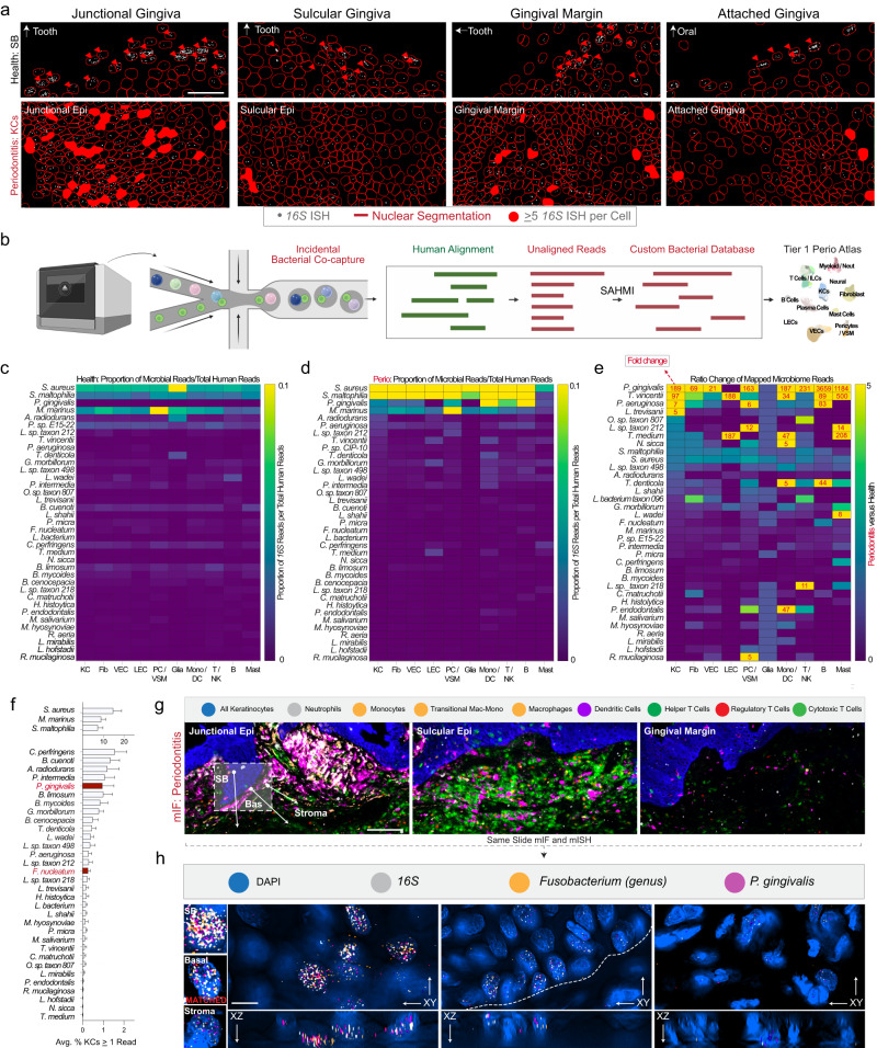
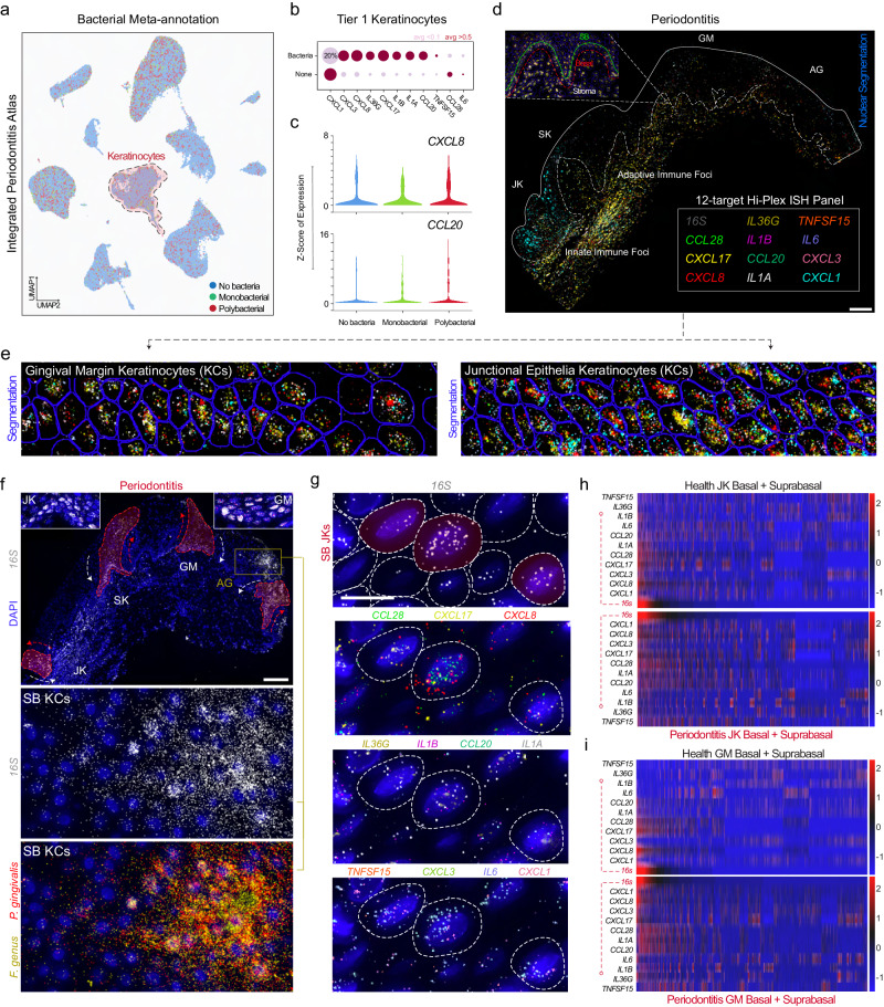
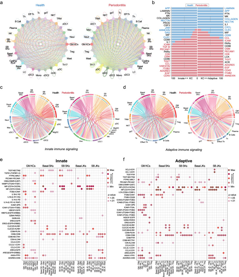
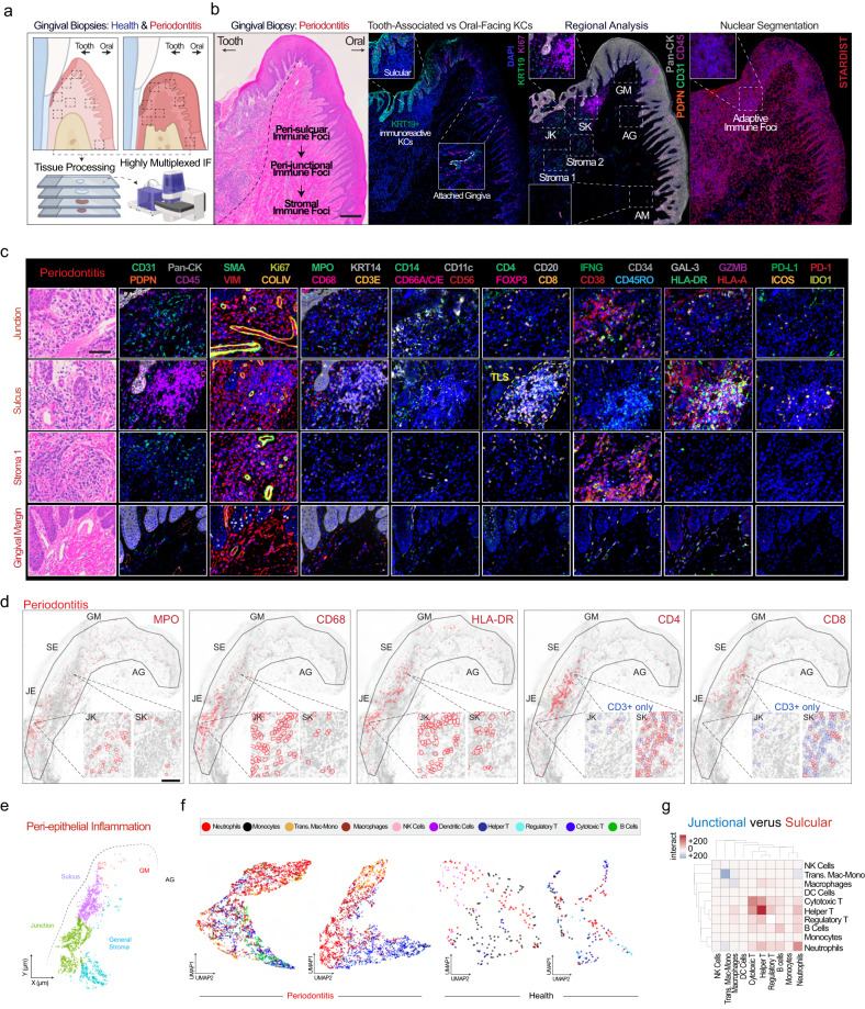
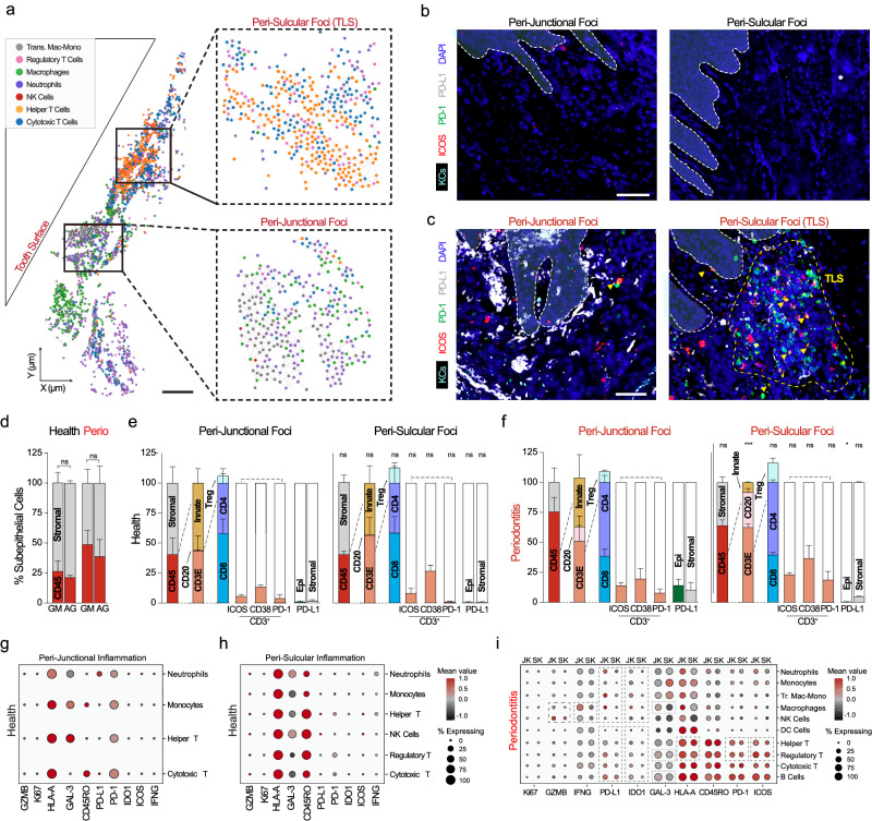
Periodontitis affects billions of people worldwide. To address relationships of periodontal niche cell types and microbes in periodontitis, we generated an integrated single-cell RNA sequencing (scRNAseq) atlas of human periodontium (34-sample, 105918-cell), including sulcular and junctional keratinocytes (SK/JKs). SK/JKs displayed altered differentiation states and were enriched for effector cytokines in periodontitis. Single-cell metagenomics revealed 37 bacterial species with cell-specific tropism. Fluorescence in situ hybridization detected intracellular 16 S and mRNA signals of multiple species and correlated with SK/JK proinflammatory phenotypes in situ. Cell-cell communication analysis predicted keratinocyte-specific innate and adaptive immune interactions. Highly multiplexed immunofluorescence (33-antibody) revealed peri-epithelial immune foci, with innate cells often spatially constrained around JKs. Spatial phenotyping revealed immunosuppressed JK-microniches and SK-localized tertiary lymphoid structures in periodontitis. Here, we demonstrate impacts on and predicted interactomics of SK and JK cells in health and periodontitis, which requires further investigation to support precision periodontal interventions in states of chronic inflammation.


Conflict of interest statement

The authors had access to the study data and reviewed and approved the final manuscript. Although the authors view each of these as noncompeting financial interests, K.M.B., Q.T.E., B.F.M., D.P., T.W., A.H., I.S., J.L., and S.A.T. are all active members of the Human Cell Atlas; furthermore, K.M.B. is a scientific advisor at Arcato Laboratories; I.S. a consultant for L’Oréal Research and Innovation; and S.A.T. has consulted for Roche and Genentech and is a scientific advisor for Biogen, GlaxoSmithKline, and Foresite Labs. All other authors declare no competing interests.

Figures

Fig. 1
Fig. 1. An integrated periodontitis atlas reveals important oral keratinocyte population roles in immune signaling.
a Specialized tissues support human teeth, including the periodontium, consisting of 1) gingiva (blue/box: epithelia; stroma), 2) periodontal ligament, and 3) mineralized tissues (cementum; alveolar bone). b Four single-cell RNA sequencing (scRNAseq) datasets were reprocessed for broad cell class comparison between studies. c The gingival epithelial attachment is a specialized transitional epithelium example, changing from non-keratinized alveolar mucosa (AM; if present), to keratinized attached gingiva (AG), altering expression profiles at the gingival margin (GM), then specializing in gingival sulcus and junctional epithelial keratinocytes (SK/JKs). d Each study was first integrated using Harmony and assigned Tier 1 cell type annotations. e Harmonized tier annotation was performed between epithelial, stromal, endothelial, neural, and immune cell populations. f Integrated UMAP and cell assignments and g cell signatures (Supplementary Data 1) were generated. Epithelial cells (blue) are highlighted. The entire dataset was uploaded to publicly-available CELLxGENE (cellxgene.cziscience.com/). SKs and JKs were grouped in the Tier 3 analysis as co-expressing Keratin 14 (KRT14) and Keratin 19 (KRT19). h Pseudobulk analysis of some differentially expressed genes (DEGs) in periodontitis using all keratinocytes (Tier 3 annotations) in volcano plots; full list, Supplementary Data 1. i Using CellPhoneDB, all Tier 3 cell types were analyzed for inferred receptor-ligand interactions; most frequent–bottom left. SK/JKs appear uniquely expressive of effector cytokines/other ligands compared to other keratinocytes (Supplementary Data 2). Abbreviations: ILCs Innate Lymphoid Cells, KCs Keratinocytes, VECs Vascular Endothelial Cells, VSM Vascular Smooth Muscle, LECs Lymphatic Endothelial Cells, Neut Neutrophils, Mast Muc, MM Masticatory (Keratinized) Mucosa, Lining Muc, LM Lining (Non-Keratinized) Mucosa, SB Suprabasal (Differentiated) Keratinocytes, Fib Fibroblast, AECs Arterial Endothelial Cells, PCV Postcapillary Venule, VECs Venule Endothelial Cells, Mac/Mono Macrophage/Monocytes, cDCs Conventional Dendritic Cells, pDC Plasmacytoid Dendritic Cells, Tc Cytotoxic T Cells, gdT Gamma Delta T Cells, Treg Regulatory T Cells, MAIT Mucosal Associated Invariant T Cells, Th Helper T Cells. Illustration from (a) created with BioIcons (image hosted at https://bioicons.com; tooth icon by Servier https://smart.servier.com/, licensed under CC-BY 3.0 Unported https://creativecommons.org/licenses/by/3.0/); illustration from (c) created with BioRender (https://www.biorender.com/). n = 34-sample, 105918-cells.
Fig. 2
Fig. 2. Gingival keratinocyte diversity is molecularly defined, spatially distinct, and preserved in vitro.
a To validate keratinocyte heterogeneity, healthy human gingival tissues were preserved on the tooth surface after extraction and fixed. b IHC revealed the gradual transition from tooth-facing JKs and SKs to GM, AG, and AM keratinocytes using KRT19; c KRT19-high and -low epithelial stem cells proliferate in the basal layer in health. d All keratinocytes (KRT14+) were subclustered from the integrated periodontitis meta-atlas (30 samples, 8584 cells) and assigned annotations (e) based on Louvain clustering. Cell signatures for these populations are plotted and included in Supplementary Data 1. f Using these signatures, we used a custom 12-plex ISH panel to reveal heterogeneity in keratinocyte populations (AG, GM, and JK here; SK and AM as in Fig. 1). Markers such as CXCL14 and NEAT1 marked the AG basal epithelium in the opposite pattern of KRT19 protein of the SK/JK cells; ODAM, RHCG, IL18, and SAA1/2 marked SK and JK cells in sequencing and in situ (See Supplementary Fig. 3 for sulcular, marginal, and alveolar mucosa imaging). g Primary human gingival keratinocytes were cultured over multiple passages. KRT19-high (marked by+) basal and larger suprabasal keratinocytes are found in mixed populations at (h) first passage and over (i) multiple passages. j Using RNA ISH and additional markers, cell subpopulations that were defined in vivo such as AG (LGR6+) and SK/JK (FDCSP+) can be identified in vitro, suggesting a heterogeneous 2D model of tooth-facing and oral-facing keratinocytes can be utilized for future assays and that these markers are more likely cell identities than cell states. Abbreviations: P Passage; see Fig. 1 legend. Sequential sections from samples were used (n = 3 health). Scale bars: b 100 μm; c 50 μm; f 25 μm; i, j 10 μm. Illustration from (g) created with BioRender.com. For this figure, n = 30-sample, 8554-cell for scRNAseq; n = 3 for tissues; n = 2 for unique primary cell lines.
Fig. 3
Fig. 3. Increased proinflammatory profiles coincide with altered differentiation patterns of tooth-associated keratinocytes.
a Due to single-cell annotation and in situ validation (Fig. 2; Supplementary Fig. 3), a draft model of the oral-to-tooth transition zone in humans is presented, with basal and suprabasal keratinocyte markers uniquely identifying the alveolar mucosa (AM), attached gingiva (AG), gingival margin (GM), sulcular epithelium (SK) and junctional epithelium (JK). b These markers allowed for KRT19-high keratinocyte (KC) cell subclustering for the first time (2504 cells in total), including gingival margin keratinocytes (GM), sulcular keratinocytes (SK), and junctional keratinocytes (JK). c A more granular draft (Tier 4) annotation of epithelial cells of the gingival attachment. d Assaying differentially expressed genes in periodontitis, SKs and JKs only share about a quarter of upregulated genes. JKs displayed nearly 125 unique upregulated genes in diseased cells. e Further analysis of basal and suprabasal (differentiating) SK and JK keratinocytes revealed unique cell signatures between basal and suprabasal cell types. This full list is included in Supplementary Data 1. fi To understand SK and JK developmental progression and cell state alterations, we used partitioned-based graph abstraction (PAGA). f Examining the basal to suprabasal transition, JKs display altered gene expressed comparing health to disease cell types, including broader expression of KRT17 and more expression of JUND, COL17A1, and CDH3. Key differentiation genes such as SPRR family members were also downregulated. g JKs displayed robust cell signaling and inflammatory phenotypes, which appear exacerbated in disease states. h In SKs, differentiation-related genes were uniquely expressed compared to JKs in health but also appeared altered in disease along the basal to suprabasal trajectory. i SKs appeared generally less reactive compared to JKs in disease, with lower overall expression of effector molecules such as CXCL1, CXCL8, IL1A, IL1B, and IL1RN. Abbreviations: Merk Merkel Cells, LC langerhans cells, Mela melanocytes, Muc mucosa, SB suprabasal Keratinocytes; for others, see Fig. 1 legend. For this figure, n = 30-sample, 2504-cell for scRNAseq.
Fig. 4
Fig. 4. Single-cell metagenomic reanalysis of keratinocytes reveals periopathogen signals concentrated in tooth-associated keratinocytes.
a Using 16S rRNA FISH, we segmented cells using StarDist in health and disease tissues. We found bacterial signals primarily focused on the most terminally differentiated suprabasal keratinocytes across each region of the oral- and tooth-associated keratinocytes. In disease, epithelial barrier integrity appeared compromised: we observed more bacterial-associated stromal and epithelial stem cells—especially in SKs and JKs. b Using a modified single-cell analysis of host-microbiome interactions (SAHMI) pipeline and a custom Kraken2 database, we used unmapped reads from our integrated single-cell periodontitis meta-atlas. c Using a broad Tier 1 annotation of cell types revealed 37 distinct species captured from inside of or membrane-bound to Keratinocytes (KC), Fibroblasts (Fib), Vascular Endothelial (VEC), Lymphatic Endothelial (LEC), Pericyte/Vascular Smooth Muscle (PC/VSM), Glial (Glia), Monocyte/Dendritic (Mono/DC), T/Natural Killer (T/NK), B (B), and Mast Cells (Mast). Using microbial per averaged total reads per human health cell class, we found low read counts across most bacterial species. d In periodontitis, we found large associated read shifts, often in well-known periodontal pathogen species (“periopathogen”; i.e., P. gingivalis, T. vincentii, P. aeruginosa [P. sp. CIP-10]). e Performing a ratio analysis of (d) over (c), we found dramatic increases in many bacteria—especially in known periopathogens. f Focusing on all keratinocytes, we found variable bacterial numbers (0.1%-15% of all KCs). g Utilizing broad cell classification of our multiplex immunofluorescence data (mIF; Figs. 7, 8), we showed the innate versus adaptive immune disease foci differ between 16S-high regions in situ. h Using the same mIF slides and targets predicted from our SAHMI pipeline, we applied in situ hybridization against 16S and two common periopathogen mRNA (P. gingivalis, fimA; Fusobacterium, fadA). We found polybacterial 16S and mRNA signals in some epithelial stem cells and their progeny using Nyquist-optimized, three-dimensional imaging. Abbreviations: Fig. 1 legend. Scale bars: a 100 μm; g 50 μm; h 10 μm. Samples from Figs. 7 and 8 were used (n = 6, 3 health, 3 periodontitis). Illustration from (b) created with BioRender.com. n = 30-sample, 8554-cell for scRNAseq; n = 3 for tissues.
Fig. 5
Fig. 5. Polybacterial interactions with gingival keratinocytes are highly specific in situ and can be niche- and disease-state agnostic.
a Metagenomic “reannotation” of the integrated periodontitis atlas showed most cells have no bacterial reads. Some (~15%) have 1 read (Monobacterial); a minority have 2 or greater reads (Polybacterial). b Disease-agnostic analysis of keratinocyte immune signatures revealed chemokine and interleukin signatures potentially related to bacterial signal—even in health. This was visualized using (b) dot plots and (c) violin plots. d 12-plex custom ISH panel of 11 human effector cytokine mRNA targets and 16S were overlaid using Warpy. e All 11 cytokines are shown simultaneously in GM and JK. Without 16S overlaid, each cytokine had a distinct patterning. Some i.e., IL1B, were broadly expressed in epithelia and stroma. Others i.e., CXCL3 and CXCL8 appeared to be cell-specific and enriched in JK over GM keratinocytes. f There appeared to be polybacterial patterns in disease that spread to all epithelial regions, including in terminally differentiated attached gingiva keratinocytes i.e., keratinized mucosa. Epithelial stem cell 16S signal was found in each region (insets). g Even in regions with sparse 16S signal (intracellular, red overlay), phenotypes appeared highly specific to cells with the highest 16S per-cell counts. h, i Considering keratinocytes at a cell-specific level, we found that 16S alone is positively associated with most cytokines in health and disease states whether in (h) JK or (i) GM. Assessing both JKs and GM, all keratinocytes were plotted on a normalized heatmap relative to 16S expression. We quantified that CXCL8, CXCL17, CCL20CCL28, IL1A, and IL1B are associated with microbial burden in healthy GM keratinocytes. In JKs, nearly all cytokines were positively associated with microbial burden in heath, suggesting that some bacteria may have cell-specific effects in vivo. Scale bars: (a, d) 100 μm (insets; 50 μm); (b, c) 25 μm. Abbreviations: ISH In situ hybridization; also see Fig. 1 legend. Illustration from (a) created with BioRender.com. Imaging was performed on sequential sections of samples used in Figs. 4, 7, and 8. For this figure, n = 34-sample, 105918-cell for scRNAseq; n = 6 for tissues: 3 health, 3 periodontitis.
Fig. 6
Fig. 6. Cell–cell communication between keratinocytes and immune cells is predicted to occur through innate and adaptive cell-specific programs.
a CellChat was used to understand cell signaling pathways in health and disease considering tooth-associated keratinocytes (basal versus suprabasal; SB and junctional versus sulcular keratinocytes; JK/SKs). Circle plots highlight the significant receptor-ligand interactions between any cell populations, including same-cell type signaling interactions (i.e., JK-JK, SK-SK, etc.). The proportion of interactions increased across more detailed immune cell type annotations (Tier 4 annotations; see Supplementary Fig. 6). b Relative information flow in health and disease showed a preference for cell adhesion (NECTIN, COLLAGEN, JAM, LAMININ) and other pathways such as APP, CXCL, and MIF pathways. In disease, more preference for cell signaling pathways is preferred, such as TGFB, TIGIT, CCL, CD45, and EGF. Value of 0 red signifies the pathway is not enriched in periodontitis; value of 100 red signifies highly enriched. Innate (c) and adaptive (d) immune cell communication was measured gene-by-gene using a chord diagram for visualizing cell-cell communication. e, f Dot plots showed upregulated signaling pathways in periodontitis at the level of predicted receptor–ligand interactions (y-axis) based on tooth-associated keratinocytes (x-axis). Innate cells appeared to potentially interact with junctional keratinocytes (JK) via CD99-CD99, CD99-PILRA, GAS6-AXL, and MIF-(CD74 + CXCR4/CD44). Adaptive cells appeared to potentially interact via similar pathways. Unique to adaptive cells-JK signaling include XCL2-XCR1; unique to innate cells, AREG-EGFR. Abbreviations: Cycle; KCs Cycling Keratinocytes, Spin Spinous Layer, Granular Granular Layer, Inter Intermediate Layer, Super Superficial; Merk Merkel Cells, Mela Melanocytes, LC Langerhans Cells, MigDC Migratory Dendritic Cells, Mast Mast Cells, also see Fig. 1 legend. Illustration from (a) created with BioRender.com. For this figure, n = 34-sample, 46835-cell for scRNAseq.
Fig. 7
Fig. 7. Spatial proteomics reveals that peri-epithelial immune microenvironments are uniquely enriched in innate immune populations nearest to junctional keratinocytes.
a The orientation of periodontal tissues is critical to show tooth-facing (sulcular, junctional epithelial keratinocytes; SK/JKs) and oral-facing (gingival margin, GM; attached gingiva, AG; and alveolar mucosa, AM) attachments for highly multiplexed immunofluorescence (mIF) assays of periodontitis. b By doing this in sequential sections, we first confirmed orientation and noted highly localized inflammatory profiles near tooth-facing epithelial keratinocytes. As discovered in the initial analysis (Fig. 2), tooth-facing SKs and JKs uniquely express Keratin 19 (KRT19) in every cell type, highlighting the transition zone. An initial analysis of Tier 1 cell assignments using mIF (PhenoCycler Fusion; Akoya Biosciences) revealed adaptive immune foci concentrated near SKs and more diverse, innate immune populated foci near JKs. Cell segmentation was performed using StarDist. c The 33-antibody assay revealed more heterogeneity at the cell type and cell state level, including peri-sulcular tertiary lymphoid structures (TLS, yellow) defined by T cell, B cell, and dendritic cell mixed aggregates. Antibodies are grouped and zoomed-in regions from a periodontitis sample are featured. d Spatial analysis of peri-epithelial regions was broken into four specific regions as before, highlighting the innate (MPO, CD68, HLA-DR) to adaptive (CD4, CD8) cell transition. e Segmented immune cells were assigned identities in health and disease across the four regions. f Periodontitis displayed more diverse heterogeneity considering the whole tissue. g However, cell–cell interactions among immune cells revealed diverse enrichment of immune cell types in peri-junctional and peri-sulcular immune foci in periodontitis. Abbreviations: Antibodies (see Methods); also see Fig. 1 legend. Scale bars: (b, d) 250 μm, (c) 50 μm. Illustration from (a) created with BioRender.com. For this figure, n = 6 for tissues: 3 health, 3 periodontitis).
Fig. 8
Fig. 8. Spatial analyses of peri-junctional and peri-sulcular microniches display regionalized and distinct immunophenotypes.
a Network diagram analysis of only immune-immune cell predicted interactions highlighted regional differences in immune cell aggregation in periodontitis. Both cell identities and their cell states are spatially distinct in periodontitis, with the peri-junctional immune cells expressing more immune exhaustion phenotypes than (b) health, contributed by both epithelial cells and stromal-resident cells in (c) periodontitis. Peri-sulcular immune cells, including tertiary lymphoid structures (TLS) more often found localized near this zone, contained mixed active (ICOS+) and exhausted (PD-1+) populations in periodontitis, even when compared to peri-junctional foci. d Manual thresholding was performed to show individual marker heterogeneity along the peri-epithelial niches. The cell assignment algorithm for this study is shown in Supplementary Fig. 2. e Single marker analysis of CD45+ cells revealed general increases in both GM and AG stroma. f Comparison of cell identities and cell states showed minimal differences between peri-junctional and peri-sulcular niches in health. While both peri-junctional and peri-sulcular immune infiltrate increased in disease, peri-junctional foci were biased toward more innate and relatively more CD4+ T cells compared to peri-sulcular niches, which were biased toward adaptive immune cells (T and B cells). Both junctional stromal and JKs also expressed more PD-L1 compared to sulcular cell types. Dot plots from total spatial analysis of immune cell states in health considering (g) peri-junctional and (h) peri-sulcular microenvironments. i In periodontitis, differences were quantified between both regions. Abbreviations: Antibodies (see Methods); also see Fig. 1 legend. ns not significant; TLS tertiary lymphoid structure, JK junctional keratinocytes, SK sulcular keratinocytes. Scale bars: (a) 100 μm (b, c) 50 μm. For this figure, n = 6 for tissues: 3 health, 3 periodontitis. Two-sided t tests and chi-square tests were used. *, **, and *** signify less than p < 0.05 when comparing healthy JKs to SKs and diseased JKs to SKs.

Similar articles

Cited by

References

    1. Chen MX, Zhong YJ, Dong QQ, Wong HM, Wen YF. Global, regional, and national burden of severe periodontitis, 1990–2019: an analysis of the Global Burden of Disease Study 2019. J. Clin. Periodontol. 2021;48:1165–1188. doi: 10.1111/jcpe.13506. - DOI - PubMed
    1. Jepsen S, et al. Periodontal manifestations of systemic diseases and developmental and acquired conditions: consensus report of workgroup 3 of the 2017 world workshop on the classification of periodontal and peri‐implant diseases and conditions. J. Clin. Periodontol. 2018;45:S219–S229. doi: 10.1111/jcpe.12951. - DOI - PubMed
    1. Marchesan JT, et al. Flossing is associated with improved oral health in older adults. J. Dent. Res. 2020;99:1047–1053. doi: 10.1177/0022034520916151. - DOI - PMC - PubMed
    1. Byrd, K. M., Gulati, A. The ‘gum-gut’ axis in inflammatory bowel diseases: a hypothesis-driven review of associations and advances. Front. Immunol.12, 620124 (2021). - PMC - PubMed
    1. Beck JD, Papapanou PN, Philips KH, Offenbacher S. Periodontal medicine: 100 years of progress. J. Dent. Res. 2019;98:1053–1062. doi: 10.1177/0022034519846113. - DOI - PubMed

MeSH terms