. 2024 Jun 14;15(1):5051.
doi: 10.1038/s41467-024-49101-7.

Adhesion pilus retraction powers twitching motility in the thermoacidophilic crenarchaeon Sulfolobus acidocaldarius

Affiliations

Adhesion pilus retraction powers twitching motility in the thermoacidophilic crenarchaeon Sulfolobus acidocaldarius

Arthur Charles-Orszag et al. Nat Commun. .

Abstract

Type IV pili are filamentous appendages found in most bacteria and archaea, where they can support functions such as surface adhesion, DNA uptake, aggregation, and motility. In most bacteria, PilT-family ATPases disassemble adhesion pili, causing them to rapidly retract and produce twitching motility, important for surface colonization. As archaea do not possess PilT homologs, it was thought that archaeal pili cannot retract and that archaea do not exhibit twitching motility. Here, we use live-cell imaging, automated cell tracking, fluorescence imaging, and genetic manipulation to show that the hyperthermophilic archaeon Sulfolobus acidocaldarius exhibits twitching motility, driven by retractable adhesion (Aap) pili, under physiologically relevant conditions (75 °C, pH 2). Aap pili are thus capable of retraction in the absence of a PilT homolog, suggesting that the ancestral type IV pili in the last universal common ancestor (LUCA) were capable of retraction.


Conflict of interest statement

The authors declare no competing interests.

Figures

Fig. 1
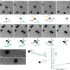
Fig. 1. Automated tracking of type IV pili mutants in S. acidocaldarius at high temperature.
ac Differential interference contrast (DIC) live-cell imaging of indicated S. acidocaldarius strains at 75 °C. Shown are selected stills representing noticeable steps of glass-adhered cells over a five-minute observation window. Overlaid tracks were automatically obtained with TrackMate 7 after machine-trained detection of the cells with Weka. d–f Worm plots of 15–30 representative tracks for each indicated strain. g Track displacement measured over five minutes for each indicated strain. h Distribution of individual track displacements in the indicated strains. Tracks with a displacement under 2 µm correspond to non-motile cells. i Track persistence ratio in the indicated strains. Gray scatter dot plots correspond to the total number of cells analyzed (N = 200 WT, 222 ∆aapF, 284 ∆arlJ, 171 ∆upsE, 245 ∆upsEarlJ). In g and i, superimposed yellow circles represent average values from each of n = 3 independent experiments and error bars represent the mean ± SEM of those three biological replicates. P values were calculated on the replicate means using a one-way ANOVA test with a Tukey’s correction for multiple comparisons with a single pooled variance. j Mean square displacement (MSD) analysis of all tracks in WT and ΔaapF strains. k Distribution of slope value α in MSD analysis of all tracks in WT, ΔaapF and ΔarlJ strains. Source data are provided as a Source Data file.
Fig. 2
Fig. 2. Super-resolution fluorescence imaging of S. acidocaldarius adhesion pili dynamics at high temperature.
Surface proteins of ∆upsE∆arlJ S. acidocaldarius cells were labeled non-specifically with AlexaFluor 568 NHS-ester. Glass-adhered cells were imaged in Structured Illumination Microscopy (iSIM) every 500 ms. Shown are selected stills from representative movies. a Aap pili mediate dynamic adhesion and twitching motility in S. acidocaldarius. b Pilus retraction mediate dynamic cell-cell interactions in S. acidocaldarius. c Single Aap pilus retraction events were used to measure pilus retraction speed. Retraction speed is given as the difference between final (Lfinal) and initial (Lini) pilus length over the difference between tfinal and tini. d Mean retraction speed in single-pilus retraction events in 19 cells. Superimposed yellow circles represent average values from each of n = 3 independent experiments. Error bars represent the mean ± SEM of those three biological replicates. e Retraction speed is not correlated with pilus length. Scale bars, 2 µm. Source data are provided as a Source Data file.
Fig. 3
Fig. 3. Automated tracking of S. acidocaldarius Aap mutants.
a Map of the Aap pilus operon in S. acidocaldarius. b Representative examples of tracks generated for indicated Aap mutants. Arrowheads denote long aligned tracks generated by detaching cells. Scale bars = 5 µm. c Track displacement measured over five minutes for each indicated strain (ΔaapA: N = 418, ΔaapB: N = 302, ΔaapX: N = 427). d Distribution of individual track displacements in the indicated strains. Tracks with a displacement <2 µm were defined as non-motile. e Track persistence ratio in the indicated strains. Superimposed yellow circles represent sample means from each of n = 3 (ΔaapA, ΔaapX) or n = 2 (ΔaapB) independent experiments. Error bars represent the mean ± SEM of those biological replicates for ΔaapA and ΔaapX. Source data are provided as a Source Data file.
Fig. 4
Fig. 4. Automated tracking in other Sulfolobales species.
a Track displacement and persistence ratio measured over five minutes for each indicated. b Distribution of individual track displacements in the indicated species. Tracks with a displacement under 2 µm correspond to the percentage of non-motile cells. c Track persistence ratio in the indicated species. Data represents N = 836 Saccharolobus solfataricus P2 cells, N = 180 Sulfolobus islandicus REY15A cells and N = 344 Sulfolobus islandicus M16.4 cells in n = 1 experiment. Source data are provided as a Source Data file.

Update of

References

    1. Pohlschroder M, Ghosh A, Tripepi M, Albers S-V. Archaeal type IV pilus-like structures–evolutionarily conserved prokaryotic surface organelles. Curr. Opin. Microbiol. 2011;14:357–363. doi: 10.1016/j.mib.2011.03.002. - DOI - PubMed
    1. Berry J-L, Pelicic V. Exceptionally widespread nanomachines composed of type IV pilins: the prokaryotic Swiss Army knives. FEMS Microbiol Rev. 2015;39:134–154. doi: 10.1093/femsre/fuu001. - DOI - PMC - PubMed
    1. Chaudhury P, Quax TEF, Albers S-V. Versatile cell surface structures of archaea. Mol. Microbiol. 2018;107:298–311. doi: 10.1111/mmi.13889. - DOI - PubMed
    1. van Wolferen M, et al. Species-specific recognition of sulfolobales mediated by UV-inducible pili and S-Layer glycosylation patterns. mBio. 2020;11:e03014–e03019. - PMC - PubMed
    1. Bonazzi D, et al. Intermittent pili-mediated forces fluidize Neisseria meningitidis aggregates promoting vascular colonization. Cell. 2018;174:143–155.e16. doi: 10.1016/j.cell.2018.04.010. - DOI - PubMed

MeSH terms