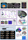
Advances in fetal and neonatal neuroimaging and everyday exposures
- PMID: 38877283
- PMCID: PMC11624138
- DOI: 10.1038/s41390-024-03294-1
Advances in fetal and neonatal neuroimaging and everyday exposures
Abstract
The complex, tightly regulated process of prenatal brain development may be adversely affected by "everyday exposures" such as stress and environmental pollutants. Researchers are only just beginning to understand the neural sequelae of such exposures, with advances in fetal and neonatal neuroimaging elucidating structural, microstructural, and functional correlates in the developing brain. This narrative review discusses the wide-ranging literature investigating the influence of parental stress on fetal and neonatal brain development as well as emerging literature assessing the impact of exposure to environmental toxicants such as lead and air pollution. These 'everyday exposures' can co-occur with other stressors such as social and financial deprivation, and therefore we include a brief discussion of neuroimaging studies assessing the effect of social disadvantage. Increased exposure to prenatal stressors is associated with alterations in the brain structure, microstructure and function, with some evidence these associations are moderated by factors such as infant sex. However, most studies examine only single exposures and the literature on the relationship between in utero exposure to pollutants and fetal or neonatal brain development is sparse. Large cohort studies are required that include evaluation of multiple co-occurring exposures in order to fully characterize their impact on early brain development. IMPACT: Increased prenatal exposure to parental stress and is associated with altered functional, macro and microstructural fetal and neonatal brain development. Exposure to air pollution and lead may also alter brain development in the fetal and neonatal period. Further research is needed to investigate the effect of multiple co-occurring exposures, including stress, environmental toxicants, and socioeconomic deprivation on early brain development.
© 2024. The Author(s).
Conflict of interest statement
Competing interests: The authors declare no competing interests.
Figures


Similar articles
-
Epidemiologic evidence of relationships between reproductive and child health outcomes and environmental chemical contaminants.J Toxicol Environ Health B Crit Rev. 2008 May;11(5-6):373-517. doi: 10.1080/10937400801921320. J Toxicol Environ Health B Crit Rev. 2008. PMID: 18470797 Review.
-
Environmental exposures and fetal growth: the Haifa pregnancy cohort study.BMC Public Health. 2018 Jan 12;18(1):132. doi: 10.1186/s12889-018-5030-8. BMC Public Health. 2018. PMID: 29329571 Free PMC article.
-
Social Susceptibility to Multiple Air Pollutants in Cardiovascular Disease.Res Rep Health Eff Inst. 2021 Jul;2021(206):1-71. Res Rep Health Eff Inst. 2021. PMID: 36004603 Free PMC article.
-
Combined Impacts of Prenatal Environmental Exposures and Psychosocial Stress on Offspring Health: Air Pollution and Metals.Curr Environ Health Rep. 2020 Jun;7(2):89-100. doi: 10.1007/s40572-020-00273-6. Curr Environ Health Rep. 2020. PMID: 32347455 Free PMC article. Review.
-
Neuroimaging is a novel tool to understand the impact of environmental chemicals on neurodevelopment.Curr Opin Pediatr. 2014 Apr;26(2):230-6. doi: 10.1097/MOP.0000000000000074. Curr Opin Pediatr. 2014. PMID: 24535497 Free PMC article. Review.
Cited by
-
Machine-learning based prediction of future outcome using multimodal MRI during early childhood.Semin Fetal Neonatal Med. 2024 Nov;29(2-3):101561. doi: 10.1016/j.siny.2024.101561. Epub 2024 Nov 7. Semin Fetal Neonatal Med. 2024. PMID: 39528363 Review.
-
MRI-Based Structural Development of the Human Newborn Hypothalamus.bioRxiv [Preprint]. 2025 Jun 21:2025.06.20.660741. doi: 10.1101/2025.06.20.660741. bioRxiv. 2025. PMID: 40667207 Free PMC article. Preprint.
References
-
- Maternal Mental Health Alliance. “All About Maternal Mental Health”. https://maternalmentalhealthalliance.org/about-maternal-mental-health/ Last accessed 9th February 2024.
-
- World Health Organization. “Air Pollution”. https://www.who.int/health-topics/air-pollution Last accessed 9th February 2024
Publication types
MeSH terms
Substances
LinkOut - more resources
Full Text Sources

