. 2024 Jun 14;7(1):682.
doi: 10.1038/s42003-024-06280-9.

Left ventricular trabeculation in Hominidae: divergence of the human cardiac phenotype

Affiliations

Left ventricular trabeculation in Hominidae: divergence of the human cardiac phenotype

Bryony A Curry et al. Commun Biol. .

Abstract

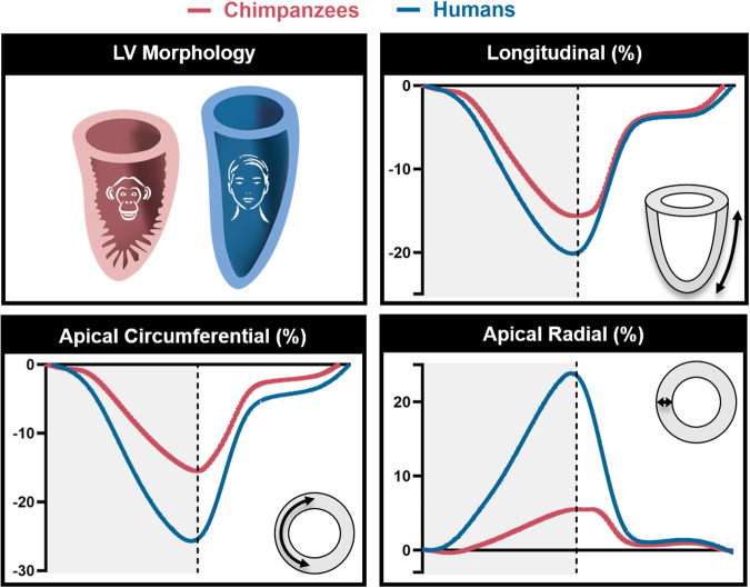
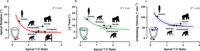
Although the gross morphology of the heart is conserved across mammals, subtle interspecific variations exist in the cardiac phenotype, which may reflect evolutionary divergence among closely-related species. Here, we compare the left ventricle (LV) across all extant members of the Hominidae taxon, using 2D echocardiography, to gain insight into the evolution of the human heart. We present compelling evidence that the human LV has diverged away from a more trabeculated phenotype present in all other great apes, towards a ventricular wall with proportionally greater compact myocardium, which was corroborated by post-mortem chimpanzee (Pan troglodytes) hearts. Speckle-tracking echocardiographic analyses identified a negative curvilinear relationship between the degree of trabeculation and LV systolic twist, revealing lower rotational mechanics in the trabeculated non-human great ape LV. This divergent evolution of the human heart may have facilitated the augmentation of cardiac output to support the metabolic and thermoregulatory demands of the human ecological niche.


Conflict of interest statement

The authors declare no competing interests.

Figures

Fig. 1
Fig. 1. Comparison of left ventricular trabeculation in great apes.
The bullseye plots represent the trabecular:compact (T:C) ratio for each segment of the left ventricle. The outer layer of the bullseye plots represents the basal segments, the middle and innermost layers represent the midpapillary and apical segments of the left ventricle, respectively. Red segments correspond to an average T:C ratio of >2; orange segments correspond to an average T:C ratio >1.5–2; yellow segments correspond to an average T:C ratio of >1–1.5; green segments correspond to an average T:C ratio of >0.5–1; blue segments correspond to an average T:C ratio of <0.05. Echocardiographic images of the parasternal short-axis at the apex are shown at end-diastole. *No data were available for the basal or midpapillary segments in the orangutans due to artifact from laryngeal air sacs. †These T:C ratios compare favorably with other reports in healthy human cohorts, ranging from 0.2 to 0.9,. ‡Anatomical labels have been provided in accordance with the conventional guidelines for cardiac chamber quantification by the American Society of Echocardiography and European Association of Cardiovascular Imaging. However, we note that this clinical convention does not align with the recognized anatomical approach and may result in confusion across disciplines—see ref. for further clarification.
Fig. 2
Fig. 2. Graphical representation of the trabecular:compact (T:C) ratio for each segment of the left ventricle in chimpanzees.
The outer layer of the bullseye plots represents the basal segments, the middle and innermost layers represent the midpapillary and apical segments of the left ventricle, respectively. Red segments correspond to an average T:C ratio of >2; orange segments correspond to an average T:C ratio >1.5–2; yellow segments correspond to an average T:C ratio of >1–1.5; green segments correspond to an average T:C ratio of >0.5–1; blue segments correspond to an average T:C ratio of <0.05. Infant age class includes individuals of ≤4 years of age, juveniles between 5–7 years of age, sub-adults between 8-11 years of age and adults ≥12 years of age.
Fig. 3
Fig. 3. Comparison of left ventricular morphology and mechanical indices of ventricular function between chimpanzees and humans.
Shortening along the long axis of the left ventricle (i.e., longitudinal strain) and deformation at the apex (i.e., apical circumferential and apical radial strain) were averaged across a mixed-sex, adult cohort of chimpanzees (n = 136) and represented in maroon. Blue reflects the deformation patterns of a mixed-sex, adult human cohort (n = 34). Dashed line represents aortic valve closure. Gray shading to the left of dashed line represents systole and white represents diastole.
Fig. 4
Fig. 4. Relationship between markers of left ventricular (LV) function and apical trabeculation in the extant Hominidae taxon.
a Peak LV systolic apical rotation, shown in red, in a mixed-sex, adult cohort of humans (male n = 18, female n = 16), chimpanzees (male n = 59, female n = 51), bonobos (male n = 2, female n = 4), gorillas (male n = 4, female n = 6) and orangutans (male n = 10, female n = 6). b Peak LV systolic twist, shown in green, and (c) peak diastolic untwisting velocity, shown in blue, in a cohort of humans (male n = 18, female n = 16), chimpanzees (male n = 47, female n = 43), bonobos (male n = 1, female n = 3) and gorillas (male n = 4, female n = 5). Analyses of LV twist and untwisting velocity were not possible in all individuals, nor any of the orangutans due to artifacts from laryngeal air sacs, hence the reduced sample size. The exponential plateau curve is shown, with the 95% confidence bands represented by the dotted line. The mean and standard error are shown in black for each species.

References

    1. Stephenson A, Adams JW, Vaccarezza M. The vertebrate heart: an evolutionary perspective. J. Anat. 2017;231:787–797. doi: 10.1111/joa.12687. - DOI - PMC - PubMed
    1. Rowlatt U. Functional morphology of the heart in mammals. Am. Zool. 1968;8:221–229. doi: 10.1093/icb/8.2.221. - DOI - PubMed
    1. Shave RE, et al. Selection of endurance capabilities and the trade-off between pressure and volume in the evolution of the human heart. Proc. Natl. Acad. Sci. USA. 2019;116:19905–19910. doi: 10.1073/pnas.1906902116. - DOI - PMC - PubMed
    1. Jensen B, et al. The hypertrabeculated (noncompacted) left ventricle is different from the ventricle of embryos and ectothermic vertebrates. Biochim. Biophys. Acta. 2016;1863:1696–1706. doi: 10.1016/j.bbamcr.2015.10.018. - DOI - PubMed
    1. Sedmera D. Function and form in the developing cardiovascular system. Cardiovasc. Res. 2011;91:252–259. doi: 10.1093/cvr/cvr062. - DOI - PubMed

Publication types