Review
. 2024 Jun 14;14(1):89.
doi: 10.1186/s13613-024-01325-y.

Impaired angiotensin II signaling in septic shock

Affiliations
Review

Impaired angiotensin II signaling in septic shock

Adrien Picod et al. Ann Intensive Care. .

Abstract

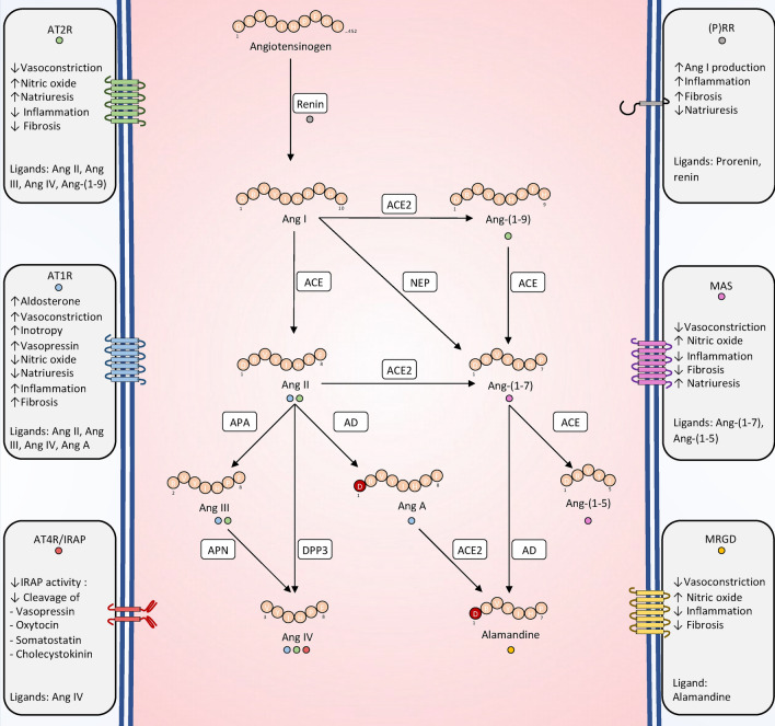
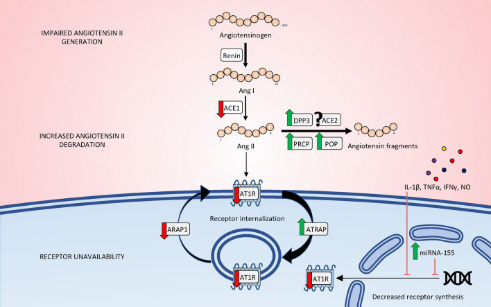
Recent years have seen a resurgence of interest for the renin-angiotensin-aldosterone system in critically ill patients. Emerging data suggest that this vital homeostatic system, which plays a crucial role in maintaining systemic and renal hemodynamics during stressful conditions, is altered in septic shock, ultimately leading to impaired angiotensin II-angiotensin II type 1 receptor signaling. Indeed, available evidence from both experimental models and human studies indicates that alterations in the renin-angiotensin-aldosterone system during septic shock can occur at three distinct levels: 1. Impaired generation of angiotensin II, possibly attributable to defects in angiotensin-converting enzyme activity; 2. Enhanced degradation of angiotensin II by peptidases; and/or 3. Unavailability of angiotensin II type 1 receptor due to internalization or reduced synthesis. These alterations can occur either independently or in combination, ultimately leading to an uncoupling between the renin-angiotensin-aldosterone system input and downstream angiotensin II type 1 receptor signaling. It remains unclear whether exogenous angiotensin II infusion can adequately address all these mechanisms, and additional interventions may be required. These observations open a new avenue of research and offer the potential for novel therapeutic strategies to improve patient prognosis. In the near future, a deeper understanding of renin-angiotensin-aldosterone system alterations in septic shock should help to decipher patients' phenotypes and to implement targeted interventions.

Keywords: Angiotensin II; Angiotensin-converting enzyme; Circulatory failure; Dipeptidyl peptidase 3; Neprilysin; Renin–angiotensin–aldosterone system; Sepsis; Septic shock.


Conflict of interest statement

The Cardiovascular Markers in Stress Conditions (MASCOT) Research Group is supported by a research grant from 4TEEN4 Pharmaceuticals GmbH, which allowed salary support for two co-authors (AP, MG). AM received speaker’s honoraria from Abbott, Novartis, Orion, Roche, and Servier, and fees as a member of the advisory board and/or steering committee from Cardiorentis, Adrenomed, MyCartis, Neurotronik, and Sphingotec. PP received travel and consultancy reimbursement from Adrenomed, SphingoTec, 4TEEN4, AM-Pharma, Baxter, and EBI. The remaining authors have nothing to disclose.

Figures

Fig. 1
Fig. 1
Overview of the RAAS. All angiotensin peptides originate from the sequential proteolytic cleavage of angiotensinogen. For clarity, only peptides with a firmly established biological function are depicted. The interactions of a given peptide with its receptor(s) are illustrated by the color-coded dot(s): blue (AT1R), green (AT2R), red (AT4R/IRAP), gray (PRR), purple (MAS) or yellow (MGRD). In addition to the ligand-receptor interactions depicted, a potential binding of Ang-(1–7) to AT1R at elevated concentrations has been reported. However, the biological significance of this interaction remains uncertain. Ang angiotensin, ACE angiotensin-converting enzyme, ACE2 angiotensin-converting enzyme 2, NEP neprilysin, DPP3 dipeptidyl peptidase 3, APA aminopeptidase A, APN aminopeptidase N, AD aspartate decarboxylase, (P)RR (pro-)renin receptor, AT1R angiotensin II, type 1 receptor, AT2R angiotensin II, type 2 receptor, AT4R angiotensin IV receptor; MRGD MAS-related G-protein-coupled D receptor
Fig. 2
Fig. 2
Regulation of renin secretion. Juxtaglomerular cells release renin through exocytosis in response to various stimuli. Systemic factors such as blood pressure, salt intake, and activation of the sympathetic nervous system exert their effects, in part, through the mediators described here. At the cellular level, all molecular mediators that stimulate renin release do so by elevating intracellular cAMP concentration. Conversely, any stimuli associated with intracellular calcium accumulation inhibit renin release. cGMP can either stimulate or inhibit renin secretion depending on its mode of production. Activation of sGC by NO inhibits PDE3 and thus stimulates renin release through cAMP accumulation. Conversely, activation of mGC-A by ANP or BNP inhibits renin release. SNS sympathetic nervous system, NO nitric oxide, AC adenylate cyclase, mGC-A membrane guanylate cyclase A, sGC soluble guanylate cyclase, PDE3/4 phosphodiesterase 3/4, ATP adenosine triphosphate, cAMP cyclic adenosine monophosphate, GTP guanosine triphosphate, cGMP cyclic guanosine monophosphate
Fig. 3
Fig. 3
Glomerular hemodynamics in health and circulatory failure. Basal state (A). During circulatory failure characterized by low cardiac output (e.g., cardiogenic shock), reflex myogenic vasodilation of the afferent arteriole and vasoconstriction of the efferent arteriole tend to maintain PGC and therefore GFR. When these mechanisms are insufficient to maintain PGC, GFR falls (B). Conversely, during vasodilatory shock with high cardiac output (e.g., septic shock), preferential vasodilation of the efferent arteriole is sufficient to explain the fall in PCG and therefore GFR decrease despite a high RBF (C). Additionally, reopening of peri-glomerular shunts could participate in the RBF–GFR decoupling observed during sepsis-associated acute kidney injury (D). RBF renal blood flow, PGC Glomerular capillary pressure, GFR Glomerular filtration rate
Fig. 4
Fig. 4
Mechanisms of impaired Ang II–AT1R signaling in septic shock. Impaired angiotensin II–AT1R signaling occur at multiple levels, including: 1. Impaired generation of angiotensin II, possibly attributable to defects in angiotensin-converting enzyme activity; 2. Enhanced degradation of angiotensin II by peptidases such as circulating DPP3, POP or PRCP; and/or 3. Unavailability of the AT1R receptor due to internalization or reduced synthesis under the influence of pro-inflammatory cytokines, NO or miRNA-155. Importantly, the angiotensin fragments that originate from angiotensin II cleavage could also mediates their own biological effects (e.g., angiotensin (1–7) via ACE2, POP or PRCP; angiotensin IV via DPP3). ACE angiotensin-converting enzyme, DPP3 dipeptidyl peptidase 3, ACE2 angiotensin-converting enzyme 2, POP prolyl olygopeptidase, PRCP prolyl carboxypeptidase, AT1R angiotensin II, type 1 receptor, ARAP1 AT1R-associated protein 1, ATRAP AT1R-associated protein. IL-1β interleukin-1β, TNFα tumor necrosis factor α, IFN interferon γ, NO nitric oxide

Similar articles

Cited by

References

    1. Singer M, Deutschman CS, Seymour CW, Shankar-Hari M, Annane D, Bauer M, et al. The third international consensus definitions for sepsis and septic shock (Sepsis-3) JAMA. 2016;315(8):801–810. doi: 10.1001/jama.2016.0287. - DOI - PMC - PubMed
    1. Vincent JL, De Backer D. Circulatory shock. N Engl J Med. 2013;369(18):1726–1734. doi: 10.1056/NEJMra1208943. - DOI - PubMed
    1. Evans L, Rhodes A, Alhazzani W, Antonelli M, Coopersmith CM, French C, et al. Surviving sepsis campaign: international guidelines for management of sepsis and septic shock 2021. Intensive Care Med. 2021;47(11):1181–1247. doi: 10.1007/s00134-021-06506-y. - DOI - PMC - PubMed
    1. Khanna A, English SW, Wang XS, Ham K, Tumlin J, Szerlip H, et al. Angiotensin II for the treatment of vasodilatory shock. N Engl J Med. 2017;377(5):419–430. doi: 10.1056/NEJMoa1704154. - DOI - PubMed
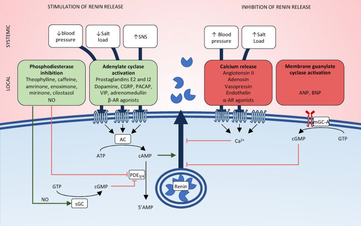
    1. Schweda F, Kurtz A. Regulation of renin release by local and systemic factors. Rev Physiol Biochem Pharmacol. 2011;161:1–44. - PubMed

LinkOut - more resources