. 2024 Jun 14;24(1):552.
doi: 10.1186/s12870-024-05249-4.

Revealing the Arabidopsis AtGRP7 mRNA binding proteome by specific enhanced RNA interactome capture

Affiliations

Revealing the Arabidopsis AtGRP7 mRNA binding proteome by specific enhanced RNA interactome capture

Marlene Reichel et al. BMC Plant Biol. .

Abstract

Background: The interaction of proteins with RNA in the cell is crucial to orchestrate all steps of RNA processing. RNA interactome capture (RIC) techniques have been implemented to catalogue RNA- binding proteins in the cell. In RIC, RNA-protein complexes are stabilized by UV crosslinking in vivo. Polyadenylated RNAs and associated proteins are pulled down from cell lysates using oligo(dT) beads and the RNA-binding proteome is identified by quantitative mass spectrometry. However, insights into the RNA-binding proteome of a single RNA that would yield mechanistic information on how RNA expression patterns are orchestrated, are scarce.

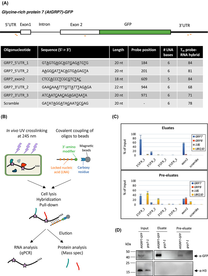
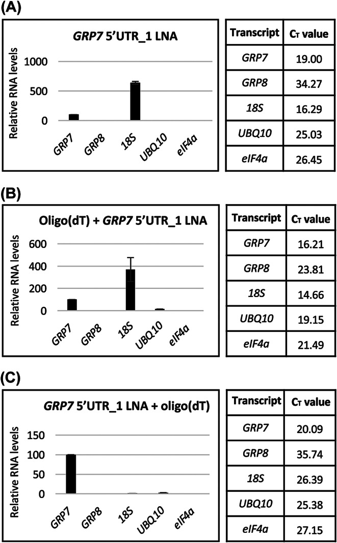
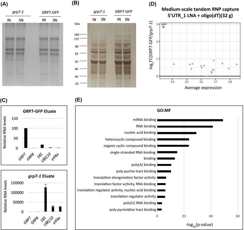
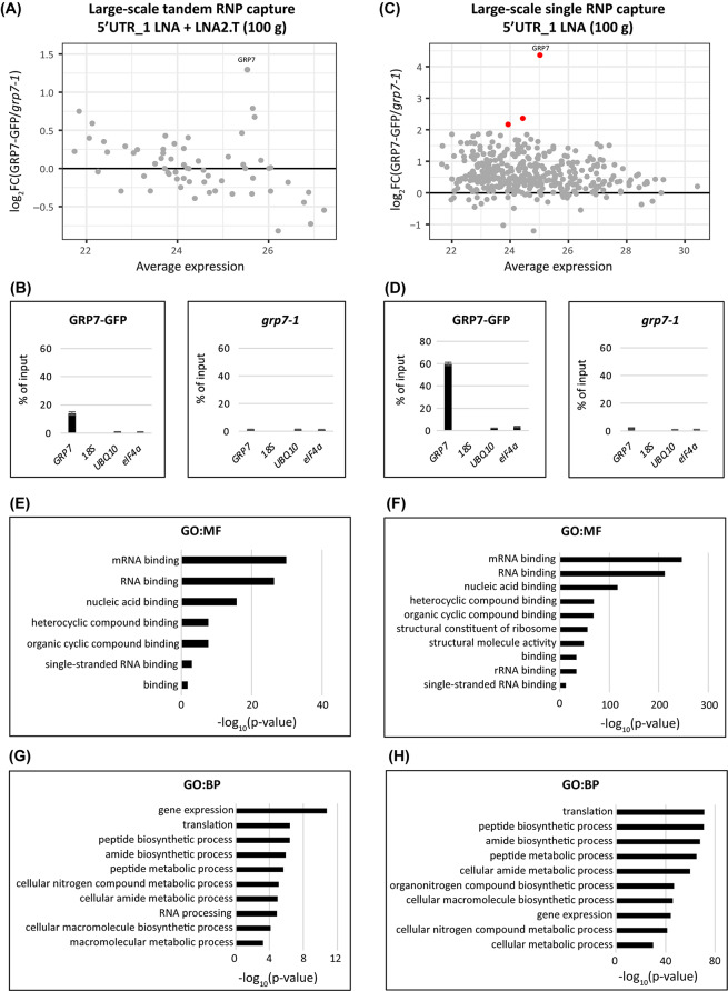
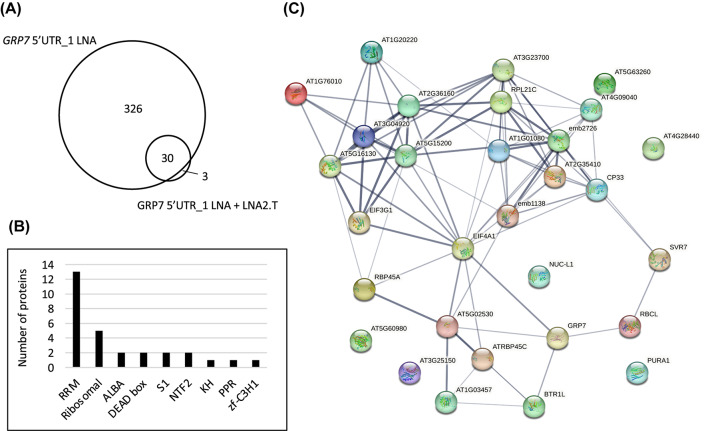
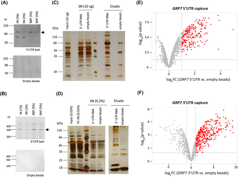
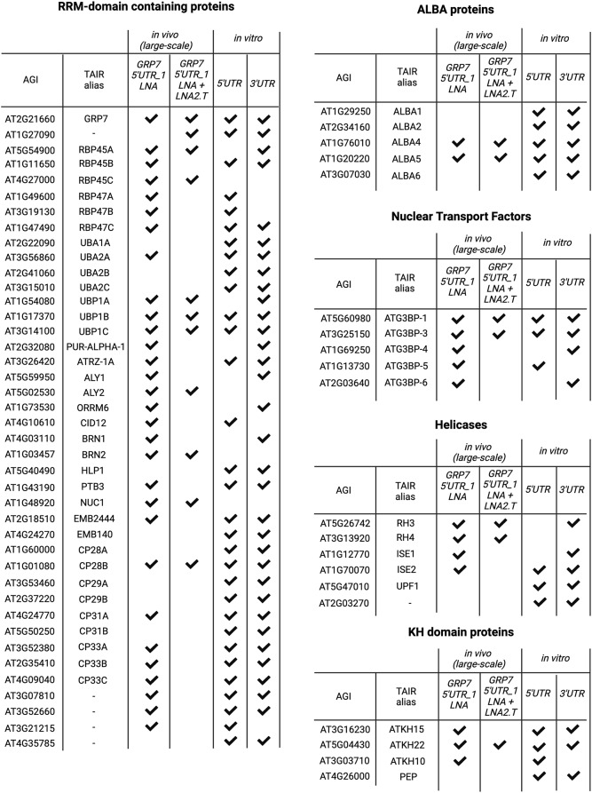
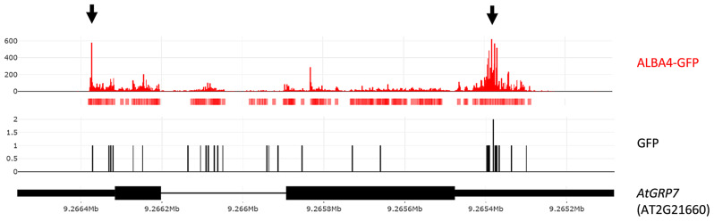
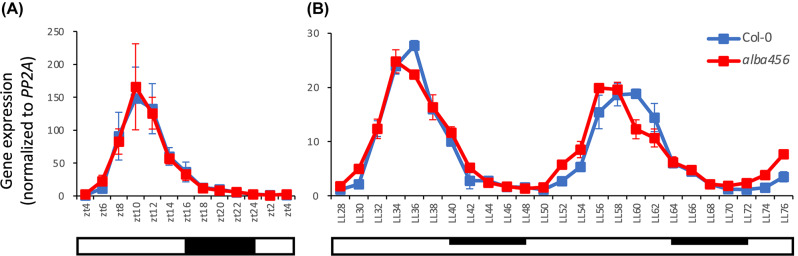
Results: Here, we explored RIC in Arabidopsis to identify proteins interacting with a single mRNA, using the circadian clock-regulated Arabidopsis thaliana GLYCINE-RICH RNA-BINDING PROTEIN 7 (AtGRP7) transcript, one of the most abundant transcripts in Arabidopsis, as a showcase. Seedlings were treated with UV light to covalently crosslink RNA and proteins. The AtGRP7 transcript was captured from cell lysates with antisense oligonucleotides directed against the 5'untranslated region (UTR). The efficiency of RNA capture was greatly improved by using locked nucleic acid (LNA)/DNA oligonucleotides, as done in the enhanced RIC protocol. Furthermore, performing a tandem capture with two rounds of pulldown with the 5'UTR oligonucleotide increased the yield. In total, we identified 356 proteins enriched relative to a pulldown from atgrp7 mutant plants. These were benchmarked against proteins pulled down from nuclear lysates by AtGRP7 in vitro transcripts immobilized on beads. Among the proteins validated by in vitro interaction we found the family of Acetylation Lowers Binding Affinity (ALBA) proteins. Interaction of ALBA4 with the AtGRP7 RNA was independently validated via individual-nucleotide resolution crosslinking and immunoprecipitation (iCLIP). The expression of the AtGRP7 transcript in an alba loss-of-function mutant was slightly changed compared to wild-type, demonstrating the functional relevance of the interaction.

Conclusion: We adapted specific RNA interactome capture with LNA/DNA oligonucleotides for use in plants using AtGRP7 as a showcase. We anticipate that with further optimization and up scaling the protocol should be applicable for less abundant transcripts.

Keywords: Arabidopsis; Enhanced RNA interactome capture; LNA oligonucleotides; iCLIP.


Conflict of interest statement

The authors declare no competing interests.

Figures

Fig. 1
Fig. 1
Principle of the AtGRP7 RNA interactome capture. (A) Scheme of the AtGRP7-GFP mRNA and details of the antisense LNA/DNA mixmer oligonucleotides tested for capture of the AtGRP7 transcript. LNA nucleotides are underlined. The probe position is indicated relative to the TAIR cDNA sequence. (B) Principle of the specific RNP capture. 14-day-old seedlings are crosslinked with 254 nm UV light to establish covalent bonds between RNA and proteins. The LNA oligonucleotides are coupled to carboxylated magnetic beads via a primary amine attached to a C6 linker at their 3’end. Cell lysates are incubated with the bead-coupled oligonucleotides to pull down proteins interacting with the AtGRP7 RNA. RNA-protein complexes are eluted and either subjected to RNA analysis via RT-qPCR or to protein analysis via mass spectrometry. (C) Capture efficiency and specificity with the different LNA/DNA mixmer oligonucleotides. Captures were performed with UV crosslinked AtGRP7::AtGRP7-GFP (grp7-1) seedlings and RNA levels of AtGRP7, AtGRP8, 18 S rRNA and UBIQUITIN 10 were measured in the eluates (top panel) and pre-eluates (bottom panel) by RT-qPCR using the primers listed in Additional file 1. Transcript levels are expressed relative to the transcript level in the input. (D) Immunoblot analysis of AtGRP7::AtGRP7-GFP grp7-1 and grp7-1 control plants subjected to RNP capture with 5’UTR_1 LNA oligonucleotides. The lysate (input), pre-eluate, and eluate fractions were probed with the α-GFP antibody (top) or the α-Histone H3 antibody (bottom)
Fig. 2
Fig. 2
Optimization of the capture efficiency for the LNA 5’UTR_1 probe by tandem capture with oligo(dT). Relative RNA levels in the eluates after a single round of RNP capture with LNA 5’UTR_1 (A), tandem capture with oligo(dT) followed by the LNA 5’UTR_1 probe (B), and tandem capture with the LNA 5’UTR_1 probe followed by oligo(dT) capture (C). The AtGRP7 level is set to 100%
Fig. 3
Fig. 3
Mass spectrometry analysis of proteins bound to the AtGRP7 transcript after medium-scale tandem capture. (A) Agarose-formaldehyde gel electrophoresis of total RNA in the lysate (input) and the supernatant after probe hybridization (SN) in AtGRP7-GFP grp7-1 plants and grp7-1 control plants. (B) Silver staining of total protein in the lysate (input) and the supernatant after probe hybridization (SN) in the AtGRP7-GFP grp7-1 plants and grp7-1 control plants. The positions of the molecular weight markers are indicated. (C) Relative RNA levels of AtGRP7, AtGRP8, 18 S rRNA, UBIQUITIN10, and eIF4α RNA in the eluates of AtGRP7-GFP grp7-1 plants (top) and grp7-1 control plants (bottom). (D) MA plot of identified proteins displaying the relation between log2 fold-change and the average expression (as log2 TMT signal). The AtGRP7 protein was significantly enriched (red dot). (E) GO term analysis of the molecular function of the proteins identified
Fig. 4
Fig. 4
Mass spectrometry of proteins binding to the AtGRP7 transcript after large-scale capture. MA plot of identified proteins (A, C), relative RNA levels of AtGRP7, 18 S rRNA, UBIQUITIN 10, and eIF4α in the eluates of AtGRP7-GFP grp7-1 plants (left) and grp7-1 control plants (right) (B, D) and enriched GO terms of identified proteins (E and G, F and G) after large-scale tandem capture with two consecutive rounds of hybridization with the 5’UTR_1 LNA oligo followed by LNA2.T capture, or after large-scale single capture with two consecutive rounds of hybridization with the 5’UTR_1 LNA oligo only. Proteins with a log2 fold-change  2) are indicated by the red dots
Fig. 5
Fig. 5
Properties of proteins with a positive fold-change after large-scale RNP capture. (A) Venn diagram showing the overlap between the proteins identified in the large-scale single capture with two consecutive rounds of hybridization with the 5’UTR_1 LNA oligo and the large-scale tandem capture with hybridization with 5’UTR_1 LNA oligo followed by LNA2.T capture. (B) Number of proteins annotated with classical RNA-binding domains among the 30 common proteins. (C) STRING network analysis of the 30 common proteins
Fig. 6
Fig. 6
Identification of AtGRP7 binding proteins by in vitro pulldowns. (A, B) Coupling efficiency of biotinylated 5’UTR bait (A) and 3’UTR bait (B) to magnetic streptavidin beads. Aliquots of the RNA isolated from the input (IN), supernatant after coupling (SN) and eluates from the beads before (BBP) and after pulldown (BAP) were analyzed on 12.5% urea PAGE gels. Empty beads were used as controls. The arrows indicate the in vitro transcripts of the sizes expected for the 5’UTR (A) or 3’UTR (B), respectively. The additional bands likely represent additional conformations. (C, D) Silver staining of proteins recovered after in vitro captures with 5’UTR bait (C) and 3’UTR bait (D). Aliquots of the input, supernatant and eluates of the respective coupled and empty beads were separated on a 12% SDS-Page and subjected to silver staining. (E, F) Volcano plots of proteins identified by in vitro capture with the 5’UTR bait (E) or 3’UTR bait (F). Significantly enriched proteins (log2 fold change  1, p-value < 0.05) are indicated by the red dots
Fig. 7
Fig. 7
Overview of prominent protein groups identified by in vivo and in vitro pulldowns of AtGRP7 interactors
Fig. 8
Fig. 8
In vivo binding of ALBA4 to the AtGRP7 transcript. Binding sites of ALBA4::ALBA4-GFP in alba456 to AtGRP7 determined by iCLIP [73]. iCLIP peaks of ALBA4 are shown in red and peaks of a GFP control sample are shown in black. The red boxes below the ALBA4-GFP iCLIP reads denote called peaks. Prominent ALBA4 peaks are highlighted by arrows. The AtGRP7 gene model is shown at the bottom. Black boxes: exons; narrow black boxes: untranslated regions; line: intron
Fig. 9
Fig. 9
Impact of altered ALBA protein levels on AtGRP7 transcript oscillations. Col-0 wild type plants and the alba456 triple mutant were grown in long day conditions (16 h light/8 h dark) for 12 days before transfer to constant light (LL). Seedlings were harvested at 2-h intervals throughout the light-dark cycle (A) and from 28 h to 76 h in LL (B). AtGRP7 transcript levels were analyzed by RT-qPCR and normalized to PP2A. Error bars represent the standard deviation of two biological replicates. Open bar: constant light; dark bar: (subjective) night

Similar articles

References

    1. Singh G, Pratt G, Yeo GW, Moore MJ. The clothes make the mRNA: past and Present trends in mRNP Fashion. Annu Rev Biochem. 2015;84(1):325–54. doi: 10.1146/annurev-biochem-080111-092106. - DOI - PMC - PubMed
    1. Glisovic T, Bachorik JL, Yong J, Dreyfuss G. RNA-binding proteins and post-transcriptional gene regulation. FEBS Lett. 2008;582(14):1977–86. doi: 10.1016/j.febslet.2008.03.004. - DOI - PMC - PubMed
    1. Mateos JL, Staiger D. Toward a systems view on RNA-binding proteins and associated RNAs in plants: guilt by association. Plant Cell. 2023;35(6):1708–26. doi: 10.1093/plcell/koac345. - DOI - PMC - PubMed
    1. Castello A, Fischer B, Eichelbaum K, Horos R, Beckmann B, Strein C, Davey Norman E, Humphreys David T, Preiss T, Steinmetz L, et al. Insights into RNA Biology from an Atlas of mammalian mRNA-Binding proteins. Cell. 2012;149(6):1393–406. doi: 10.1016/j.cell.2012.04.031. - DOI - PubMed
    1. Baltz AG, Munschauer M, Schwanhäusser B, Vasile A, Murakawa Y, Schueler M, Youngs N, Penfold-Brown D, Drew K, Milek M, et al. The mRNA-Bound proteome and its global occupancy Profile on protein-coding transcripts. Mol Cell. 2012;46(5):674–90. doi: 10.1016/j.molcel.2012.05.021. - DOI - PubMed

MeSH terms

LinkOut - more resources