

α-Ketoglutarate alleviates osteoarthritis by inhibiting ferroptosis via the ETV4/SLC7A11/GPX4 signaling pathway
- PMID: 38877424
- PMCID: PMC11177415
- DOI: 10.1186/s11658-024-00605-6
α-Ketoglutarate alleviates osteoarthritis by inhibiting ferroptosis via the ETV4/SLC7A11/GPX4 signaling pathway
Abstract
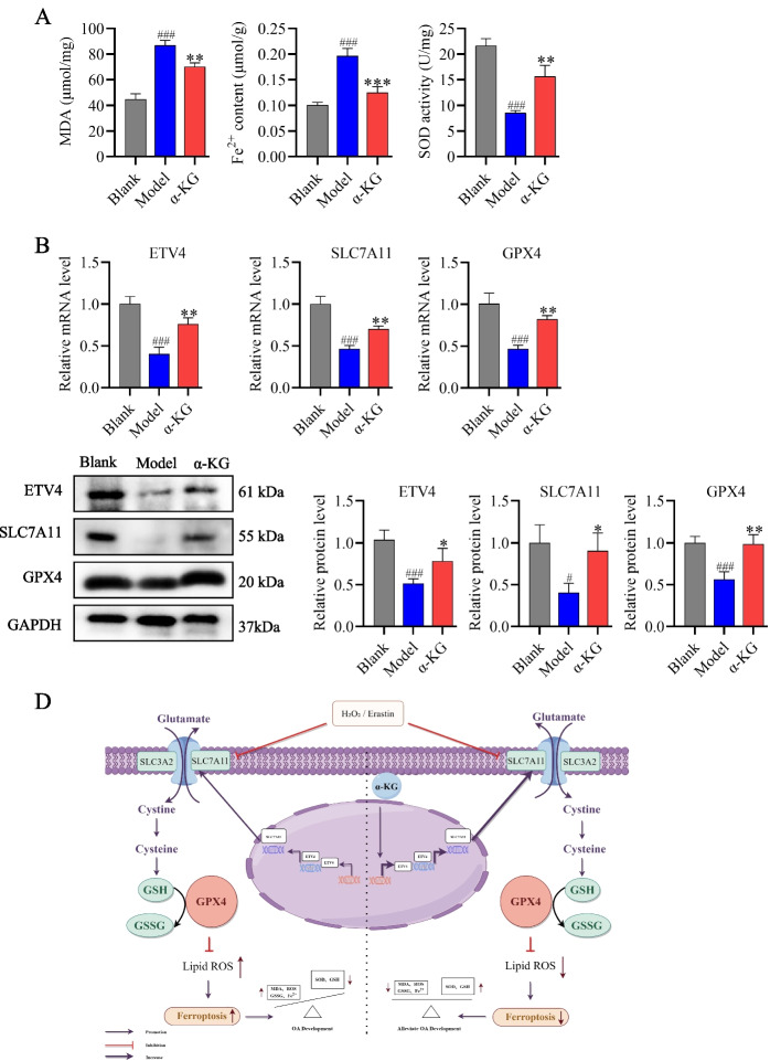
Osteoarthritis (OA) is the most common degenerative joint disorder that causes disability in aged individuals, caused by functional and structural alterations of the knee joint. To investigate whether metabolic drivers might be harnessed to promote cartilage repair, a liquid chromatography-mass spectrometry (LC-MS) untargeted metabolomics approach was carried out to screen serum biomarkers in osteoarthritic rats. Based on the correlation analyses, α-ketoglutarate (α-KG) has been demonstrated to have antioxidant and anti-inflammatory properties in various diseases. These properties make α-KG a prime candidate for further investigation of OA. Experimental results indicate that α-KG significantly inhibited H2O2-induced cartilage cell matrix degradation and apoptosis, reduced levels of reactive oxygen species (ROS) and malondialdehyde (MDA), increased superoxide dismutase (SOD) and glutathione (GSH)/glutathione disulfide (GSSG) levels, and upregulated the expression of ETV4, SLC7A11 and GPX4. Further mechanistic studies observed that α-KG, like Ferrostatin-1 (Fer-1), effectively alleviated Erastin-induced apoptosis and ECM degradation. α-KG and Fer-1 upregulated ETV4, SLC7A11, and GPX4 at the mRNA and protein levels, decreased ferrous ion (Fe2+) accumulation, and preserved mitochondrial membrane potential (MMP) in ATDC5 cells. In vivo, α-KG treatment inhibited ferroptosis in OA rats by activating the ETV4/SLC7A11/GPX4 pathway. Thus, these findings indicate that α-KG inhibits ferroptosis via the ETV4/SLC7A11/GPX4 signaling pathway, thereby alleviating OA. These observations suggest that α-KG exhibits potential therapeutic properties for the treatment and prevention of OA, thereby having potential clinical applications in the future.
Keywords: ETV4/SLC7A11/GPX4 signaling pathway; Ferroptosis; Osteoarthritis; α-ketoglutarate.
© 2024. The Author(s).
Conflict of interest statement
The authors declare that they have no competing interests.
Figures


Similar articles
-
Icariin inhibits chondrocyte ferroptosis and alleviates osteoarthritis by enhancing the SLC7A11/GPX4 signaling.Int Immunopharmacol. 2024 May 30;133:112010. doi: 10.1016/j.intimp.2024.112010. Epub 2024 Apr 17. Int Immunopharmacol. 2024. PMID: 38636375
-
The role and possible mechanism of the ferroptosis-related SLC7A11/GSH/GPX4 pathway in myocardial ischemia-reperfusion injury.BMC Cardiovasc Disord. 2024 Oct 1;24(1):531. doi: 10.1186/s12872-024-04220-3. BMC Cardiovasc Disord. 2024. PMID: 39354361 Free PMC article.
-
Folic Acid Ameliorates Neuronal Ferroptosis in Aging by Up-Regulating SLC7A11-GSH-GPX4 Antioxidant Pathway and Increasing Cystine Levels.Int J Mol Sci. 2025 Jul 11;26(14):6669. doi: 10.3390/ijms26146669. Int J Mol Sci. 2025. PMID: 40724917 Free PMC article.
-
The role of ferroptosis in osteoarthritis: Progress and prospects.Biochem Biophys Res Commun. 2024 Nov 12;733:150683. doi: 10.1016/j.bbrc.2024.150683. Epub 2024 Sep 10. Biochem Biophys Res Commun. 2024. PMID: 39293333 Review.
-
Novel insights into the role of ferroptosis in temporomandibular joint osteoarthritis and knee osteoarthritis.Int J Med Sci. 2025 Apr 9;22(9):2119-2131. doi: 10.7150/ijms.107057. eCollection 2025. Int J Med Sci. 2025. PMID: 40303500 Free PMC article. Review.
Cited by
-
Exogenous Alpha-Ketoglutaric Acid Alleviates the Rabbit Dermal Papilla Cell Oxidative Damage Caused by Hydrogen Peroxide Through the ERK/Nrf2 Signaling Pathway.Antioxidants (Basel). 2025 Apr 11;14(4):455. doi: 10.3390/antiox14040455. Antioxidants (Basel). 2025. PMID: 40298808 Free PMC article.
-
Paeonol ameliorates ferroptosis and inflammation in chondrocytes through AMPK/Nrf2/GPX4 pathway.Front Pharmacol. 2025 Mar 7;16:1526623. doi: 10.3389/fphar.2025.1526623. eCollection 2025. Front Pharmacol. 2025. PMID: 40124777 Free PMC article.
-
Inhibition of Heat Shock Protein 90β by Catalpol: A Potential Therapeutic Approach for Alleviating Inflammation-Induced Cartilage Injuries in Osteoarthritis.Adv Sci (Weinh). 2025 Jul;12(26):e2503909. doi: 10.1002/advs.202503909. Epub 2025 Apr 25. Adv Sci (Weinh). 2025. PMID: 40277849 Free PMC article.
-
Novel insights into the role of metabolic disorder in osteoarthritis.Front Endocrinol (Lausanne). 2024 Dec 18;15:1488481. doi: 10.3389/fendo.2024.1488481. eCollection 2024. Front Endocrinol (Lausanne). 2024. PMID: 39744183 Free PMC article. Review.
-
PSAT1 impairs ferroptosis and reduces immunotherapy efficacy via GPX4 hydroxylation.Nat Chem Biol. 2025 Sep;21(9):1420-1432. doi: 10.1038/s41589-025-01887-3. Epub 2025 Apr 25. Nat Chem Biol. 2025. PMID: 40281343
References
MeSH terms
Substances
Grants and funding
- No. U22A20367/the National Natural Science Foundation Regional Innovation and Development Joint Fund
- No. 2023DJ04/the Innovation and Entrepreneurship Talent Project of Jilin Province
- Grant number 20210304001YY/Science and Technology Major Project of Jilin Province
- 20200201406JC/Key Laboratory in Science and Technology Development Project of Suzhou
- No. 82305274/Innovative Research Group Project of the National Natural Science Foundation of China
LinkOut - more resources
Full Text Sources
Medical
