. 2024 Jun 14;21(1):139.
doi: 10.1186/s12985-024-02411-0.

Evaluation of novel Epstein-Barr virus-derived antigen formulations for monitoring virus-specific T cells in pediatric patients with infectious mononucleosis

Affiliations

Evaluation of novel Epstein-Barr virus-derived antigen formulations for monitoring virus-specific T cells in pediatric patients with infectious mononucleosis

Franziska Fischer et al. Virol J. .

Abstract

Background: Infection with the Epstein-Barr virus (EBV) elicits a complex T-cell response against a broad range of viral proteins. Hence, identifying potential differences in the cellular immune response of patients with different EBV-associated diseases or different courses of the same disorder requires interrogation of a maximum number of EBV antigens. Here, we tested three novel EBV-derived antigen formulations for their ability to reactivate virus-specific T cells ex vivo in patients with EBV-associated infectious mononucleosis (IM).

Methods: We comparatively analyzed EBV-specific CD4+ and CD8+ T-cell responses to three EBV-derived antigen formulations in 20 pediatric patients during the early phase of IM: T-activated EBV proteins (BZLF1, EBNA3A) and EBV-like particles (EB-VLP), both able to induce CD4+ and CD8+ T-cell responses ex vivo, as well as an EBV-derived peptide pool (PP) covering 94 well-characterized CD8+ T-cell epitopes. We assessed the specificity, magnitude, kinetics, and functional characteristics of EBV-specific immune responses at two sequential time points (v1 and v2) within the first six weeks after IM symptom onset (Tonset).

Results: All three tested EBV-derived antigen formulations enabled the detection of EBV-reactive T cells during the early phase of IM without prior T-cell expansion in vitro. EBV-reactive CD4+ and CD8+ T cells were mainly mono-functional (CD4+: mean 64.92%, range 56.15-71.71%; CD8+: mean 58.55%, range 11.79-85.22%) within the first two weeks after symptom onset (v1) with IFN-γ and TNF-secreting cells representing the majority of mono-functional EBV-reactive T cells. By contrast, PP-reactive CD8+ T cells were primarily bi-functional (>60% at v1 and v2), produced IFN-γ and TNF and had more tri-functional than mono-functional components. We observed a moderate correlation between viral load and EBNA3A, EB-VLP, and PP-reactive CD8+ T cells (rs = 0.345, 0.418, and 0.356, respectively) within the first two weeks after Tonset, but no correlation with the number of detectable EBV-reactive CD4+ T cells.

Conclusions: All three EBV-derived antigen formulations represent innovative and generic recall antigens suitable for monitoring EBV-specific T-cell responses ex vivo. Their combined use facilitates a thorough analysis of EBV-specific T-cell immunity and allows the identification of functional T-cell signatures linked to disease development and severity.

Keywords: Epstein-Barr virus; Immune monitoring; Infectious mononucleosis; Pediatric patients; T-cell response.


Conflict of interest statement

UP received personal fees from Abbott, Abbvie, Arbutus, Gilead, GSK, J & J, MSD, Roche, Sanofi, Sobi, and Vaccitech. UP is a cofounder and share-holder of SCG Cell Therapy. The lab of UP received grants from SCG Cell Therapy. The remaining authors declare no competing interests.

Figures

Fig. 1
Fig. 1
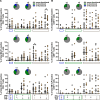
Total cytokine responses of EBV-reactive CD4+ and CD8+ T cells. Depicted are the frequencies of EBV-reactive CD4+ and CD8+ T cells upon stimulation of PBMC with T-activated EBNA3A and BZLF1 proteins, EBV-like particles (EB-VLP), and a synthetic EBV-derived peptide pool (PP) at visit 1 (v1) (A, B) (grey dots) and visit 2 (v2) (C, D) (brown dots). Total cytokine responses were calculated by adding together the frequencies of EBV-reactive IFN-γ, IL-2, and/or TNF-positive T cells within the CD4+ or CD8+ T-cell populations. Dots represent the patient samples analyzed (n = 20), the horizontal line marks the median, dashed lines the 25th and 75th percentiles. For reasons of clarity, the x-axis was moved to -0.05. v1: 2-14 days after symptom onset; v2: 23-39 days after symptom onset
Fig. 2
Fig. 2
Individual EBV-specific CD4+ and CD8+ T-cell responses at visits 1 and 2. Stacked bars depict the frequencies of EBV-reactive CD4+ and CD8+ T cells upon stimulation of PBMC from 20 IM patients with T-activated EBNA3A and BZLF1 proteins, EBV-like particles (EB-VLP), and a synthetic EBV-derived peptide pool (PP) at visit 1 (v1) (A, B) and visit 2 (v2) (C, D). Total cytokine responses were calculated by adding together the frequencies of EBV-reactive IFN-γ, IL-2, and/or TNF-positive T cells within the CD4+ or CD8+ T-cell population. v1: 2-14 days after symptom onset; v2: 23-39 days after symptom onset
Fig. 3
Fig. 3
Functionality of EBV-reactive CD4+ and CD8+ T cells at visit 1 and 2. The colored dots and black lines depict individual frequencies and median frequencies, respectively, of antigen-reactive CD4+ (A) or CD8+ (B) T cells characterized by the indicated combination of cytokines (IFN-γ, IL-2, TNF) at visit 1 (v1) (grey dots) and visit 2 (v2) (brown dots) in samples from 20 patients each. The depicted pies show the average proportion of antigen-reactive mono- (cells producing only one of the cytokines), bi- (cells producing two of the respective cytokines), and tri-functional (cells producing all three cytokines simultaneously) CD4+ (A) and CD8+ (B) T cells at visits 1 and 2. v1: 2-14 days after symptom onset; v2: 23-39 days after symptom onset. * p < 0.05
Fig. 4
Fig. 4
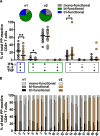
Functionality of PP-reactive CD8+ T cells at visit 1 and 2. (A) Frequencies of PP-reactive CD8+ T cells in 20 IM patients at visit 1 (v1, grey dots) and visit 2 (v2, brown dots) are depicted for each functional combination of the three cytokines (IFN-γ, IL-2, TNF) analyzed. The colored dots and black lines represent individual frequencies and median frequencies of antigen reactive CD8+ T cells, respectively. The depicted pies show the average proportion of antigen-reactive mono- (cells producing only one of the cytokines), bi- (cells producing two of the respective cytokines), and tri-functional (cells producing all three cytokines simultaneously) CD8+ T cells at visits 1 and 2. * p < 0.05, ** p < 0.01. B Individual proportion of mono-, bi-, and tri-functional PP-reactive CD8+ T cells in the 20 IM patients at v1 and v2

Similar articles

Cited by

References

    1. Longnecker RM, Kieff E, Cohen JI. Epstein-Barr virus. In: Knipe DM, Howley PM, editors. Fields Virology. Philadelphia: Wolters Kluwer Health/Lippincott Williams & Wilkins; 2013. pp. 1898–1959.
    1. Damania B, Kenney SC, Raab-Traub N. Epstein-Barr virus: Biology and clinical disease. Cell. 2022;185(20):3652–3670. doi: 10.1016/j.cell.2022.08.026. - DOI - PMC - PubMed
    1. Balfour HH, Jr., Dunmire SK, Hogquist KA. Hogquist, Infectious mononucleosis. Clin Transl Immunology. 2015;4(2):e33. doi: 10.1038/cti.2015.1. - DOI - PMC - PubMed
    1. Dunmire SK, Verghese PS, Balfour HH., Jr Primary Epstein-Barr virus infection. J Clin Virol. 2018;102:84–92. doi: 10.1016/j.jcv.2018.03.001. - DOI - PubMed
    1. Long HM, Meckiff BJ, Taylor GS. The T-cell Response to Epstein-Barr Virus-New Tricks From an Old Dog. Front Immunol. 2019;10:2193. doi: 10.3389/fimmu.2019.02193. - DOI - PMC - PubMed

Publication types