

Diffusion Tensor Imaging in Diagnosing and Evaluating Degenerative Cervical Myelopathy: A Systematic Review and Meta-Analysis
- PMID: 38877604
- PMCID: PMC11572101
- DOI: 10.1177/21925682241263792
Diffusion Tensor Imaging in Diagnosing and Evaluating Degenerative Cervical Myelopathy: A Systematic Review and Meta-Analysis
Abstract
Study design: Systematic review.
Objective: Degenerative cervical myelopathy (DCM) is a common spinal cord disorder necessitating surgery. We aim to explore how effectively diffusion tensor imaging (DTI) can distinguish DCM from healthy individuals and assess the relationship between DTI metrics and symptom severity.
Methods: We included studies with adult DCM patients who had not undergone decompressive surgery and implemented correlation analyses between DTI parameters and severity, or compared healthy controls and DCM patients.
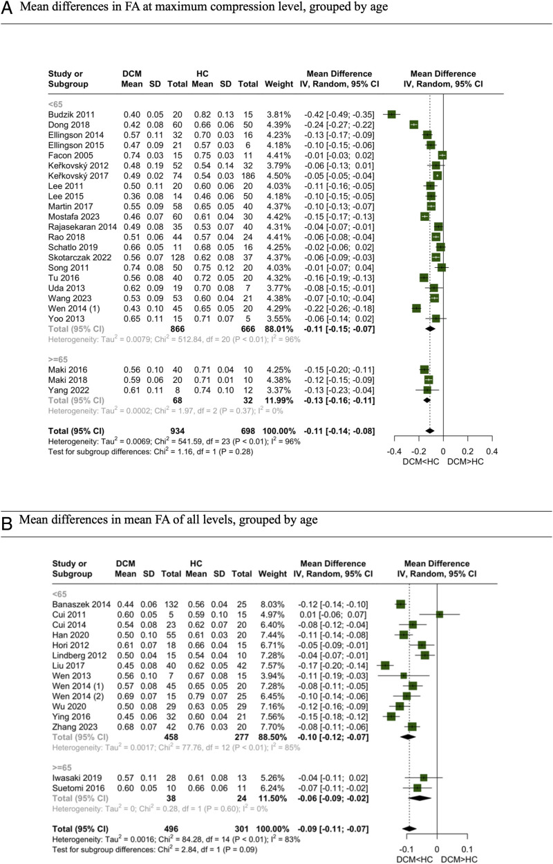
Results: 57 studies were included in our meta-analysis. At the maximal compression (MC) level, fractional anisotropy (FA) exhibited lower values in DCM patients, while apparent diffusion coefficient (ADC), mean diffusivity (MD), and radial diffusivity (RD) were notably higher in the DCM group. Moreover, our investigation into the diagnostic utility of DTI parameters disclosed high sensitivity, specificity, and area under the curve values for FA (.84, .80, .83 respectively) and ADC (.74, .84, .88 respectively). Additionally, we explored the correlation between DTI parameters and myelopathy severity, revealing a significant correlation of FA (.53, 95% CI:0.40 to .65) at MC level with JOA/mJOA scores.
Conclusion: Current guidelines for DCM suggest decompressive surgery for both mild and severe cases. However, they lack clear recommendations on which mild DCM patients might benefit from conservative treatment vs immediate surgery. ADC's role here could be pivotal, potentially differentiating between healthy individuals and DCM. While it may not correlate with symptom severity, it might predict surgical outcomes, making it a valuable imaging biomarker for clearer management decisions in mild DCM.
Keywords: degenerative cervical myelopathy; diagnosis; diffusion tensor imaging; meta-analysis.
Conflict of interest statement
Declaration of Conflicting InterestsThe author(s) declared no potential conflicts of interest with respect to the research, authorship, and/or publication of this article.
Figures


Similar articles
-
Longitudinal diffusion tensor imaging of patients with degenerative cervical myelopathy following decompression surgery.J Clin Neurosci. 2020 Apr;74:194-198. doi: 10.1016/j.jocn.2019.05.018. Epub 2019 Jun 11. J Clin Neurosci. 2020. PMID: 31201048
-
Value of conventional MRI and diffusion tensor imaging parameters in predicting surgical outcome in patients with degenerative cervical myelopathy.J Back Musculoskelet Rehabil. 2018;31(3):525-532. doi: 10.3233/BMR-170972. J Back Musculoskelet Rehabil. 2018. PMID: 29332032
-
Feasibility of diffusion tensor imaging in cervical spondylotic myelopathy using MUSE sequence.Spine J. 2024 Aug;24(8):1352-1360. doi: 10.1016/j.spinee.2024.03.015. Epub 2024 Mar 29. Spine J. 2024. PMID: 38556218
-
The role of diffusion tensor imaging and fractional anisotropy in the evaluation of patients with idiopathic normal pressure hydrocephalus: a literature review.Neurosurg Focus. 2016 Sep;41(3):E12. doi: 10.3171/2016.6.FOCUS16192. Neurosurg Focus. 2016. PMID: 27581308 Review.
-
Clinical and Research MRI Techniques for Assessing Spinal Cord Integrity in Degenerative Cervical Myelopathy-A Scoping Review.Biomedicines. 2022 Oct 18;10(10):2621. doi: 10.3390/biomedicines10102621. Biomedicines. 2022. PMID: 36289883 Free PMC article.
Cited by
-
Degenerative cervical myelopathy: timing of surgery.EFORT Open Rev. 2025 Jun 2;10(6):403-415. doi: 10.1530/EOR-2025-0070. EFORT Open Rev. 2025. PMID: 40459154 Free PMC article.
References
LinkOut - more resources
Full Text Sources
Miscellaneous
