Observational Study
. 2024 Aug;154(2):435-446.
doi: 10.1016/j.jaci.2024.03.029. Epub 2024 Jun 13.

Patients taking benralizumab, dupilumab, or mepolizumab have lower postvaccination SARS-CoV-2 immunity

Affiliations
Observational Study

Patients taking benralizumab, dupilumab, or mepolizumab have lower postvaccination SARS-CoV-2 immunity

Martin C Runnstrom et al. J Allergy Clin Immunol. 2024 Aug.

Abstract

Background: Biologic therapies inhibiting the IL-4 or IL-5 pathways are very effective in the treatment of asthma and other related conditions. However, the cytokines IL-4 and IL-5 also play a role in the generation of adaptive immune responses. Although these biologics do not cause overt immunosuppression, their effect in primary severe acute respiratory syndrome coronavirus 2 (SARS-CoV-2) immunization has not been studied completely.

Objective: Our aim was to evaluate the antibody and cellular immunity after SARS-CoV-2 mRNA vaccination in patients on biologics (PoBs).

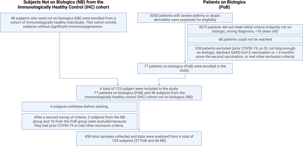
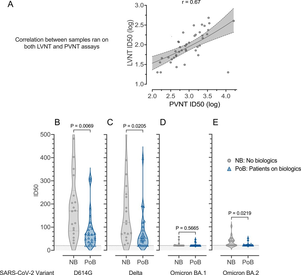
Methods: Patients with severe asthma or atopic dermatitis who were taking benralizumab, dupilumab, or mepolizumab and had received the initial dose of the 2-dose adult SARS-CoV-2 mRNA vaccine were enrolled in a prospective, observational study. As our control group, we used a cohort of immunologically healthy subjects (with no significant immunosuppression) who were not taking biologics (NBs). We used a multiplexed immunoassay to measure antibody levels, neutralization assays to assess antibody function, and flow cytometry to quantitate Spike-specific lymphocytes.

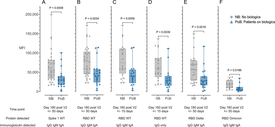
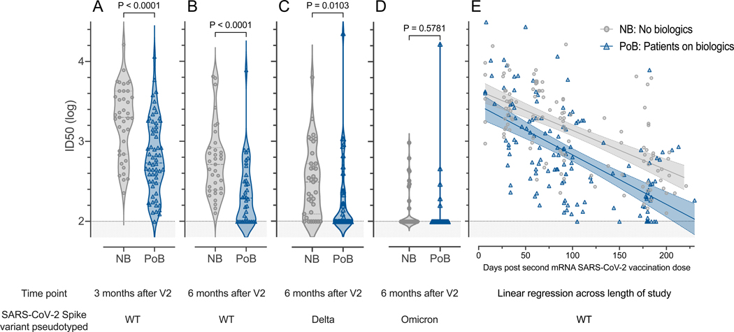
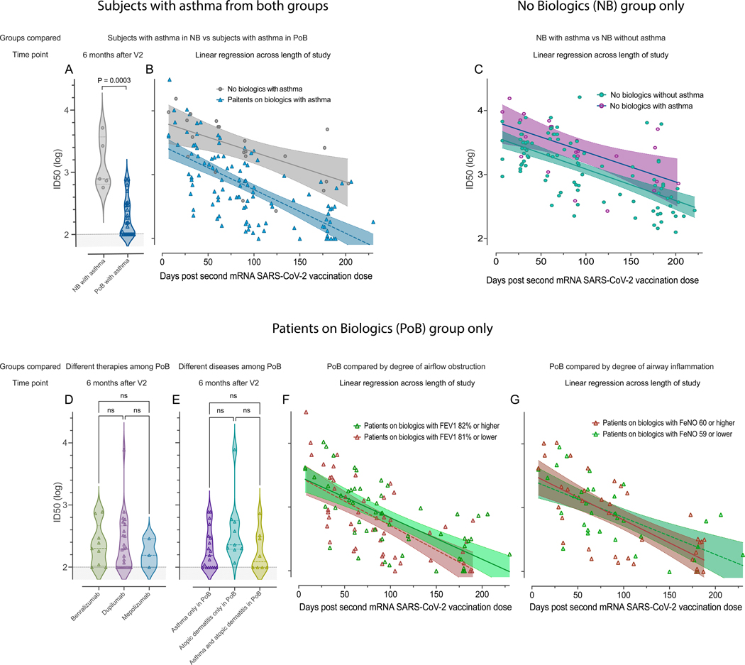
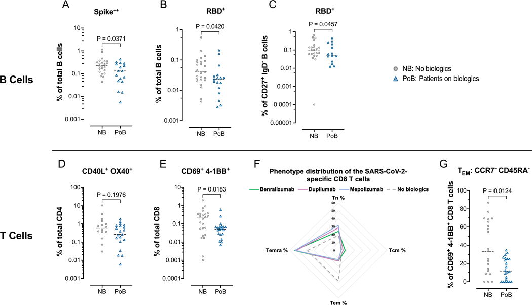
Results: We analyzed blood from 57 patients in the PoB group and 46 control subjects from the NB group. The patients in the PoB group had lower levels of SARS-CoV-2 antibodies, pseudovirus neutralization, live virus neutralization, and frequencies of Spike-specific B and CD8 T cells at 6 months after vaccination. In subgroup analyses, patients with asthma who were taking biologics had significantly lower pseudovirus neutralization than did subjects with asthma who were not taking biologics.

Conclusion: The patients in the PoB group had reduced SARS-CoV-2-specific antibody titers, neutralizing activity, and virus-specific B- and CD8 T-cell counts. These results have implications when considering development of a more individualized immunization strategy in patients who receive biologic medications blocking IL-4 or IL-5 pathways.

Keywords: Asthma biologics; COVID-19; SARS-CoV-2; antibody neutralization; benralizumab; dupilumab; mRNA vaccines; memory B cells; memory T cells; mepolizumab.


Conflict of interest statement

Disclosure statement Supported by the National Institutes of Health (NIH)/National Institute of Allergy and Infectious Diseases (NIAID) (grants NIH/NHLBI T32HL116271 [to M.C.R., P.A.L., and N.S.]; the National Center for Advancing Translational Sciences of the NIH (UL1TR002378 [to N.S.]; P01AI125180 [to I.S. and F.E.L.]; U54CA260563 [to I.S. and F.E.L.]; R01AI121252 [to F.E.L.]; R01AI172254 [to F.E.L.]; U01AI141993 [to F.E.L.]; and NIH P51OD011132, 1U54CA260563, and the NIH/NIAID Centers of Excellence for Influenza Research and Response (under contract 75N93021C00017 to Emory University). This work does not necessarily represent the views of the US government or Department of Veterans Affairs. Disclosure of potential conflict of interest: N. S. Haddad is employed by MicroB-plex, Inc, but did not receive any payments for this article. C. Swenson receives compensation and consulting fees from Insmed, Inc, that are unrelated to this article. F. Holguin is a member of the adjudication committee of the ASPEN (A Study to Assess the Efficacy, Safety, and Tolerability of Brensocatib in Participants With Non-Cystic Fibrosis Bronchiectasis) trial at Insmed, Inc. J. D. Roback received funding from an NIH grant in the past 36 months. I. Sanz receives royalties from BLI INC for plasma cell survival media; consulting fees from GSK, Pfizer, Kayverna, Johnson & Johnson, Celgene, Bristol-Myer Squibb, and Visterra; and honoraria for presentations from Yale and Harvard Universities. In addition, I. Sanz has a patent on plasma cell survival media. F. Eun-Hyung Lee receives or has received research grants from Genentech and the Gates Foundation; royalties from BLI INC for plasma cell survival media; consulting fees from Be Bio Pharma; honoraria for presentations at the University of Pennsylvania, the University of Cincinnati, and the Gerontological Advanced Practice Nurses Association; has patents on plasma cell survival media and media of elaborated newly synthesized antibodies (MENSA); and is the founder and owner of MicroB-plex, Inc. The rest of the authors declare that they have no relevant conflicts of interest.

Figures

Figure 1.
Figure 1.. Diagram of the enrollment of patients on biologics (PoB) and subjects not on biologics (NB) from the immunologically healthy control (IHC) cohort in our prospective, observational study.
COVID-19, Coronavirus disease 2019; IS, immunosuppression; SARS-CoV-2, severe acute respiratory syndrome coronavirus 2.
Figure 2.
Figure 2.. Antibody binding is correlated with antibody levels.
(A) Combined IgA, IgG, and IgM immunoglobulins binding to WT SARS-CoV-2 Spike 1 protein measured at day 180 after V2 +/− 50 days. (B) Combined IgA, IgG, and IgM immunoglobulins binding to WT SARS-CoV-2 RBD protein measured at day 180 after V2 +/− 50 days. (C) Combined IgA, IgG, and IgM immunoglobulins binding to WT SARS-CoV-2 RBD protein measured at day 180 after V2 +/− 15 days. (D) Immunoglobulin IgG only binding to WT SARS-CoV-2 RBD protein measured at day 180 after V2 +/− 15 days. (E) Combined IgA, IgG, and IgM immunoglobulins binding to the Delta-variant SARS-CoV-2 RBD protein measured at day 180 after V2 +/− 50 days. (F) Combined IgA, IgG, and IgM immunoglobulins binding to the Omicron-variant SARS-CoV-2 RBD protein measured at day 180 after V2 +/− 50 days. NB, control subjects not on biologics; PoB, patients on biologics include all subjects treated with benralizumab, dupilumab or mepolizumab; IgA, Immunoglobulin A; IgM, Immunoglobulin M; IgG, Immunoglobulin G; WT, wild-type; SARS-CoV-2, severe acute respiratory syndrome coronavirus 2; mRNA, messenger ribonucleic acid; RBD, receptor-binding domain; MFI, median fluorescence intensity; V2, second mRNA SARS-CoV-2 vaccination dose.
Figure 3.
Figure 3.. Pseudovirus neutralization.
All comparisons were performed between subjects not on biologics (NB) and patients on biologics (PoB) (A) Pseudovirus neutralization of WT SARS-CoV-2, comparison between three months after V2. (B) Pseudovirus neutralization of WT SARS-CoV-2 six months after V2. (C) Pseudovirus neutralization of Delta-variant SARS-CoV-2 six months after V2. (D) Pseudovirus neutralization of Omicron-variant BA.1 SARS-CoV-2 six months after V2. (E) Linear regression of data points from each group (NB and PoB) across the entire study follow-up period. Their 95% confidence intervals are represented by the shaded area. For all subfigures: the shaded area under 2 represents the lower limit of detection of the assay. NB, control subjects not on biologics; PoB, patients on biologics include all subjects treated with benralizumab, dupilumab and mepolizumab; V2, dose two of the SARS-CoV-2 mRNA vaccine; WT, wild-type; SARS-CoV-2, severe acute respiratory syndrome coronavirus 2; mRNA, messenger ribonucleic acid; ID50, 50% inhibitory dilution.
Figure 4.
Figure 4.. Live virus neutralization.
Samples collected between days 69 and 105 after the second mRNA SARS-CoV-2 vaccination (A) Correlation between live-virus neutralization and pseudovirus neutralization for samples run on both assays. (B) Live-virus neutralization of D614G-variant SARS-CoV-2. (C) Live-virus neutralization of Delta-variant SARS-CoV-2. (D) Live virus neutralization of Omicron-variant BA.1 SARS-CoV-2. (E) Live virus neutralization of Omicron-variant BA.2 SARS-CoV-2. For all subfigures: the shaded area under 20 represents the lower limit of detection of the LVNT assay. NB, control subjects not on biologics; PoB, patients on biologics include all subjects treated with benralizumab, dupilumab, and mepolizumab; SARS-CoV-2, severe acute respiratory syndrome coronavirus 2; WT, wild-type; mRNA, messenger ribonucleic acid; ID50, 50% inhibitory dilution; PVNT, pseudovirus neutralization; LVNT, live virus neutralization.
Figure 5.
Figure 5.. Subgroup analyses.
Pseudovirus neutralization of WT SARS-CoV-2, comparison between subjects with asthma in the NB group and subjects with asthma in the PoB group (A) six months after V2 and (B) across the entire study follow-up period. (C) Pseudovirus neutralization of WT SARS-CoV-2, comparison between subjects with and without asthma in the no biologics control group across the entire study follow-up period. (D) Pseudovirus neutralization of WT SARS-CoV-2, comparison between different therapies (benralizumab, dupilumab, or mepolizumab) among all PoB six months after V2. (E) Pseudovirus neutralization of WT SARS-CoV-2, comparison between diseases (asthma, atopic dermatitis, or both) among all PoB six months after V2. (F) Pseudovirus neutralization of WT SARS-CoV-2 of patients with asthma on biologics stratified by the degree of airway obstruction across the entire study follow-up period. In green: patients with asthma on biologics who have an FEV1 of 82% of predicted or higher. In red: patients with asthma on biologics who have an FEV1 of 81% of predicted or lower. (G) Pseudovirus neutralization of WT SARS-CoV-2 of patients with asthma on biologics stratified by degree of airway inflammation across the entire study follow-up period. In red: patient with asthma on biologics who have a FeNO of 60ppb or higher. In green: patients with asthma on biologics who have a FeNO of 59 or lower. For subfigures A, D, and E: the shaded area under 2 represents the lower limit of detection of the assay. For subfigures B, C, F, and G: the diagonal lines represent the linear regression of data points from each group and their 95% confidence intervals are represented by the shaded area. NB, control subjects not on biologics; PoB, patients on biologics include all subjects treated with benralizumab, dupilumab and mepolizumab; V2, dose two of the SARS-CoV-2 mRNA vaccine; WT, wild-type; SARS-CoV-2, severe acute respiratory syndrome coronavirus 2; mRNA, messenger ribonucleic acid; ID50, 50% inhibitory dilution; ns, non-significant p>0.05; FEV1, forced expiratory volume in the first second; FeNO, fractional exhaled nitric oxide.
Figure 6.
Figure 6.. Flow cytometry assessment of SARS-CoV-2-specific B and T lymphocytes.
All data points in this figure are from days 130–250 after V2 (A-C) Fluorescently-conjugated Spike and RBD proteins in a tetramer conformation were used to identify ex-vivo SARS-CoV-2-specific B cells. All Spike-specific cell populations were identified using double-labeling of fluorochromes. (A) Percentages of Spike positive cells of total B cells. (B) Percentages of RBD positive cells of total B cells. (C) Percentages of RBD positive cells of switched memory B cells (CD27+, IgD). (D-G) Ex-vivo PBMCs from NB and PoB were incubated for 24-hours with Spike peptides, with Staphylococcal enterotoxin B as a positive control, or with no antigens as a negative control. SARS-CoV-2 specific T cells were quantified using markers of activation (CD40L and OX40 for CD4 T cells, and CD69 and 4–1BB for CD8 T cells). SARS-CoV-2 specific values are calculated and plotted as the experimental condition with Spike peptides minus the experimental condition without peptides (negative control). (D) Percentages of CD40L+ OX40+ cells of total CD4 T cells. (E) Percentages of CD69+ 4–1BB+ cells of total CD8 T cells. (F) Radar plot showing the phenotypic distribution of SARS-CoV-2-specific CD8 T cells. (G) Comparisons of TEM percentages of the SARS-CoV-2 specific CD8 T cells. PBMC, peripheral blood mononuclear cells; NB, control subjects not on biologics; PoB, patients on biologics include all subjects treated with benralizumab, dupilumab and mepolizumab; V2, dose two of the SARS-CoV-2 mRNA vaccine; WT, wild-type; SARS-CoV-2, severe acute respiratory syndrome coronavirus 2; RBD, receptor-binding domain; mRNA, messenger ribonucleic acid; TEM, effector memory CCR7 CD45RA; TCM, central memory CCR7+ CD45RA; TEMRA, terminally differentiated effector CCR7 CD45RA+, TN, naïve CCR7+ CD45RA+.

References

    1. Polack FP, Thomas SJ, Kitchin N, Absalon J, Gurtman A, Lockhart S, et al. Safety and Efficacy of the BNT162b2 mRNA Covid-19 Vaccine. New England Journal of Medicine. 2020;383(27):2603–15. - PMC - PubMed
    1. Baden LR, El Sahly HM, Essink B, Kotloff K, Frey S, Novak R, et al. Efficacy and Safety of the mRNA-1273 SARS-CoV-2 Vaccine. New England Journal of Medicine. 2021;384(5):403–16. - PMC - PubMed
    1. Levin EG, Lustig Y, Cohen C, Fluss R, Indenbaum V, Amit S, et al. Waning Immune Humoral Response to BNT162b2 Covid-19 Vaccine over 6 Months. New England Journal of Medicine. 2021;385(24):e84. - PMC - PubMed
    1. Nguyen DC, Lamothe PA, Woodruff MC, Saini AS, Faliti CE, Sanz I, et al.. COVID-19 and plasma cells: Is there long-lived protection? Immunol Rev. 2022. - PMC - PubMed
    1. Wang Z, Schmidt F, Weisblum Y, Muecksch F, Barnes CO, Finkin S, et al. mRNA vaccine-elicited antibodies to SARS-CoV-2 and circulating variants. Nature. 2021;592(7855):616–22. - PMC - PubMed

Publication types

MeSH terms