

Effect of hot water maceration, rehydration, and soft tissue presence on 3D geometry of bone
- PMID: 38878109
- PMCID: PMC11953162
- DOI: 10.1007/s12024-024-00845-0
Effect of hot water maceration, rehydration, and soft tissue presence on 3D geometry of bone
Abstract
Purpose: In forensic medicine, maceration is often essential for examining bone surfaces, serving purposes such as identifying cut marks, making geometric measurements, and determining the victim's age. While hot water maceration removes soft tissue effectively, it is known to cause bone surface shrinkage. This raises the question of whether this effect is permanent or if it can be partially reversed through rehydration, considering the presence of soft tissue.
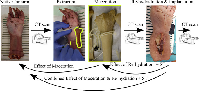
Methods: Computed tomography (CT) scans were conducted on the radii of 20 paired human anatomic forearm specimens. Subsequently, the radii were extracted, macerated in 60 °C water, CT-scanned in an air environment, rehydrated, re-implanted into the forearms, and CT-scanned again.
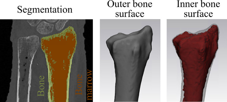
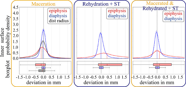
Results: Maceration resulted in a mean shrinkage of 0.12 mm on the outer bone surface. This shrinkage was nearly fully recoverable for the diaphysis after rehydration and accounting for soft tissue surrounding the bone. In contrast, the epiphysis showed permanent shrinkage, likely due to the loss of small bone fragments. Analysis of the inner bone surface indicated a smaller effect, but with significant standard deviations, especially for the epiphysis, possibly related to the less well-defined nature of the inner bone surface.
Conclusion: The epiphyseal surface of hot water-macerated bone will, on average, be approximately 0.15 mm deflated and cannot retain the original surface. On the other hand, the diaphyseal surface is less affected and can be nearly completely restored after rehydration and accounting for soft tissue surrounding the bone.
Keywords: 3D model; Bone; Computed tomography; Dimensional accuracy; Forensic anthropology; Maceration; Rehydration.
© 2024. The Author(s).
Conflict of interest statement
Declarations. Ethical declaration: The study was approved by the Ethics Committee of the Medical University of Vienna (EK-Nr: 2003/2019). Competing interests: The authors have no relevant financial or non-financial interests to disclose.
Figures


References
-
- Weber G, Bookstein F. Virtual Anthropology: A new interdisciplinary field of science, in: 2011: pp. 1–36. 10.1007/978-3-211-49347-2_1.
-
- Winskog C, Persson A, Ljung P, Ynnerman A, Lundström C. Full body virtual autopsies using a state-of-the-art volume rendering Pipeline. IEEE Trans Vis Comput Graph. 2006;12:869–76. 10.1109/TVCG.2006.146. - PubMed
-
- Okuda T, Shiotani S, Sakamoto N, Kobayashi T. Background and current status of postmortem imaging in Japan: short history of autopsy imaging (Ai). Forensic Sci Int. 2013;225:3–8. 10.1016/j.forsciint.2012.03.010. - PubMed
-
- Malfroy Camine L, Varlet V, Campana L, Grabherr S, Moghaddam N. The big puzzle: a critical review of virtual re-association methods for fragmented human remains in a DVI context’. Forensic Sci Int. 2022;330:111033. 10.1016/j.forsciint.2021.111033. - PubMed
-
- Krentz BV, Alamo L, Grimm J, Dédouit F, Bruguier C, Chevallier C, Egger C, Da Silva LFF, Grabherr S. Performance of post-mortem CT compared to autopsy in children. Int J Legal Med. 2016;130:1089–99. 10.1007/s00414-016-1370-z. - PubMed
MeSH terms
Substances
LinkOut - more resources
Full Text Sources
