Activation of androgen receptor-expressing neurons in the posterior medial amygdala is associated with stress resistance in dominant male hamsters
- PMID: 38878493
- PMCID: PMC11330741
- DOI: 10.1016/j.yhbeh.2024.105577
Activation of androgen receptor-expressing neurons in the posterior medial amygdala is associated with stress resistance in dominant male hamsters
Abstract
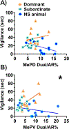
Social stress is a negative emotional experience that can increase fear and anxiety. Dominance status can alter the way individuals react to and cope with stressful events. The underlying neurobiology of how social dominance produces stress resistance remains elusive, although experience-dependent changes in androgen receptor (AR) expression is thought to play an essential role. Using a Syrian hamster (Mesocricetus auratus) model, we investigated whether dominant individuals activate more AR-expressing neurons in the posterior dorsal and posterior ventral regions of the medial amygdala (MePD, MePV), and display less social anxiety-like behavior following social defeat stress compared to subordinate counterparts. We allowed male hamsters to form and maintain a dyadic dominance relationship for 12 days, exposed them to social defeat stress, and then tested their approach-avoidance behavior using a social avoidance test. During social defeat stress, dominant subjects showed a longer latency to submit and greater c-Fos expression in AR+ cells in the MePD/MePV compared to subordinates. We found that social defeat exposure reduced the amount of time animals spent interacting with a novel conspecific 24 h later, although there was no effect of dominance status. The amount of social vigilance shown by dominants during social avoidance testing was positively correlated with c-Fos expression in AR+ cells in the MePV. These findings indicate that dominant hamsters show greater neural activity in AR+ cells in the MePV during social defeat compared to their subordinate counterparts, and this pattern of neural activity correlates with their proactive coping response. Consistent with the central role of androgens in experience-dependent changes in aggression, activation of AR+ cells in the MePD/MePV contributes to experience-dependent changes in stress-related behavior.
Keywords: Aggression; Androgen receptor; Medial amygdala; Proactive coping; Social defeat; Social dominance; Social vigilance.
Copyright © 2024 Elsevier Inc. All rights reserved.
Conflict of interest statement
Declaration of competing interest The authors declare no competing financial interests.
Figures


Similar articles
-
Dominance status modulates activity in medial amygdala cells with projections to the bed nucleus of the stria terminalis.Behav Brain Res. 2023 Sep 13;453:114628. doi: 10.1016/j.bbr.2023.114628. Epub 2023 Aug 12. Behav Brain Res. 2023. PMID: 37579818 Free PMC article.
-
Gonadal steroid hormone receptors in the medial amygdala contribute to experience-dependent changes in stress vulnerability.Psychoneuroendocrinology. 2021 Jul;129:105249. doi: 10.1016/j.psyneuen.2021.105249. Epub 2021 May 3. Psychoneuroendocrinology. 2021. PMID: 33971475 Free PMC article.
-
Maintenance of dominance status is necessary for resistance to social defeat stress in Syrian hamsters.Behav Brain Res. 2014 Aug 15;270:277-86. doi: 10.1016/j.bbr.2014.05.041. Epub 2014 May 27. Behav Brain Res. 2014. PMID: 24875769 Free PMC article.
-
Parent-training programmes for improving maternal psychosocial health.Cochrane Database Syst Rev. 2004;(1):CD002020. doi: 10.1002/14651858.CD002020.pub2. Cochrane Database Syst Rev. 2004. Update in: Cochrane Database Syst Rev. 2012 Jun 13;(6):CD002020. doi: 10.1002/14651858.CD002020.pub3. PMID: 14973981 Updated.
-
The Black Book of Psychotropic Dosing and Monitoring.Psychopharmacol Bull. 2024 Jul 8;54(3):8-59. Psychopharmacol Bull. 2024. PMID: 38993656 Free PMC article. Review.
Cited by
-
Testosterone differentially modulates the display of agonistic behavior and dominance over opponents before and after adolescence in male Syrian hamsters.Front Behav Neurosci. 2025 Jul 10;19:1603862. doi: 10.3389/fnbeh.2025.1603862. eCollection 2025. Front Behav Neurosci. 2025. PMID: 40708844 Free PMC article.
-
Distinguishing neural ensembles in the infralimbic cortex that regulate stress vulnerability and coping behavior.Neurobiol Stress. 2025 Mar 20;36:100720. doi: 10.1016/j.ynstr.2025.100720. eCollection 2025 May. Neurobiol Stress. 2025. PMID: 40230624 Free PMC article.
References
-
- Albers HE, Huhman KL, & Meisel RL (2002). 6—Hormonal Basis of Social Conflict and Communication. In Pfaff DW, Arnold AP, Fahrbach SE, Etgen AM, & Rubin RT (Eds.), Hormones, Brain and Behavior (pp. 393–433). Academic Press. 10.1016/B978-012532104-4/50008-1 - DOI
-
- Amat J, Paul E, Zarza C, Watkins LR, & Maier SF (2006). Previous Experience with Behavioral Control over Stress Blocks the Behavioral and Dorsal Raphe Nucleus Activating Effects of Later Uncontrollable Stress: Role of the Ventral Medial Prefrontal Cortex. Journal of Neuroscience, 26(51), 13264–13272. 10.1523/JNEUROSCI.3630-06.2006 - DOI - PMC - PubMed
Publication types
MeSH terms
Substances
Grants and funding
LinkOut - more resources
Full Text Sources
Medical
Research Materials

