

Ferumoxytol-enhanced MRI assessment of venous Thrombus resolution and macrophage content in a murine deep vein thrombosis model
- PMID: 38878741
- PMCID: PMC11239555
- DOI: 10.1016/j.thromres.2024.109063
Ferumoxytol-enhanced MRI assessment of venous Thrombus resolution and macrophage content in a murine deep vein thrombosis model
Abstract
Background: Imaging evaluation of acute deep vein thrombosis (DVT) or post-thrombotic syndrome (PTS) in animal or clinical models is limited to anatomical assessment of the location and extent of thrombi. We hypothesize that Fe-MRI, used to evaluate macrophage content in other inflammatory diseases, can be useful to evaluate the thromboinflammatory features after DVT over time.
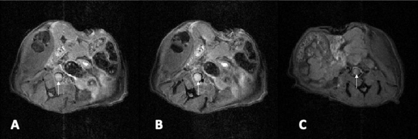
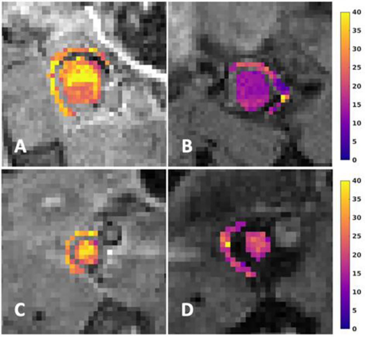
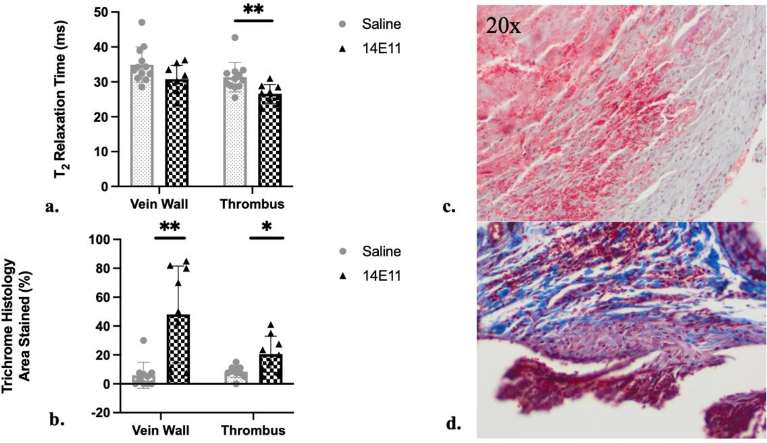
Methods: Nineteen wild-type CD-1 mice underwent surgical IVC ligation to induce DVT. Mice received either saline or 5 mg/kg of 14E11, a Factor XI inhibitor, before the procedure. Fe-MRI was performed on days 6-7 after ligation to evaluate thrombus volume, perfusion, and macrophage content via T2-weighted images. Mice were euthanized at days 3-15 after surgery. The thrombi and adjacent vein walls were excised, weighed, formalin-fixed, and paraffin-embedded for immunohistological analysis. Specimens were stained with specific antibodies to evaluate macrophage content, collagen deposition, neovascularization, and recanalization. Significance was determined using the Mann-Whitney U or Student's t-test.
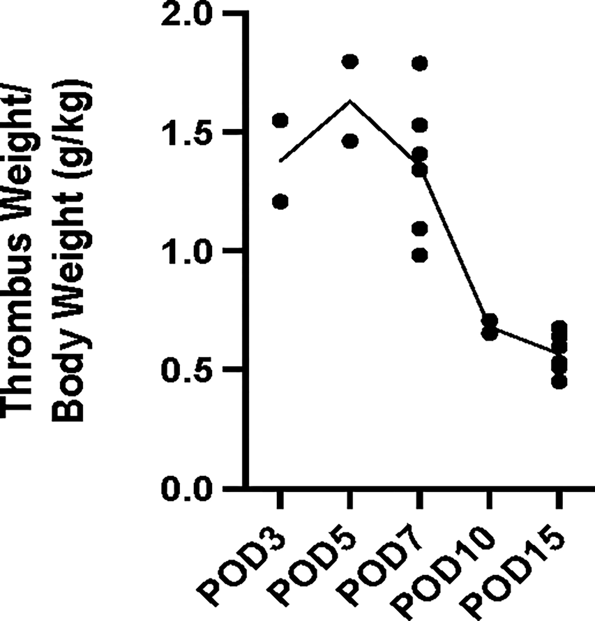
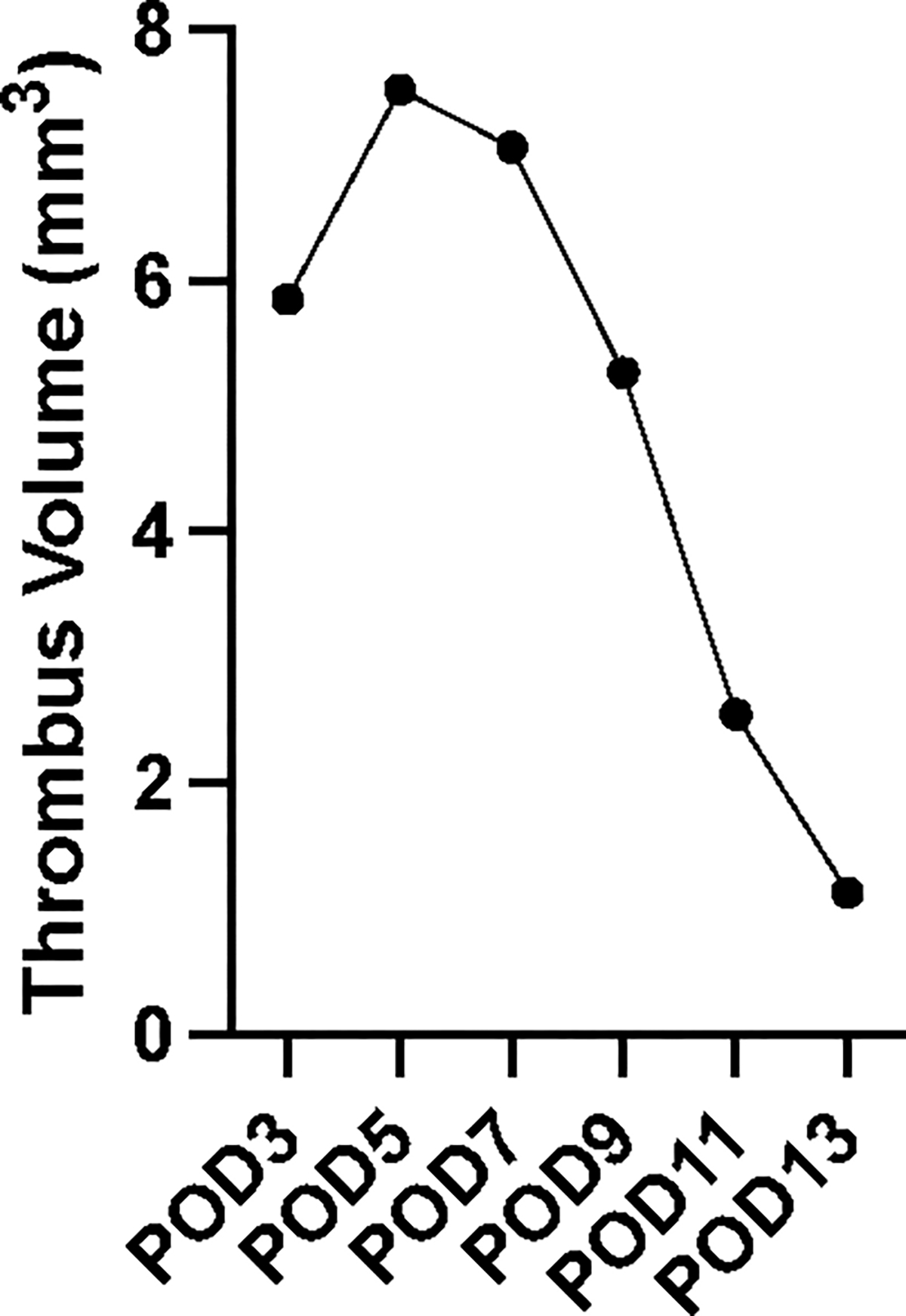
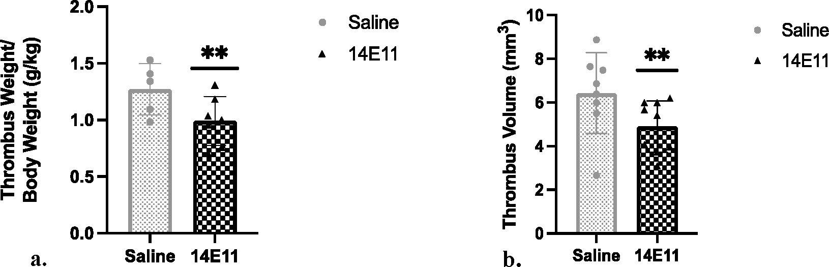
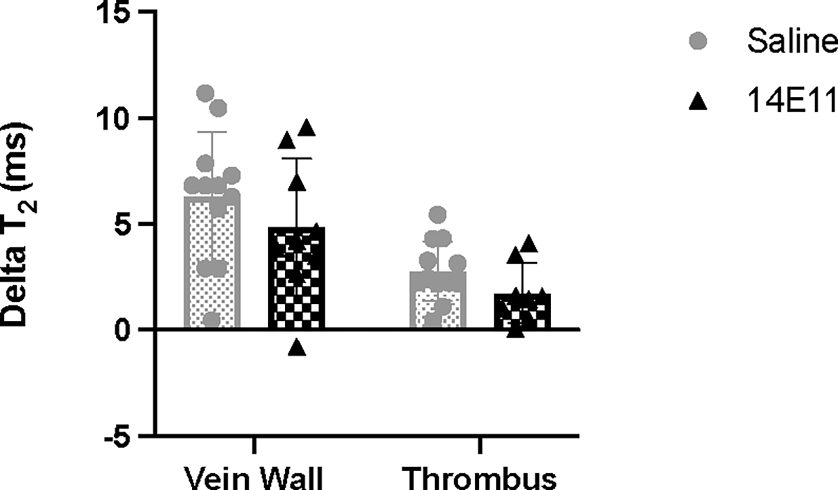
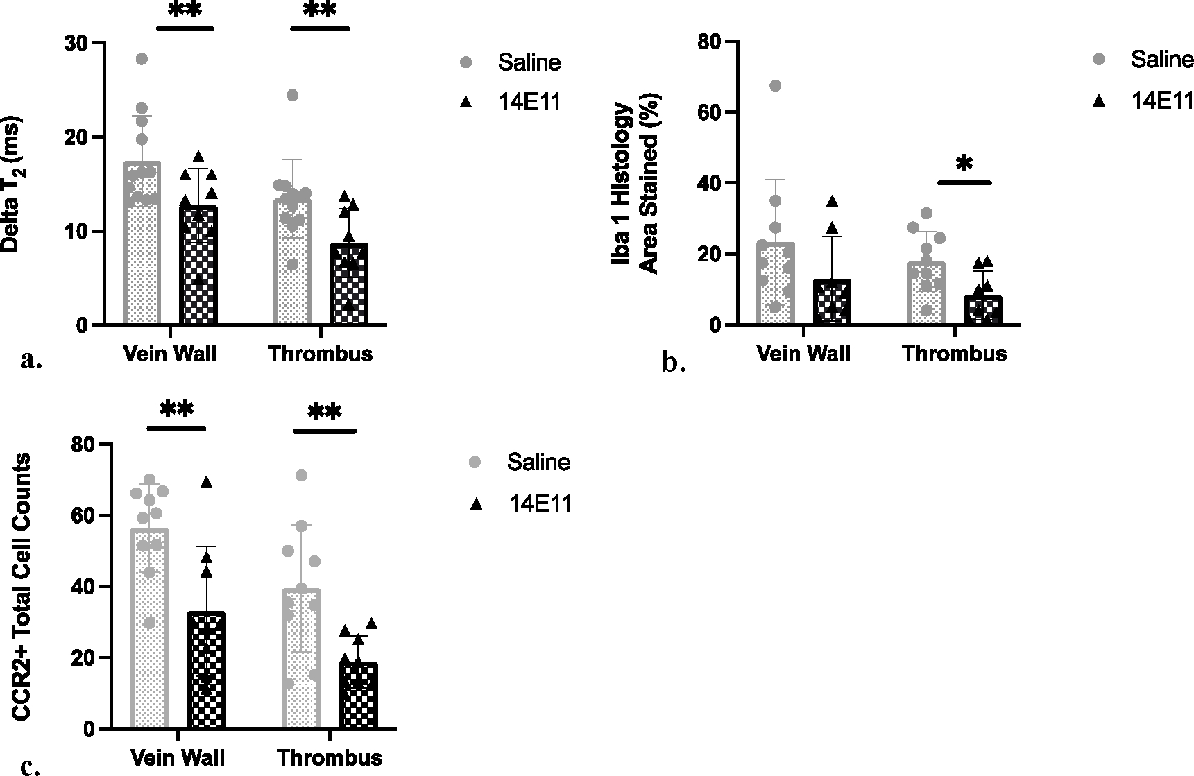
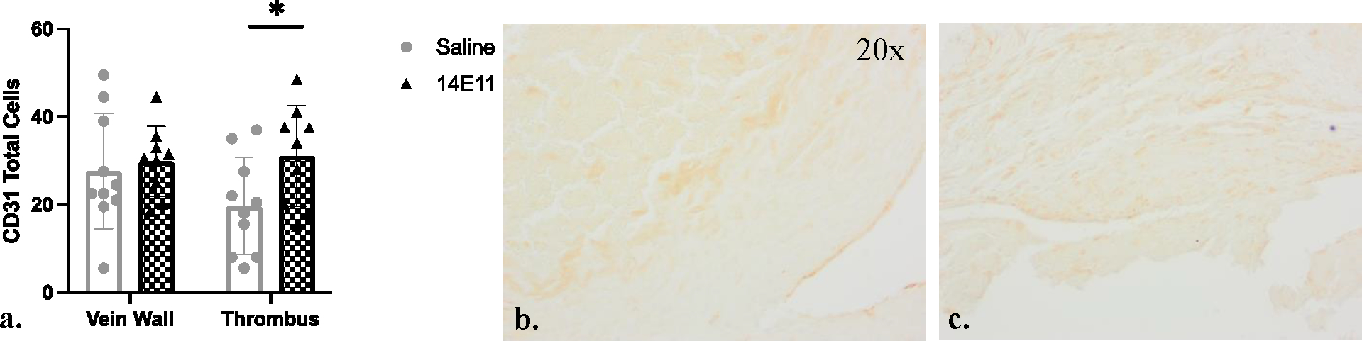
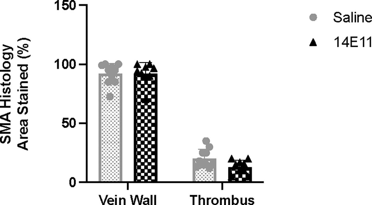
Results: After IVC-ligation in control mice, thrombus weights decreased by 59 % from day 3 to 15. Thrombus volumes peaked on day 5 before decreasing by 85 % by day 13. FXI inhibition led to reduced macrophage content in both thrombi (p = .008) and vein walls (p = .01), decreased thrombus volume (p = .03), and decreased thrombus mass (p = .01) compared to control mice. CCR2+ staining corroborated these findings, showing significantly reduced macrophage presence in the thrombi (p = .002) and vein wall (p = .002).
Conclusions: Fe-MRI T2 relaxation times can be used to characterize and quantify post-thrombotic changes of perfusion, macrophage content, and thrombus volume over time in a surgical mouse model of venous thrombosis. This approach could lead to better quantification of in vivo inflammation correlating monocyte and macrophage content within resolving thrombi and veins and may serve as a useful tool for research and clinically in the evaluation of the post-thrombotic environment.
Keywords: FXI inhibition; Ferumoxytol; Magnetic resonance; Mouse; thrombus resolution.
Copyright © 2024 Elsevier Ltd. All rights reserved.
Conflict of interest statement
Declaration of competing interest The authors declare that they have no known competing financial interests or personal relationships that could have appeared to influence the work reported in this paper.
Figures
Similar articles
-
Evaluating the theranostic potential of ferumoxytol when combined with radiotherapy in a mammary dual tumor mouse model.Med Phys. 2025 Jul;52(7):e17888. doi: 10.1002/mp.17888. Epub 2025 May 21. Med Phys. 2025. PMID: 40400098 Free PMC article.
-
Thrombolysis for acute deep vein thrombosis.Cochrane Database Syst Rev. 2016 Nov 10;11(11):CD002783. doi: 10.1002/14651858.CD002783.pub4. Cochrane Database Syst Rev. 2016. Update in: Cochrane Database Syst Rev. 2021 Jan 19;1:CD002783. doi: 10.1002/14651858.CD002783.pub5. PMID: 27830895 Free PMC article. Updated.
-
Home versus in-patient treatment for deep vein thrombosis.Cochrane Database Syst Rev. 2018 Jan 9;1(1):CD003076. doi: 10.1002/14651858.CD003076.pub3. Cochrane Database Syst Rev. 2018. PMID: 29315455 Free PMC article.
-
Pharmacological reduction of coagulation factor XI reduces macrophage accumulation and accelerates deep vein thrombosis resolution in a mouse model of venous thrombosis.J Thromb Haemost. 2022 Sep;20(9):2035-2045. doi: 10.1111/jth.15777. Epub 2022 Jul 18. J Thromb Haemost. 2022. PMID: 35638310 Free PMC article.
-
Magnetic resonance perfusion for differentiating low-grade from high-grade gliomas at first presentation.Cochrane Database Syst Rev. 2018 Jan 22;1(1):CD011551. doi: 10.1002/14651858.CD011551.pub2. Cochrane Database Syst Rev. 2018. PMID: 29357120 Free PMC article.
References
-
- Kahn SR, et al. , The postthrombotic syndrome: evidence-based prevention, diagnosis, and treatment strategies: a scientific statement from the American Heart Association. Circulation, 2014. 130(18): p. 1636–61. - PubMed
-
- Kahn SR, Hirsch A, and Shrier I, Effect of postthrombotic syndrome on health-related quality of life after deep venous thrombosis. Arch Intern Med, 2002. 162(10): p. 1144–8. - PubMed
-
- Vedantham S, Valvular dysfunction and venous obstruction in the post-thrombotic syndrome. Thromb Res, 2009. 123 Suppl 4: p. S62–5. - PubMed
-
- Ginsberg JS, et al. , Prevention and treatment of postphlebitic syndrome: results of a 3-part study. Arch Intern Med, 2001. 161(17): p. 2105–9. - PubMed
-
- Brandjes DP, et al. , Randomised trial of effect of compression stockings in patients with symptomatic proximal-vein thrombosis. Lancet, 1997. 349(9054): p. 759–62. - PubMed
MeSH terms
Substances
Grants and funding
LinkOut - more resources
Full Text Sources
Medical
