. 2024 Jul;300(7):107478.
doi: 10.1016/j.jbc.2024.107478. Epub 2024 Jun 13.

Conservation of C4BP-binding sequence patterns in Streptococcus pyogenes M and Enn proteins

Affiliations

Conservation of C4BP-binding sequence patterns in Streptococcus pyogenes M and Enn proteins

Piotr Kolesiński et al. J Biol Chem. 2024 Jul.

Abstract

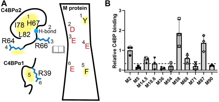
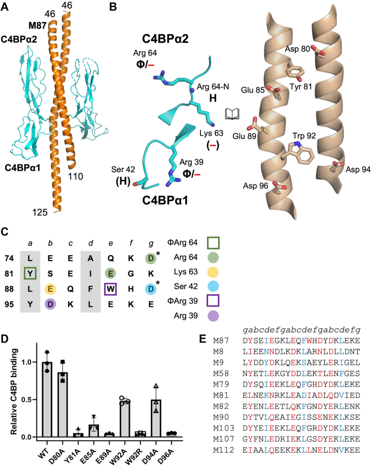
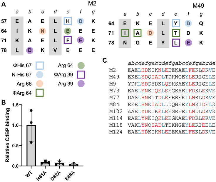
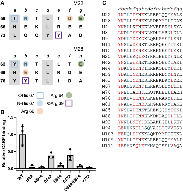
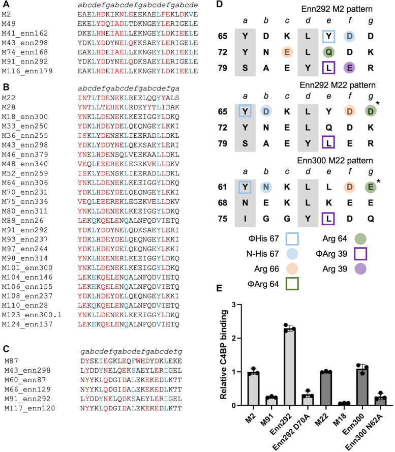
Antigenically sequence variable M proteins of the major bacterial pathogen Streptococcus pyogenes (Strep A) are responsible for recruiting human C4b-binding protein (C4BP) to the bacterial surface, which enables Strep A to evade destruction by the immune system. The most sequence divergent portion of M proteins, the hypervariable region (HVR), is responsible for binding C4BP. Structural evidence points to the conservation of two C4BP-binding sequence patterns (M2 and M22) in the HVR of numerous M proteins, with this conservation applicable to vaccine immunogen design. These two patterns, however, only partially explain C4BP binding by Strep A. Here, we identified several M proteins that lack these patterns but still bind C4BP and determined the structures of two, M68 and M87 HVRs, in complex with a C4BP fragment. Mutagenesis of these M proteins led to the identification of amino acids that are crucial for C4BP binding, enabling formulation of new C4BP-binding patterns. Mutagenesis was also carried out on M2 and M22 proteins to refine or generate experimentally grounded C4BP-binding patterns. The M22 pattern was the most prevalent among M proteins, followed by the M87 and M2 patterns, while the M68 pattern was rare. These patterns, except for M68, were also evident in numerous M-like Enn proteins. Binding of C4BP via these patterns to Enn proteins was verified. We conclude that C4BP-binding patterns occur frequently in Strep A strains of differing M types, being present in their M or Enn proteins, or frequently both, providing further impetus for their use as vaccine immunogens.

Keywords: C4BP; M protein; Streptococcus pyogenes; cross-reactivity; immunogen.


Conflict of interest statement

Conflict of interest The authors declare that they have no conflicts of interest with the contents of this article.

Figures

Figure 1
Figure 1
Binding of C4BP to M proteins.A, schematic of C4BP sites (left) and M protein amino acids (right) involved in interaction for the M2 and M22 patterns, in open book format. The M protein amino acids do not correspond to any specific M protein but show interactions common to M2, M22, M28, and M49 HVRs. Yellow shading corresponds to hydrophobic interactions. B, binding of soluble His6-MN100 protein constructs to immobilized intact C4BP, as evaluated by ELISA. Bound His6-MN100 protein constructs were detected with horseradish peroxidase-conjugated anti-His6 antibodies. Values were normalized by the value of C4BP binding to His6-M2N100. Data from three biological replicates are presented with means and SDs. The dotted line shows the normalized value for C4BP binding to His6-M5N100 plus three SDs. C4BP, C4b-binding protein; HVR, hypervariable region.
Figure 2
Figure 2
M87–C4BP interactions.A, structure of M87N100 (orange) in complex with C4BPα1-2 (cyan), in cartoon representation. Loops of C4BPα1-2 that were not modeled are shown as dotted lines. B, interaction between C4BPα1-2 (left) and M87 (right), in cartoon (main chain) or bonds (side chains) representation, shown in open book format. Carbons are cyan for C4BPα1-2 and wheat for M87; for both, nitrogens are blue and oxygens are red. Chemical character of interactions denoted as follows: Φ, hydrophobic; —, negative; and H, hydrogen bond. Contacts that exceeded hydrogen-bonding or salt-bridging limits (≤3.5 and ≤4.0 Å, respectively) have these labels in parentheses. C, heptad register of M87 74-101 (a and d positions in gray columns). Amino acids marked with an asterisk belong to the minor helix. Shapes indicate type of contact (square for hydrophobic, Φ; circle for polar), and coloring indicates which C4BP amino acid (depicted in legend to the right) was contacted. D, binding of soluble, intact His6-M87 protein constructs to immobilized intact C4BP evaluated by ELISA. Bound His6-M87 protein was detected with horseradish peroxidase-conjugated anti-His6 antibodies. Values were normalized by the value of C4BP-binding to WT His6-M87. Data from three biological replicates are presented with means and SDs. E, partial list of sequences of M proteins with M87 C4BP-binding patterns. Heptad positions are indicated above the sequences. Red indicates an observed (M87) or potential (other M proteins) C4BP-binding amino acid, and blue indicates a d heptad position. A full list is in Table S2. C4BP, C4b-binding protein.
Figure 3
Figure 3
M68–C4BP interactions.A, structure of M68 (orange) in complex with C4BPα2 (cyan), in cartoon representation. B, interaction between C4BPα1-2 (left) and M68 (right), in cartoon (main chain) or bonds (side chains) representation, shown in open book format. Carbons are cyan for C4BPα1-2 and wheat for M87; for both, nitrogens are blue and oxygens are red. Chemical character of interactions denoted as follows: Φ, hydrophobic; —, negative; and H, hydrogen bond. C, heptad register of M68 56-76 (a and d positions in gray columns). Amino acids marked without an asterisk belong to one helix, and those with an asterisk to the second helix. Shapes indicate type of contact (square for hydrophobic; circle for polar), and coloring indicates which C4BP amino acid (depicted in legend to the right) was contacted. D, binding of soluble, intact WT and mutant His6-M68 protein to immobilized intact C4BP, as evaluated by ELISA. Bound His6-M68 protein was detected with horseradish peroxidase-conjugated anti-His6 antibodies. Values were normalized by the value of C4BP binding to WT His6-M68. Data from three biological replicates are presented with means and SDs. E, M protein sequences belonging to the M68 pattern of C4BP binding. Heptad positions are indicated above the sequences. Red indicates an observed or potential C4BP-binding amino acid, and blue indicates a d heptad position. C4BP, C4b-binding protein.
Figure 4
Figure 4
M2–C4BP interactions.A, heptad register of (left) M2 (aa 57–84) and (right) M49 (aa 64–84) (a and d positions in gray columns). Shape indicates type of contact (square for hydrophobic; circle for polar), and coloring indicates which C4BP amino acid (depicted in legend at bottom) was contacted. B, binding of soluble, intact His6-M2 protein constructs to immobilized intact C4BP evaluated by ELISA. Bound His6-M2 protein was detected with horseradish peroxidase-conjugated anti-His6 antibodies. Values were normalized by the value of C4BP binding to WT His6-M2. Data from three biological replicates are presented with means and SDs. C, partial list of M protein sequences belonging to the M2 pattern of C4BP binding. Heptad positions are indicated above the sequences. Red indicates an observed or potential C4BP-binding amino acid, and blue indicates a d heptad position. A full list is in Table S2. C4BP, C4b-binding protein.
Figure 5
Figure 5
M22–C4BP interactions.A, heptad register of (top) M22 (aa 59–79) and (bottom) M28 (aa 62–82) (a and d positions in gray columns). Amino acids marked with an asterisk belong to the minor helix, and those with an asterisk to the major helix. Coloring indicates which C4BP amino acid (depicted in legend at bottom) was contacted, and shape indicates type of contact (square for hydrophobic; circle for polar). B, binding of soluble, intact His6-M22 protein constructs to immobilized intact C4BP evaluated by ELISA. Bound His6-M22 protein was detected with horseradish peroxidase-conjugated anti-His6 antibodies. Values were normalized by the value of C4BP binding to WT His6-M22. Data from three biological replicates are presented with means and SDs. C, partial list of M protein sequences belonging to the M22 pattern of C4BP binding. Heptad positions are indicated above the sequences. Red indicates an observed or potential C4BP-binding amino acid, and blue indicates a d heptad position. A full list is in Table S2. C4BP, C4b-binding protein.
Figure 6
Figure 6
Enn protein–C4BP interactions.A, partial list of Enn protein sequences belonging to the M2 pattern (M2 and M49 sequences shown at top). Heptad positions are indicated above the sequences. Red indicates an observed or potential C4BP-binding amino acid, and blue indicates a d heptad position. A full list is in Table S3. B, same as panel A, except for the M22 pattern (M22 and M28 sequences shown at top). C, same as panel A, except for the M87 pattern (M87 sequence shown at top). D, heptad register of Enn292 (aa 64–84) in the M2 and M22 patterns and Enn300 (aa 60–80) in the M22 pattern (a and d positions in gray columns). Amino acids marked with an asterisk belong to the minor helix, and those without an asterisk to the major helix. Shape indicates type of contact (square for hydrophobic; circle for polar), and coloring indicates which C4BP amino acid (depicted in legend at bottom) is predicted to be contacted. E, binding of soluble, intact His6-M or Enn protein constructs (WT or Ala-substituted) to immobilized intact C4BP evaluated by ELISA. Bound His6-M or Enn protein was detected with horseradish peroxidase-conjugated anti-His6 antibodies. Values were normalized by the value of C4BP binding to WT His6-M2 protein for Enn292 and WT His6-M22 protein for Enn300. Data from three biological replicates are presented with means and SDs. C4BP, C4b-binding protein.

Update of

Similar articles

References

    1. Barnett T.C., Bowen A.C., Carapetis J.R. The fall and rise of Group A Streptococcus diseases. Epidemiol. Infect. 2018;147:e4. - PMC - PubMed
    1. Nelson G.E., Pondo T., Toews K.A., Farley M.M., Lindegren M.L., Lynfield R., et al. Epidemiology of invasive group A streptococcal infections in the United States, 2005-2012. Clin. Infect. Dis. 2016;63:478–486. - PMC - PubMed
    1. Carapetis J.R., Beaton A., Cunningham M.W., Guilherme L., Karthikeyan G., Mayosi B.M., et al. Acute rheumatic fever and rheumatic heart disease. Nat. Rev. Dis. Primers. 2016;2 - PMC - PubMed
    1. Nitsche-Schmitz D.P., Chhatwal G.S. Host-pathogen interactions in streptococcal immune sequelae. Curr. Top. Microbiol. Immunol. 2013;368:155–171. - PubMed
    1. Carapetis J.R., Steer A.C., Mulholland E.K., Weber M. The global burden of group A streptococcal diseases. Lancet Infect. Dis. 2005;5:685–694. - PubMed

Publication types

MeSH terms