. 2024 Jul;300(7):107476.
doi: 10.1016/j.jbc.2024.107476. Epub 2024 Jun 13.

The origin of esterase activity of Parkinson's disease causative factor DJ-1 implied by evolutionary trace analysis of its prokaryotic homolog HchA

Affiliations

The origin of esterase activity of Parkinson's disease causative factor DJ-1 implied by evolutionary trace analysis of its prokaryotic homolog HchA

Aiko Watanabe et al. J Biol Chem. 2024 Jul.

Abstract

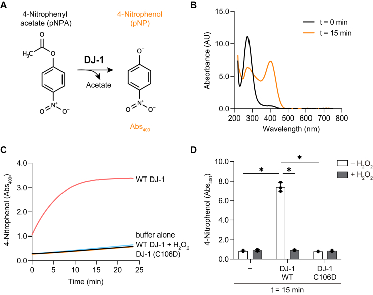
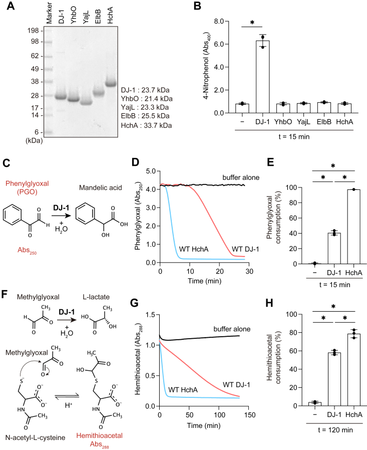
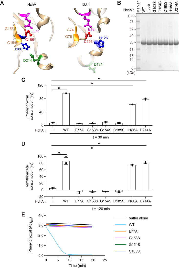
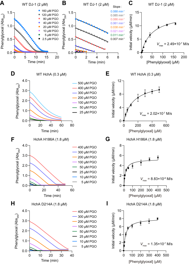
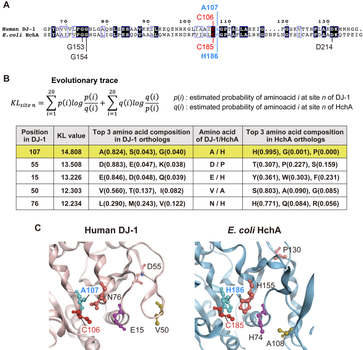
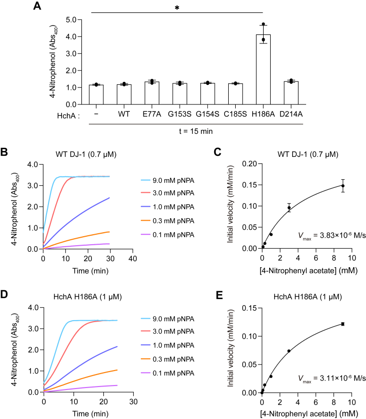
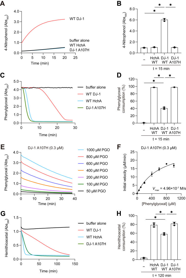
DJ-1, a causative gene for hereditary recessive Parkinsonism, is evolutionarily conserved across eukaryotes and prokaryotes. Structural analyses of DJ-1 and its homologs suggested the 106th Cys is a nucleophilic cysteine functioning as the catalytic center of hydratase or hydrolase activity. Indeed, DJ-1 and its homologs can convert highly electrophilic α-oxoaldehydes such as methylglyoxal into α-hydroxy acids as hydratase in vitro, and oxidation-dependent ester hydrolase (esterase) activity has also been reported for DJ-1. The mechanism underlying such plural activities, however, has not been fully characterized. To address this knowledge gap, we conducted a series of biochemical assays assessing the enzymatic activity of DJ-1 and its homologs. We found no evidence for esterase activity in any of the Escherichia coli DJ-1 homologs. Furthermore, contrary to previous reports, we found that oxidation inactivated rather than facilitated DJ-1 esterase activity. The E. coli DJ-1 homolog HchA possesses phenylglyoxalase and methylglyoxalase activities but lacks esterase activity. Since evolutionary trace analysis identified the 186th H as a candidate residue involved in functional differentiation between HchA and DJ-1, we focused on H186 of HchA and found that an esterase activity was acquired by H186A mutation. Introduction of reverse mutations into the equivalent position in DJ-1 (A107H) selectively eliminated its esterase activity without compromising α-oxoaldehyde hydratase activity. The obtained results suggest that differences in the amino acid sequences near the active site contributed to acquisition of esterase activity in vitro and provide an important clue to the origin and significance of DJ-1 esterase activity.

Keywords: DJ-1; PARK7; Parkinson disease (PD); catalytic triad; esterase; hydratase; oxo-aldehyde; prokaryote.


Conflict of interest statement

Conflict of interest The authors declare that they have no conflicts of interests with the contents of this article.

Figures

Figure 1
Figure 1
C106 oxidation and oxidation-mimetic mutation inhibit DJ-1 esterase activity.A, DJ-1 esterase reaction mechanism for 4-nitrophenol (pNP) formation. DJ-1 conversion of 4-nitrophenyl acetate (pNPA) to acetate and pNP was monitored via an increase in A400 by the pNP reaction product. B, DJ-1–mediated generation of pNP from pNPA with the characteristic A400. Absorbance spectra were monitored before (black) or 15 min after (orange) the start of the reaction. C, pNP generation over time. pNPA was incubated with the indicated proteins as described in the experimental procedures. WT DJ-1 was preincubated with 100 μM H2O2 for 1 h at room temperature prior to being mixed with pNPA. Representative data for three individual experiments are shown. D, A400 of pNP after 15 min incubation with or without WT DJ-1 or the C106D mutant ± H2O2 treatment (all n = 3). Bars represent the mean ± SD of three experiments. ∗p < 0.05 using two-way ANOVA with Bonferroni’s multiple comparisons test (D).
Figure 2
Figure 2
HchA possesses phenylglyoxalase and methylglyoxalase activities but not esterase activity.A, Coomassie-stained protein gel of recombinant DJ-1 homologs after purification. Gel lanes were loaded with 5 μg of recombinant DJ-1 or one of the Escherichia coli DJ-1 homologs. DJ-1 (lane 1), YhbO (lane 2), YajL (lane 3), ElbB (lane 4), and HchA (lane 5). The calculated molecular sizes of the recombinant proteins are indicated to the right of the gel. B, pNP generation from pNPA was observed with DJ-1 but not the bacterial DJ-1 homologs. DJ-1, EhbO, YajL, ElbB, or HchA protein (1 μM) was incubated with 1.6 mM pNPA for 15 min (all n = 3). C, the phenylglyoxal (PGO) hydratase reaction mechanism. PGO conversion into mandelic acid by DJ-1 can be monitored by a reduction in A250. D, PGO A250 in the presence of DJ-1 (red) or HchA (blue) over time at 37 °C. PGO was incubated with DJ-1 or HchA as described in the experimental procedures. The black line corresponds to buffer alone. E, percentage PGO consumption after 15 min incubation with either DJ-1 or HchA (all n = 3). PGO consumption values were calculated based on the difference in A250 before and after the start of the reaction. F, the methylglyoxal hydratase reaction mechanism. A288 increases following hemithioacetal formation from methylglyoxal and N-acetyl-cysteine. DJ-1 catalysis of methylglyoxal shifts the equilibrium reaction away from hemithioacetal formation with a concomitant reduction in A288. G, hemithioacetal A288 in the presence of DJ-1 (red) or HchA (blue) at 37 °C. Hemithioacetal was incubated with DJ-1 or HchA as described in the Experimental procedures. The black line corresponds to buffer alone. H, hemithioacetal A288 ratio following 120 min incubation with buffer alone, DJ-1, or HchA (all n = 3). Data shown in D and G are representative of three individual experiments. Data in B, E, and H are the mean ± SD of three experiments. ∗p < 0.05 using one-way ANOVA with Dunnett’s multiple comparisons test (B, E, and H).
Figure 3
Figure 3
HchA α-oxoaldehyde hydratase activity was abolished by E77A, G153S, G154S, and C185S mutations.A, structure of the HchA (left) and DJ-1 (right) catalytic cores. Equivalent residues between HchA and DJ-1 are highlighted by the same color. The HchA residues selected for mutation are E77 (magenta), G153 (orange), G154 (orange), C185 (red), H186 (blue), and D214 (green). B, Coomassie-stained protein gel of recombinant HchA mutants after purification. Gel lanes were loaded with 5 μg of HchA WT (lane 2), E77A (lane 3), G153S (lane 4), G154S (lane 5), C185S (lane 7), H186A (lane 7), and D214A (lane 8). C, PGO consumption after 30 min incubation with buffer alone, WT HchA, or the indicated HchA mutant proteins as described in the Experimental procedures. PGO consumption values were calculated based on the difference in A250 before and after the start of the reaction (n = 3). D, hemithioacetal consumption by WT HchA or the indicated mutants. The hemithioacetal reaction period was 120 min as described in the Experimental procedures (all n = 3). E, PGO A250 over time. Lines correspond to WT HchA (cyan) or HchA with E77A (orange), G153S (magenta), G154S (green), or C185S (dark blue) mutations. Representative data from three individual experiments are shown. Data in C and D are the mean ± SD of three experiments. ∗p < 0.05 using one-way ANOVA with Dunnett’s multiple comparisons test (C and D). PGO, phenylglyoxal.
Figure 4
Figure 4
Phenylglyoxalase enzyme kinetics of HchA H186A and HchA D214A. A, PGO consumption over time by 2 μM WT DJ-1. The substrate concentrations assayed are indicated. B, initial velocities of PGO consumption. Each reaction velocity was calculated via linear regression of the data shown in A. C, Michaelis–Menten plot of WT DJ-1–mediated PGO consumption. Values were obtained from the reaction velocities shown in B. The Vmax was calculated as 2.49 × 10−7 M/s. D, PGO consumption over time by 0.3 μM WT HchA. The substrate concentrations assayed are indicated. E, Michaelis–Menten plot of WT HchA-mediated PGO consumption. The Vmax was calculated as 2.02 × 10-7 M/s. F, PGO consumption over time by 1.8 μM HchA(H186A). The substrate concentrations assayed are indicated. G, Michaelis–Menten plot of HchA(H186A)-mediated PGO consumption. The Vmax was calculated as 8.83 × 10−8 M/s. H, PGO consumption over time by 1.8 μM HchA(D214A). The substrate concentrations assayed are indicated. I, Michaelis–Menten plot of HchA(D214A)-mediated PGO consumption. The Vmax was calculated as 1.35 × 10−7 M/s. The data shown in A, B, D, F, and H are representative data of three individual experiments. Data in C, E, G, and I are the mean ± SD of three replicates. PGO, phenylglyoxal.
Figure 5
Figure 5
Detection of amino acid sites involved in functional differentiation between DJ-1 orthologs and HchA orthologs.A, sequence alignment between human DJ-1 and Escherichia coli HchA. The alignment was generated using Clustal W and ESPript3 (http://espript.ibcp.fr). DJ-1 A107 is comparable to His186 in HchA (shown in blue font). Identical amino acids are shown in black, conserved sequences in blue boxes, catalytic cysteines in red, and amino acids mutated in HchA are indicated. B and C, top five sites with high KL values (B) and the amino acid residues with high KL values are depicted on the structures of human DJ-1 (PDB ID: 1P5F) and E. coli HchA (PDB ID: 1N57) (C). Structural visualization of DJ-1 and HchA was performed using the Molecular Operating Environment software (MOE 2020.0901) (Chemical Computing Group ULC, 910-1010 Sherbrooke St. W., Montreal, QC H3A 2R7, Canada, 2024.). KL, Kullback–Leibler; PGO, phenylglyoxal.
Figure 6
Figure 6
Latent esterase activity of HchA is induced by the H186A mutation.A, esterase activity in WT HchA or the indicated HchA mutants. pNPA and the indicated recombinant protein were incubated as described in the experimental procedures for 15 min. B, formation of pNP over time by 0.7 μM WT DJ-1. The substrate concentrations assayed are indicated. C, Michaelis–Menten plot for WT DJ-1–mediated formation of pNP. The Vmax was calculated as 3.83 × 10−6 M/s. D, formation of pNP over time by 1 μM HchA H186A. The substrate concentrations assayed are indicated. E, Michaelis–Menten plot for HchA(H186A)-mediated formation of pNP. The Vmax was calculated as 3.11 × 10−6 M/s. The data shown in B and D are representative of three individual experiments. Data in A, C, and E are the mean ± SD of three replicates. pNP, 4-nitrophenol; pNPA, 4-nitrophenyl acetate.
Figure 7
Figure 7
The A107H mutation autoinhibits DJ-1 esterase activity.A, production of 4-nitrophenol (pNP) over time. Indicated proteins and 4-nitrophenyl acetate (pNPA) were incubated as described in the Experimental procedures. B, inhibition of pNP formation by the DJ-1 A107H mutation. The enzymatic reactions were incubated for 15 min. C, reduction in PGO-dependent A250 over time. PGO was incubated with WT DJ-1 (red), WT HchA (blue), or the DJ-1(A107H) mutant (green) as described in the experimental procedures. The black line indicates the buffer control. D, PGO consumption over time. PGO was incubated for 15 min with the indicated proteins (all n = 3). E, decrease in PGO by 0.3 μM DJ-1(A107H). The substrate concentrations assayed are indicated. Initial velocities are based on the data shown. F, Michaelis–Menten plot for DJ-1(A107H)-mediated consumption of PGO (n = 3). The Vmax was calculated as 4.96 × 10−7 M/s. G, DJ-1(A107H)-mediated hemithioacetal consumption. Reaction conditions for buffer alone (black), WT DJ-1 (red), WT HchA (blue), and the DJ-1 A107H mutant (green) are as described in the Experimental procedures. H, hemithioacetal consumption following 120 min incubation with the indicated proteins (all n = 3). Data shown in A, C, and G are representative of three individual experiments. Data in B, D, and H are the mean ± SD of three experiments. PGO, phenylglyoxal.

Similar articles

Cited by

References

    1. Bonifati V., Rizzu P., van Baren M.J., Schaap O., Breedveld G.J., Krieger E., et al. Mutations in the DJ-1 gene associated with autosomal recessive early-onset parkinsonism. Science. 2003;299:256–259. - PubMed
    1. Wilson M.A. The role of cysteine oxidation in DJ-1 function and dysfunction. Antioxid. Redox Signal. 2011;15:111–122. - PMC - PubMed
    1. Kahle P.J., Waak J., Gasser T. DJ-1 and prevention of oxidative stress in Parkinson's disease and other age-related disorders. Free Radic. Biol. Med. 2009;47:1354–1361. - PubMed
    1. Taira T., Saito Y., Niki T., Iguchi-Ariga S.M., Takahashi K., Ariga H. DJ-1 has a role in antioxidative stress to prevent cell death. EMBO Rep. 2004;5:213–218. - PMC - PubMed
    1. Shendelman S., Jonason A., Martinat C., Leete T., Abeliovich A. DJ-1 is a redox-dependent molecular chaperone that inhibits alpha-synuclein aggregate formation. PLoS Biol. 2004;2:e362. - PMC - PubMed

Publication types

MeSH terms

LinkOut - more resources