. 2024 Aug:184:68-80.
doi: 10.1016/j.actbio.2024.06.013. Epub 2024 Jun 13.

Conserved and tissue-specific immune responses to biologic scaffold implantation

Affiliations

Conserved and tissue-specific immune responses to biologic scaffold implantation

Sabrina DeStefano et al. Acta Biomater. 2024 Aug.

Abstract

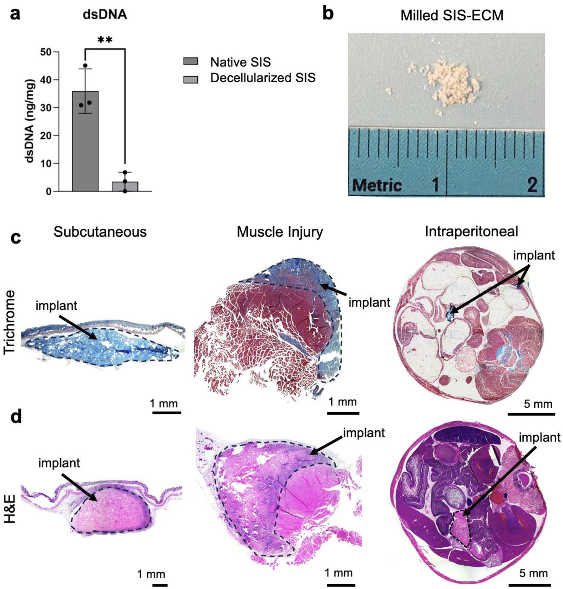
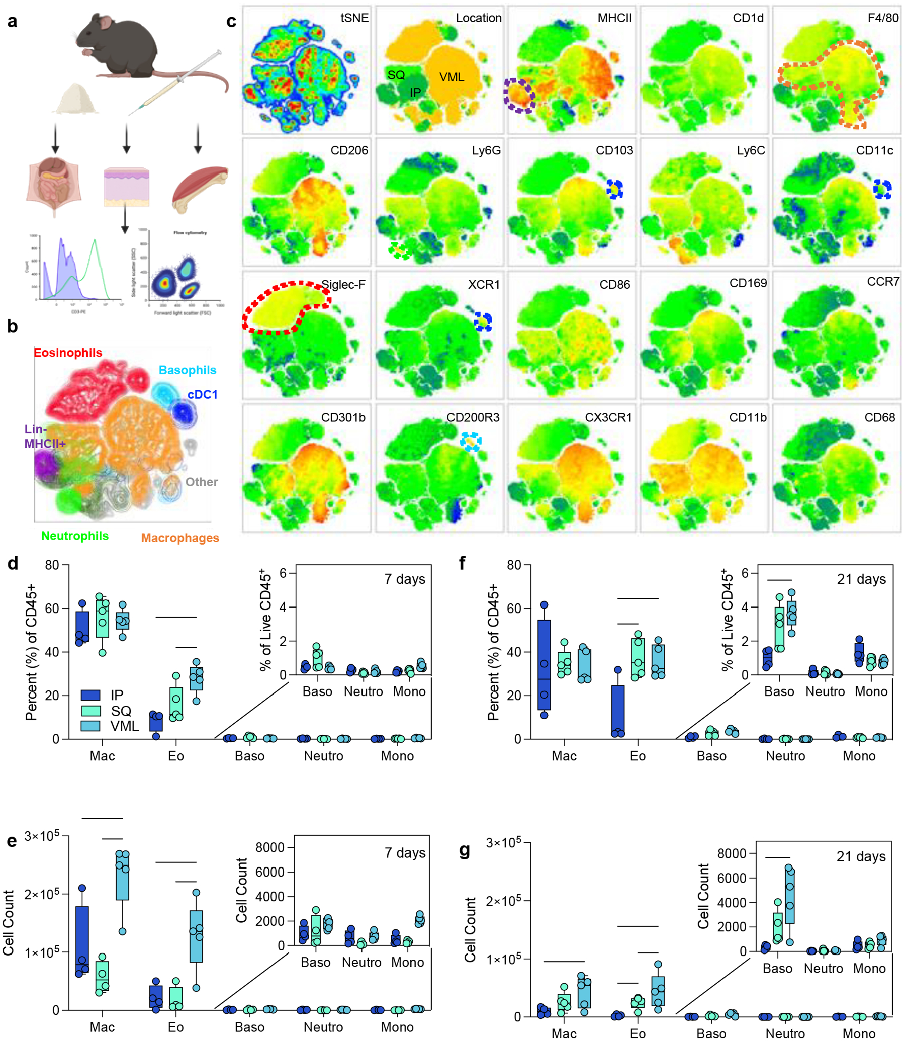
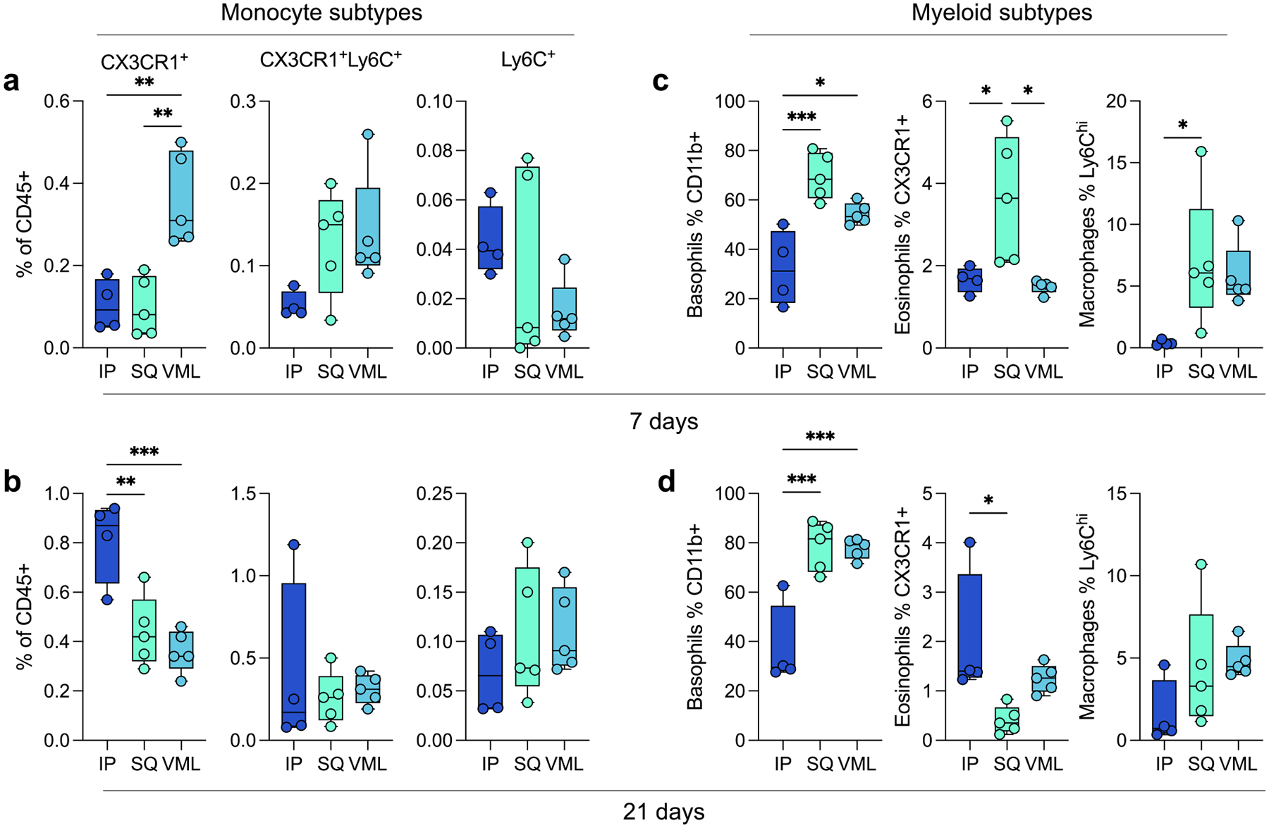
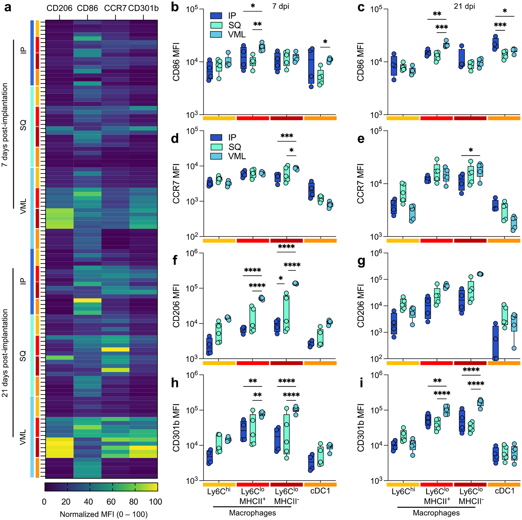
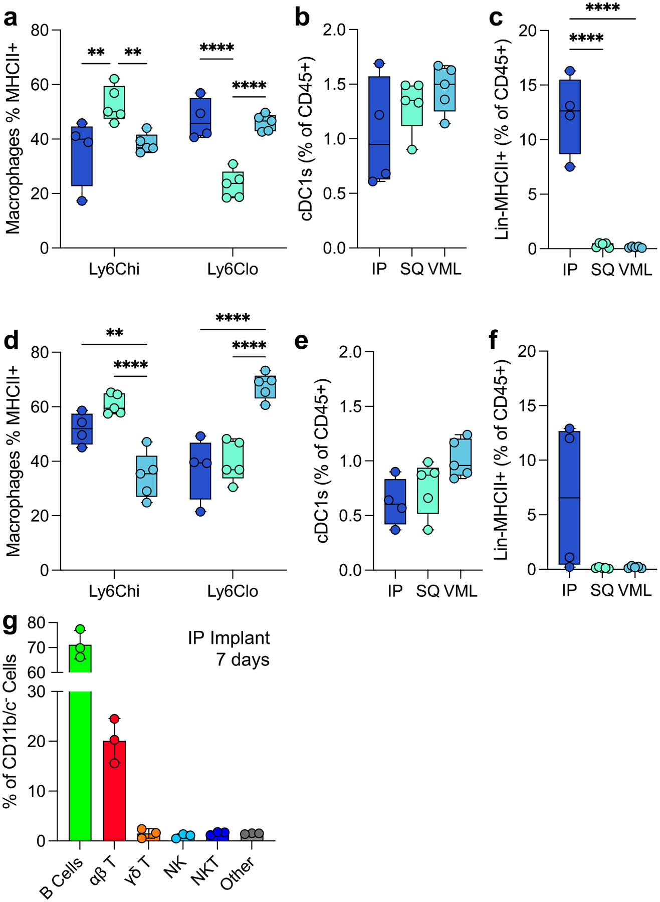
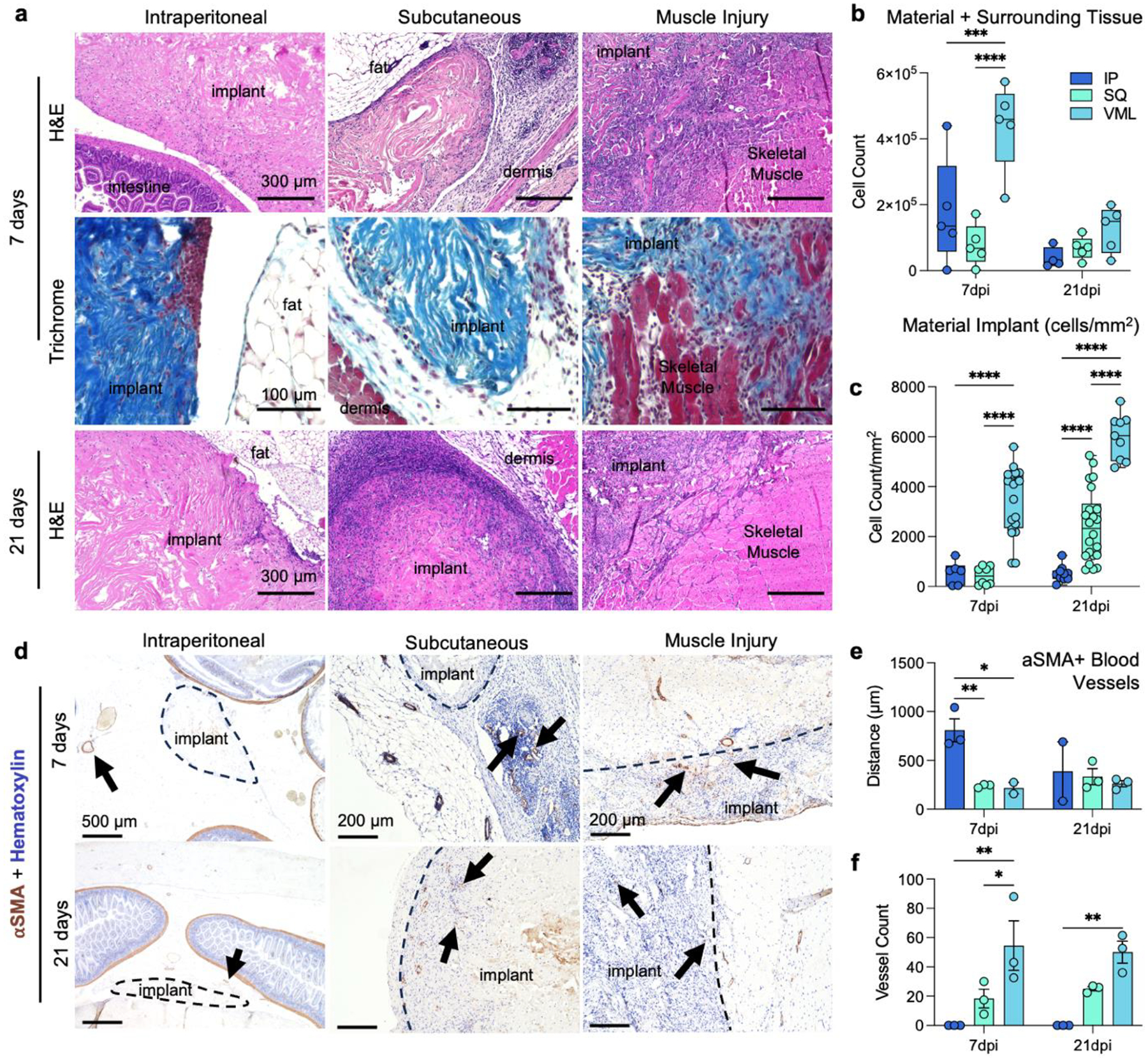
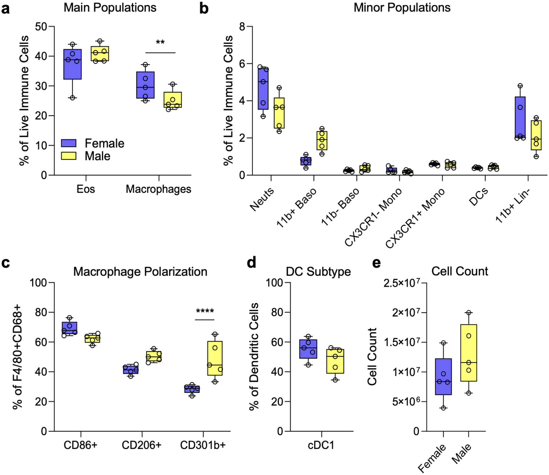
Upon implantation into a patient, any biomaterial induces a cascade of immune responses that influences the outcome of that device. This cascade depends upon several factors, including the composition of the material itself and the location in which the material is implanted. There is still significant uncertainty around the role of different tissue microenvironments in the immune response to biomaterials and how that may alter downstream scaffold remodeling and integration. In this study, we present a study evaluating the immune response to decellularized extracellular matrix materials within the intraperitoneal cavity, the subcutaneous space, and in a traumatic skeletal muscle injury microenvironment. All different locations induced robust cellular recruitment, specifically of macrophages and eosinophils. The latter was most prominent in the subcutaneous space. Intraperitoneal implants uniquely recruited B cells that may alter downstream reactivity as adaptive immunity has been strongly implicated in the outcome of scaffold remodeling. These data suggest that the location of tissue implants should be taken together with the composition of the material itself when designing devices for downline therapeutics. STATEMENT OF SIGNIFICANCE: Different tissue locations have unique immune microenvironments, which can influence the immune response to biomaterial implants. By considering the specific immune profiles of the target tissue, researchers can develop implant materials that promote better integration, reduce complications, and improve the overall outcome of the implantation process.

Keywords: Biomaterials; Extracellular matrix; Foreign body response; Immune response; Tissue immunology.


Conflict of interest statement

Declaration of competing interest The authors declare the following financial interests/personal relationships which may be considered as potential competing interests: R.L., T.B.N., and K.S. are inventors on the provisional patent application #US63/367,994 related to the information discussed in this manuscript. All other authors have nothing to declare.

Figures

FIG 1 |
FIG 1 |. Material characterization and representative images of implant locations.
(a) dsDNA content of native SIS and decellularized SIS. (b) Macro image of milled ECM powder. (c) Macro images of Masson’s trichrome staining of each implant site (Subcutaneous, Muscle Injury, Intraperitoneal) at 7 dpi. (d) Hematoxylin and eosin staining at 21 dpi.
FIG 2 |
FIG 2 |. Myeloid response to biologic scaffolds in different tissue locations.
(a) Experimental workflow (b) tSNE of manually gated immune cell (CD45+) populations in ECM scaffolds (c) Expression of myeloid phenotyping markers in different islands. SQ = subcutaneous implant; IP = intraperitoneal implant; VML = volumetric muscle loss skeletal muscle injury implant. (d) Immune cell populations as a percent of live immune cells at 7 days post-implantation (dpi). (e) Immune cell counts at 7 dpi from 50 μl implants. (f) Immune cell populations as a percent of live immune cells at 21 dpi. (g) Immune cell counts at 21 dpi. Data are range, n = 4 – 5, two-way ANOVA with Tukey posthoc. * = p < 0.05; *** = p < 0.001; **** = p < 0.0001. Schematic made with BioRender.
FIG 3 |
FIG 3 |. Myeloid subtypes vary in an implant location and time-dependent manner.
(a) Monocyte subtypes at 7 dpi. (b) Monocyte subtypes at 21 dpi. (c-d) Basophil, eosinophil, and macrophage subtypes at (c) 7 dpi and (d) 21 dpi. Data are range, n = 4 – 5, one-way ANOVA with Tukey posthoc. * = p < 0.05; ** = p < 0.01; *** = p < 0.001
FIG 4 |
FIG 4 |. Injury induces a stronger type-2 polarized immune response than non-traumatic applications.
(a) Myeloid polarization markers CD206, CD301b (M2-like), CD86, CCR7 (M1-like) over time. (b-c) CD86 median fluorescence intensity (MFI) at (b) 7 days post-implantation (dpi) and (c) 21 dpi. (d-e) CCR7 MFI at (d) 7 dpi, and (e) 21 dpi. (f-g) CD206 MFI at (f) 7 dpi and (g) 21 dpi. (h-i) CD206 MFI at (h) 7 dpi, and (i) 21 dpi. Yellow = Ly6Chi macrophages, Red = Ly6Clo MHCII+ Macrophages, Dark Red = Ly6Clo MHCII macrophages, Orange = type 1 conventional dendritic cells (cDC1s). Data are range, n = 4 – 5, two-way ANOVA with Tukey posthoc. * = p < 0.05; ** = p < 0.01; *** = p < 0.001; **** = p < 0.0001.
FIG 5 |
FIG 5 |. Variability in myeloid and lymphoid antigen-presenting cells in the tissue microenvironment.
(a) The proportion of macrophages expressing MHCII at 7 dpi. (b) The proportion of total immune cells that are cross-presenting cDC1s at 7dpi (c) The proportion of lymphoid-like Lin-MHCII+ unidentified antigen-presenting cells 7dpi. (d-f) The proportion of (d) MHCII+ macrophages, (e) cDC1s, and (f) LinMHCII+ cells at 21dpi. (g) Lymphoid profile of IP implants. Data are range (a-f) or mean ± standard deviation, n = 3 – 5, two-way ANOVA with Tukey posthoc. ** = p < 0.01; *** = p < 0.001; **** = p < 0.0001.
FIG 6 |
FIG 6 |. Histopathologic differences in cellular infiltration and vascularization of ECM implants.
(a) Top row: hematoxylin and eosin (H&E) staining of IP (intraperitoneal), SQ (subcutaneous) and VML (muscle injury) implants at 7 days post-implantation (dpi). Middle: Masson’s trichrome of tissue interface at 7dpi. Bottom row: H&E of IP, SQ, and VML implants at 21 dpi. Representative of n = 5 mice. (b) Count of cells by isolation and flow cytometry (c) Count of cells per mm2 of material only (not including surrounding tissue). (d) Immunohistochemical staining of blood vessels (αSMA) and cell nuclei (Hematoxylin) Top row: αSMA immunohistochemistry staining of VML (muscle injury) at 7 and 21 dpi. Middle: αSMA immunohistochemistry staining of SQ (subcutaneous) at 7 and 21 dpi. αSMA immunohistochemistry staining of IP (intraperitoneal) at 7 and 21 dpi. (e) Shortest distance between the middle of each blood vessel and edge of ECM material in each region at 7 and 21 dpi. (f) Number of blood vessels within ECM implant and immune infiltration zone surrounding material in each implant region at 7 and 21 dpi. n = 3, one-way ANOVA with Tukey posthoc. * = p < 0.05; ** = p < 0.01. Data are range, n = 5 mice, two-way ANOVA with Tukey posthoc. *** = p < 0.001; **** = p < 0.0001.
FIG 7 |
FIG 7 |. Sex Differences in Myeloid Cell Populations.
(a) Main (macrophages and eosinophils) immune cell populations as a percent of live immune cells at 7 dpi between female and male mice. (b) Minor immune cell populations as a percent of live CD45+ at 7 dpi between female and male mice. (c) Macrophage polarization as a percent of live F4/80+CD68+ cells at 7 dpi between female and male mice. (d). Type 1 conventional dendritic cells as a percentage of all dendritic cells across female and male mice at 7 dpi. Data are range, n = 5 mice, Student’s T-Test. ** = p < 0.01; *** = p < 0.001; **** = p < 0.0001.

Update of

Similar articles

Cited by

References

    1. Brown BN, Londono R, Tottey S, Zhang L, Kukla KA, Wolf MT, Daly KA, Reing JE, Badylak SF, Macrophage phenotype as a predictor of constructive remodeling following the implantation of biologically derived surgical mesh materials, Acta biomaterialia 8(3) (2012) 978–987. - PMC - PubMed
    1. Godwin JW, Pinto AR, Rosenthal NA, Macrophages are required for adult salamander limb regeneration, Proceedings of the National Academy of Sciences 110(23) (2013) 9415–9420. - PMC - PubMed
    1. Heredia JE, Mukundan L, Chen FM, Mueller AA, Deo RC, Locksley RM, Rando TA, Chawla A, Type 2 innate signals stimulate fibro/adipogenic progenitors to facilitate muscle regeneration, Cell 153(2) (2013) 376–388. - PMC - PubMed
    1. Goh YS, Henderson NC, Heredia JE, Red Eagle A, Odegaard JI, Lehwald N, Nguyen KD, Sheppard D, Mukundan L, Locksley RM, Eosinophils secrete IL-4 to facilitate liver regeneration, Proceedings of the National Academy of Sciences 110(24) (2013) 9914–9919. - PMC - PubMed
    1. Sadtler K, Estrellas K, Allen BW, Wolf MT, Fan H, Tam AJ, Patel CH, Luber BS, Wang H, Wagner KR, Developing a pro-regenerative biomaterial scaffold microenvironment requires T helper 2 cells, Science 352(6283) (2016) 366–370. - PMC - PubMed

Publication types

Substances

LinkOut - more resources