Review
. 2024 Jun 18;13(12):e035128.
doi: 10.1161/JAHA.123.035128. Epub 2024 Jun 15.

Role of Antitroponin Antibodies and Macrotroponin in the Clinical Interpretation of Cardiac Troponin

Affiliations
Review

Role of Antitroponin Antibodies and Macrotroponin in the Clinical Interpretation of Cardiac Troponin

Erwan Salaun et al. J Am Heart Assoc. .

Abstract

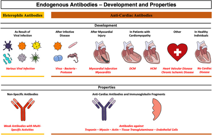
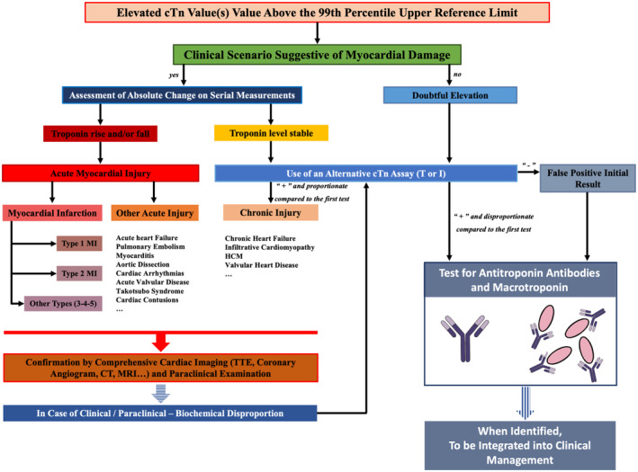
Cardiac troponin is extensively used as a biomarker in modern medicine due to its diagnostic capability for myocardial injury, as well as its predictive and prognostic value for cardiac diseases. However, heterophile antibodies, antitroponin antibodies, and macrotroponin complexes can be observed both in seemingly healthy individuals and patients with cardiac diseases, potentially leading to false positive or disproportionate elevation of cTn (cardiac troponin) assay results and introducing discrepancies in clinical interpretations with impact on medical management. In this review article, we describe the possible mechanisms of cTn release and the sources of variations in the assessment of circulating cTn levels. We also explore the pathophysiological mechanisms underlying antitroponin antibody development and discuss the influence exerted by macrotroponin complexes on the results of immunoassays. Additionally, we explore approaches to detect these complexes by presenting various clinical scenarios encountered in routine clinical practice. Finally, unsolved questions about the development, prevalence, and clinical significance of cardiac autoantibodies are discussed.

Keywords: antitroponin antibodies; biomarker; cardiac troponin; macrotroponin; myocardial infarction; myocardial injury; myocarditis.


Figures

Figure 1
Figure 1. Mechanisms of cardiac troponin release and antitroponin antibodies formation and impact of macrotroponin complex on cTn assays results and patient management.
cTn indicates cardiac troponin; hs, high‐sensitivity; ind, individual; and PEG, polyethylene glycol precipitation.
Figure 2
Figure 2. Cardiac troponin sandwich immunoassay technique and interferences.
cTn indicates cardiac troponin; and cTnI, cardiac troponin I.
Figure 3
Figure 3. Endogenous antibodies development and properties.
DCM indicates dilated cardiomyopathy; and HCM, hypertrophic cardiomyopathy.
Figure 4
Figure 4. Challenging cases for interpretation of cardiac troponins.
A, Syncope and possible macrotroponin in athlete patient. This case illustrates a false positive result of hs‐cTnI in a patient referred from a regional hospital for syncope immediately after running. The first hs‐cTnI assay performed on a Vitros 5600 instrument (Ortho Clinical Diagnostics) in the regional hospital was not increased (13 ng/L; 99th percentile URL=13 ng/L in men and 9 ng/L in women), while 6 hours later, hs‐cTnI measured using a different assay on a Dimension Vista 1500 instrument (Siemens Healthcare Diagnostics) in our hospital showed an increase at 170 ng/L (99th percentile URL=79 ng/L in men and 54 ng/L in women). Finally, the level of hs‐cTnT measured on a Cobas instrument (Roche Diagnostics, 99th percentile URL=14 ng/L) was normal and a PEG precipitation test was compatible with the presence of interference. All examinations confirmed a postexercise vasovagal syncope in a patient with athletic heart syndrome (complete description in Data S8). B, Disproportionate increase of hs‐cTnI in acute coronary disease. This panel shows the case of a 50‐year‐old man with inferior STEMI requiring an immediate PTCA of a proximal RCA and a delayed surgical myocardial revascularization for the left coronary arteries. The level of the hs‐cTnI on a Dimension Vista 1500 instrument (Siemens Healthcare Diagnostics, 99th percentile URL=79 ng/L in men and 54 ng/L in women) was very high after STEMI and CABG with slow decrease after CABG, suggesting the presence of interference, revealed by the discrepancy between cTnI and cTnT measured on a Cobas instrument (Roche Diagnostics, 99th percentile URL=14 ng/L) and a PEG test unequivocally positive (3% recovery) (complete description in Data S8). C, Suspected macrotroponin in a patient with hypertrophic cardiomyopathy. This panel highlights the probable presence of macrotroponin in a patient with HCM without obstruction (ICD since 2014). The hs‐cTnT measured on a Cobas instrument (Roche Diagnostics, 99th percentile URL=14 ng/L) was slightly increased over the years (2017: 59 ng/L; 2021: 51 ng/L; 2022: 45 ng/L) and considered related to HCM, whereas hs‐cTnI measured on a Dimension Vista 1500 instrument (Siemens Healthcare Diagnostics, 99th percentile URL=79 ng/L in men and 54 ng/L in women) was increased at a higher level and led to suspicion of NSTEMI type II in 2020 in the context of atypical chest pain. In 2022, the interference with hs‐cTnI assay was then confirmed (complete description in Data S8). 3C view indicates apical 3 cavities view; 4C view, apical 4 cavities view; Afib, atrial fibrillation; CABG, coronary artery bypass graft; cMRI, cardiac magnetic resonance imaging; CT, computed tomography; CX, circonflex artery; HCM, hypertrophic cardiomyopathy; hs‐cTn, high‐sensitivity cardiac troponin; ICD, implantable cardioverter‐defibrillator; LAD, left anterior descending artery; NSTEMI, non–ST‐segment–elevation myocardial infarction; PEG, polyethylene glycol; PSLA, parasternal long axis view; PTCA, percutaneous transluminal coronary angioplasty; RCA, right coronary artery; STEMI, ST‐segment–elevation myocardial infarction; TTE, transthoracic echocardiography; and URL, upper reference limit.
Figure 5
Figure 5. Endogenous antibodies and macrotroponin in particular conditions.
A, In a patient with cardiac amyloidosis. A 79‐year‐old man was referred to the emergency department by his family physician for atrial fibrillation with a spontaneous slow ventricular rate. He had history of suspected NSTEMI type II in 2018 and possible myocarditis in 2020, and surgery for treating bilateral carpal tunnel syndrome (additional details in Data S8). First sets of examination showed low QRS voltage in the frontal leads, an increase in NT‐proBNP level at 1225 ng/L measured with a Dimension Vista 1500 instrument (Siemens Healthcare Diagnostics, URL=300 ng/L) and hs‐cTnI result level at 629 ng/L measured with the same instrument (99th percentile URL=79 ng/L in men and 54 ng/L in women), with stable level at 6 hours and 24 hours without chest pain. As suspected, the transthoracic echocardiogram and technetium‐99m pyrophosphate scintigraphy imaging confirmed the diagnosis of cardiac transthyretin amyloidosis. Based on the current stable hs‐cTn level and the previous history of cTn elevation (suspected NSTEMI II and myocarditis), the presence of a macrotroponin complex was suspected by the absence of a significant increase in hs‐cTnT (22 ng/L) (Roche Diagnostics, 99th percentile URL=14 ng/L) and a positive PEG test (5% recovery). B, In the context of acute myocarditis. A 47‐year‐old woman was referred for acute myocarditis in January 2023. Four months before, she had acute pericarditis with posterior abundant pericardial effusion (normal LVEF and no increase in cTnI) (additional details in Data S8). She was referred for acute chest pain in the context of an inflammatory state with C‐reactive protein increased at 204 mg/L, a depressed LVEF at 25%, and an increase of cTnI at 3000 ng/L. A bolus of solumedrol was started before the admission to our center. At admission, CAG was performed and showed no significant coronary artery stenosis and LVEF was improved at 45%. The cTnI measured using a Dimension Vista 1500 instrument (Siemens Healthcare Diagnostics, 99th percentile URL=79 ng/L in men and 54 ng/L in women) and the C‐reactive protein blood level decreased during the first 4 days in our center, and cMRI confirmed signs of acute myocarditis with a quasinormalization of the LVEF. However, a small reincrease in cTnI during hospitalization was observed despite clinical and echocardiographic improvement. Interferences related to probable circulating antitroponin I antibodies and macrotroponin in the context of myocarditis was suspected and confirmed indirectly by normal hs‐cTnT results (17, 12, and 13 ng/L) (Roche Diagnostics, 99th percentile URL=14 ng/L) and the presence of a positive PEG test (8% recovery). AF indicates atrial fibrillation; ATTR, transthyretin amyloidosis; CAG, coronary angiogram; cMRI, cardiac magnetic resonance imaging; CTnI, cardiac troponin I; hs‐cTn, high‐sensitivity cardiac troponin; LVEF, left ventricular ejection fraction; NSTEMI, non–ST‐segment–elevation myocardial infarction; NT‐proBNP, N‐terminal pro‐B‐type natriuretic peptide; PEG, polyethylene glycol; and URL, upper reference limit.
Figure 6
Figure 6. Algorithm for the diagnosis of myocardial Injury considering antitroponin antibodies and macrotroponin.
CT indicates computed tomography; cTn, cardiac troponin; HCM, hypertrophic cardiomyopathy; MI, myocardial infarction; MRI, cardiac magnetic resonance imaging; and TTE, transthoracic echocardiography.

Similar articles

Cited by

References

    1. Dolci A, Panteghini M. The exciting story of cardiac biomarkers: from retrospective detection to gold diagnostic standard for acute myocardial infarction and more. Clin Chim Acta. 2006;369:179–187. doi: 10.1016/j.cca.2006.02.042 - DOI - PubMed
    1. Katrukha IA. Human cardiac troponin complex. Structure and functions. Biochemistry (Mosc). 2013;78:1447–1465. doi: 10.1134/S0006297913130063 - DOI - PubMed
    1. Raber I, McCarthy CP, Januzzi JL Jr. A test in context: interpretation of high‐sensitivity cardiac troponin assays in different clinical settings. J Am Coll Cardiol. 2021;77:1357–1367. doi: 10.1016/j.jacc.2021.01.011 - DOI - PubMed
    1. Thygesen K, Alpert JS, Jaffe AS, Chaitman BR, Bax JJ, Morrow DA, White HD. Executive group on behalf of the joint European Society of Cardiology/American College of Cardiology/American Heart Association/world heart federation task force for the universal definition of myocardial I. Fourth universal definition of myocardial infarction (2018). J Am Coll Cardiol. 2018;72:2231–2264. doi: 10.1016/j.jacc.2018.08.1038 - DOI - PubMed
    1. McCarthy CP, Raber I, Chapman AR, Sandoval Y, Apple FS, Mills NL, Januzzi JL Jr. Myocardial injury in the era of high‐sensitivity cardiac troponin assays: a practical approach for clinicians. JAMA Cardiol. 2019;4:1034–1042. doi: 10.1001/jamacardio.2019.2724 - DOI - PubMed

LinkOut - more resources