. 2024 Jun 15;30(1):89.
doi: 10.1186/s10020-024-00856-1.

Inhibition of tartrate-resistant acid phosphatase 5 can prevent cardiac fibrosis after myocardial infarction

Affiliations

Inhibition of tartrate-resistant acid phosphatase 5 can prevent cardiac fibrosis after myocardial infarction

Shujun Yang et al. Mol Med. .

Abstract

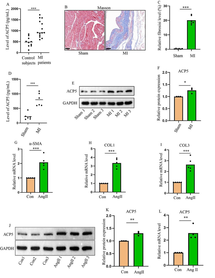
Background: Myocardial infarction (MI) leads to enhanced activity of cardiac fibroblasts (CFs) and abnormal deposition of extracellular matrix proteins, resulting in cardiac fibrosis. Tartrate-resistant acid phosphatase 5 (ACP5) has been shown to promote cell proliferation and phenotypic transition. However, it remains unclear whether ACP5 is involved in the development of cardiac fibrosis after MI. The present study aimed to investigate the role of ACP5 in post-MI fibrosis and its potential underlying mechanisms.

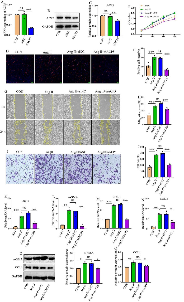
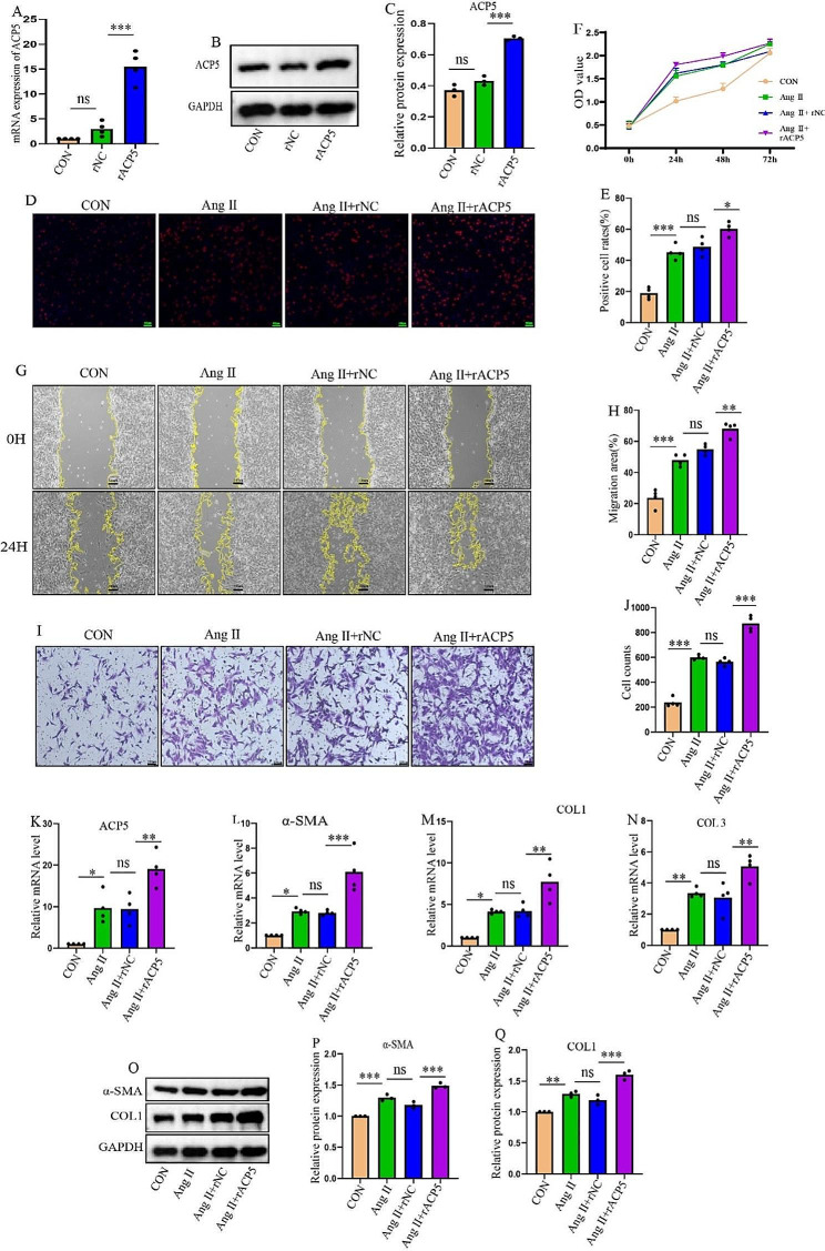
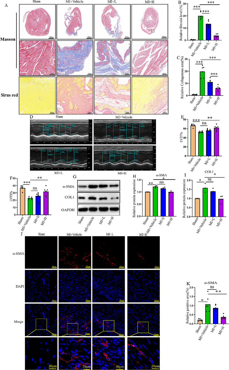
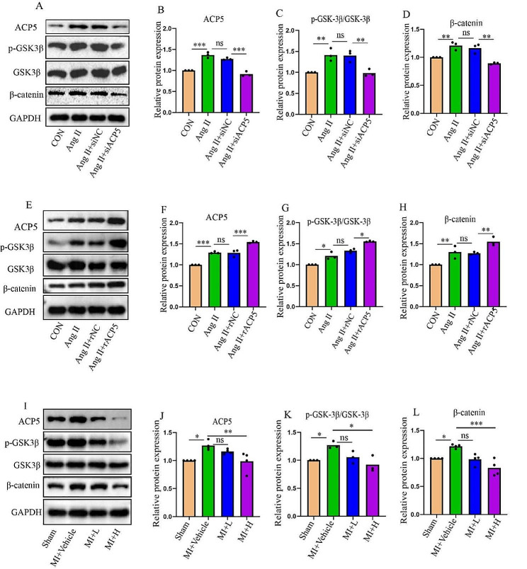
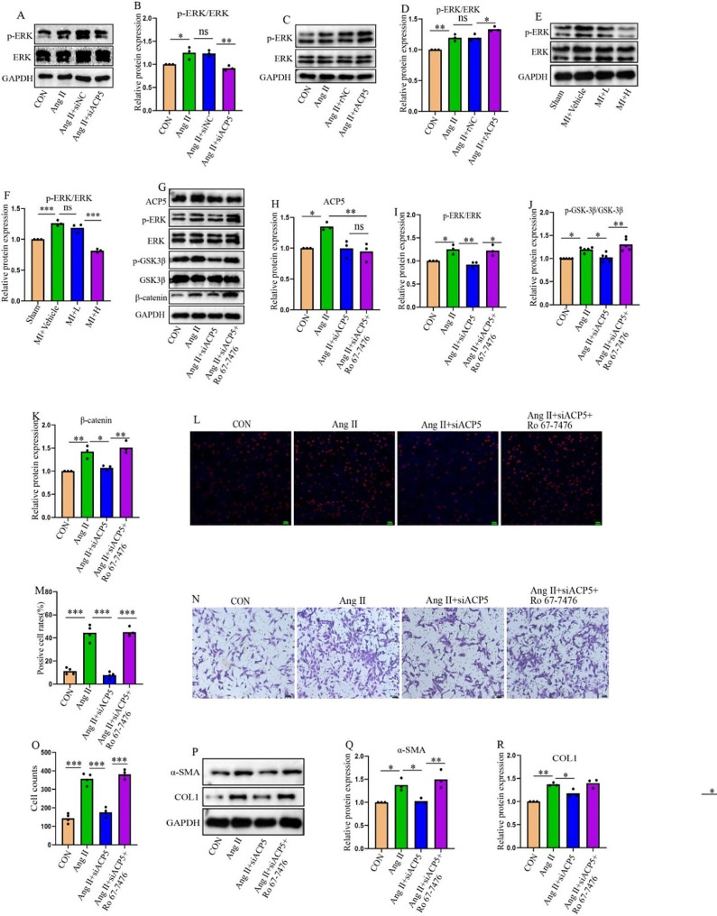
Methods: Clinical blood samples were collected to detect ACP5 concentration. Myocardial fibrosis was induced by ligation of the left anterior descending coronary artery. The ACP5 inhibitor, AubipyOMe, was administered by intraperitoneal injection. Cardiac function and morphological changes were observed on Day 28 after injury. Cardiac CFs from neonatal mice were extracted to elucidate the underlying mechanism in vitro. The expression of ACP5 was silenced by small interfering RNA (siRNA) and overexpressed by adeno-associated viruses to evaluate its effect on CF activation.

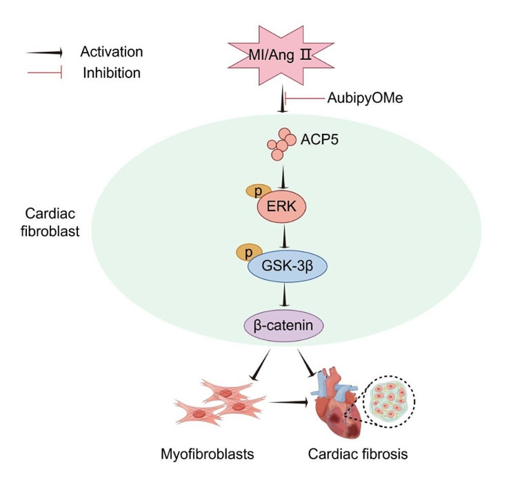
Results: The expression of ACP5 was increased in patients with MI, mice with MI, and mice with Ang II-induced fibrosis in vitro. AubipyOMe inhibited cardiac fibrosis and improved cardiac function in mice after MI. ACP5 inhibition reduced cell proliferation, migration, and phenotypic changes in CFs in vitro, while adenovirus-mediated ACP5 overexpression had the opposite effect. Mechanistically, the classical profibrotic pathway of glycogen synthase kinase-3β (GSK3β)/β-catenin was changed with ACP5 modulation, which indicated that ACP5 had a positive regulatory effect. Furthermore, the inhibitory effect of ACP5 deficiency on the GSK3β/β-catenin pathway was counteracted by an ERK activator, which indicated that ACP5 regulated GSK3β activity through ERK-mediated phosphorylation, thereby affecting β-catenin degradation.

Conclusion: ACP5 may influence the proliferation, migration, and phenotypic transition of CFs, leading to the development of myocardial fibrosis after MI through modulating the ERK/GSK3β/β-catenin signaling pathway.

Keywords: Cardiac fibroblasts (CFs); Cardiac fibrosis; Glycogen synthase kinase-3β (GSK3β)/β-catenin signaling pathway; Myocardial infarction; Tartrate-resistant acid phosphatase 5 (ACP5).


Conflict of interest statement

The authors declare no conflict of interests.

Figures

Fig. 1
Fig. 1
ACP5 expression is increased in patients with myocardial infarction, in vivo mouse models, and in vitro mouse models. (A) The expression of ACP5 in the blood of healthy individuals and postmyocardial infarction patients was detected by ELISA (control subjects: n = 10; MI patients: n = 14). (B-C) Masson staining and fibrosis ratio in each group (n = 6/group). Scale = 100 μm. (D) ACP5 expression in the blood of mice in different groups (n = 6/group). (E-F) Western blot analysis of ACP5 expression in the hearts of mice (n = 3/group). (G-I) Relative mRNA expression levels of α-SMA, COL1 and COL3 in CFs in different groups (n = 5/group). (J-K) CF Western blot analysis of ACP5 expression in different groups. (L) Relative mRNA expression levels of ACP5 in different CF groups (n = 4/group). *P < 0.05, **P < 0.01, and ***P < 0.001
Fig. 2
Fig. 2
Inhibition of ACP5 suppresses CF proliferation, migration, and transition into myofibroblasts. (A) Relative mRNA expression levels after ACP5 silencing (n = 3/group). (B-C) Western blot analysis of the silencing efficiency of ACP5 (n = 3/group). (D-E) EdU assay was used to detect the cell proliferation rate of CFs (n = 4/group); scale bar = 100 μm. (F) OD values of CFs in different groups (n = 4/group). (G-H) Cell migration was assessed by a wound-healing assay (n = 4/group); scale bar = 100 μm. (I-J) Cell migration was assessed by Transwell assays (n = 4/group); scale bar = 100 μm. (K-N) Relative mRNA expression levels of ACP5, α-SMA, COL1, and COL3 in CFs in different groups (n ≥ 3/group). (O-Q) Western blot analysis of α-SMA and COL1 (n = 3/group). *P < 0.05, **P < 0.01, ***P < 0.001, and ns: P > 0.05
Fig. 3
Fig. 3
Overexpression of ACP5 promotes CF proliferation, migration, and transition into myofibroblasts. (A) Relative mRNA expression levels of ACP5 and the overexpression efficiency (n = 4/group). (B-C) Western blot analysis of the overexpression efficiency of ACP5 (n = 3/group). (D-E) The EdU assay was used to detect the cell proliferation rate of CFs (n = 4/group); scale bar = 100 μm. (F) OD values of CFs in different groups (n = 3/group). (G-H) Cell migration was assessed by a wound-healing assay (n = 4/group); scale bar = 100 μm. (I-J) Cell migration was assessed by Transwell assays (n = 4/group); scale bar = 100 μm. (K-N) Relative mRNA expression levels of ACP5, α-SMA, COL1, and COL3 in CFs in different groups (n = 4/group). (O-Q) Western blot analysis of α-SMA and COL1 (n = 3/group). *P < 0.05, **P < 0.01, ***P < 0.001, and ns: P > 0.05
Fig. 4
Fig. 4
ACP5 inhibition suppresses fibrosis in MI mice and enhances cardiac function. (A) Masson staining and Sirius red staining in each group (n = 6/group; upper layer, scale bar = 1000 μm; middle layer, scale bar = 100 μm; lower layer, scale bar = 100 μm). (B) Masson staining of fibrosis. (C) Sirius red staining of the collagen area. (D) Representative images of echocardiography. (E-F) Echocardiographic measurements of LVEF (E) and LVFS (F) (n = 6/group). (G-I) Western blot analysis of α-SMA and COL1 in the hearts of mice in different groups (n = 3/group). (J-K) Immunofluorescence staining of α-SMA in the hearts of mice in different groups (n = 4/group); scale bar = 50 μm. (The bottom-layer image in Fig. 4J is the locally enlarged image circled in the Merge graph, bar = 20 μm). *P < 0.05, **P < 0.01, ***P < 0.001, and ns: P > 0.05
Fig. 5
Fig. 5
ACP5 affects the GSK3β/β-catenin signal transduction pathway. (A-D) Western blot analysis of the expression levels of ACP5, p-GSK3β, GSK3β, and β-catenin in ACP5-deficient CFs (n = 3/group). (E-H) Western blot analysis of the expression levels of ACP5, p-GSK3β, GSK3β, and β-catenin in CFs overexpressing ACP5 (n = 3/group). (I-L) Western blot analysis of the expression levels of ACP5, p-GSK3β, GSK3β, and β-catenin in the hearts of mice in different groups (n ≥ 3/group). *P < 0.05, **P < 0.01, ***P < 0.001, and ns: P > 0.05
Fig. 6
Fig. 6
ACP5 affects CF activation by regulating ERK. (A-B) Western blot analysis of the expression levels of p-ERK and ERK in ACP5-deficient CFs (n = 3/group). (C-D) Western blot analysis of the expression levels of p-ERK and ERK in CFs overexpressing ACP5 (n = 3/group). (E-F) Western blot analysis of the expression levels of p-ERK and ERK in the hearts of mice in different groups (n = 3/group). (G-K) Western blot analysis of the expression levels of ACP5, p-ERK, ERK, p-GSK3β, GSK3β, and β-catenin in CFs pretreated with Ro 67-7476 (an ERK agonist) (n = 3/group). (L-M) The EdU assay was used to detect the proliferation rate of CFs pretreated with Ro 67-7476 (an ERK agonist) (n = 4/group); scale bar = 100 μm. (N-O) Cell migration was assessed by Transwell assays (n = 4/group); scale bar = 100 μm. (P-R) Western blot analysis of α-SMA and COL1 in the hearts of mice in different groups (n = 3/group).*P < 0.05, **P < 0.01, ***P < 0.001, and ns: P > 0.05
Fig. 7
Fig. 7
Schematic diagram of the mechanism of ACP5 in myocardial fibrosis after MI. Under the stimulation of MI or Ang II, the increased expression of ACP5 activates the ERK/GSK3β/β-catenin signaling pathway, which promotes the transformation of CFs into myofibroblasts with more active proliferation, migration and fibrosis, leading to the onset of myocardial fibrosis. (Generated by Figdraw)

Similar articles

Cited by

References

    1. Bai X, He C, Fu B, Kong X, Bu J, Zhu K, et al. MicroRNA-877 contributes to decreased non-small cell lung cancer cell growth via the PI3K/AKT pathway by targeting tartrate resistant acid phosphatase 5 activity. Cell Cycle. 2020;19(23):3260–76. doi: 10.1080/15384101.2020.1839697. - DOI - PMC - PubMed
    1. Bergwik J, Bhongir RKV, Padra M, Adler A, Olm F, Lång P, et al. Macrophage expressed tartrate-resistant acid phosphatase 5 promotes pulmonary fibrosis progression. Immunology. 2024;171(4):583–94. doi: 10.1111/imm.13748. - DOI - PubMed
    1. Bian ZQ, Luo Y, Guo F, Huang YZ, Zhong M, Cao H. Overexpressed ACP5 has prognostic value in colorectal cancer and promotes cell proliferation and tumorigenesis via FAK/PI3K/AKT signaling pathway. Am J Cancer Res. 2019;9(1):22–35. - PMC - PubMed
    1. Caraci F, Gili E, Calafiore M, Failla M, La Rosa C, Crimi N, et al. TGF-beta1 targets the GSK-3beta/beta-catenin pathway via ERK activation in the transition of human lung fibroblasts into myofibroblasts. Pharmacol Res. 2008;57(4):274–82. doi: 10.1016/j.phrs.2008.02.001. - DOI - PubMed
    1. Castillo-Casas JM, Caño-Carrillo S, Sánchez-Fernández C, Franco D, Lozano-Velasco E. Comparative analysis of Heart Regeneration: searching for the Key to heal the heart-part II: Molecular mechanisms of Cardiac Regeneration. J Cardiovasc Dev Dis. 2023;10(9):357. doi: 10.3390/jcdd10090357. - DOI - PMC - PubMed

Substances