. 2024 Jun 15;15(6):418.
doi: 10.1038/s41419-024-06814-3.

Targeting LINC00152 activates cAMP/Ca2+/ferroptosis axis and overcomes tamoxifen resistance in ER+ breast cancer

Affiliations

Targeting LINC00152 activates cAMP/Ca2+/ferroptosis axis and overcomes tamoxifen resistance in ER+ breast cancer

Ozge Saatci et al. Cell Death Dis. .

Abstract

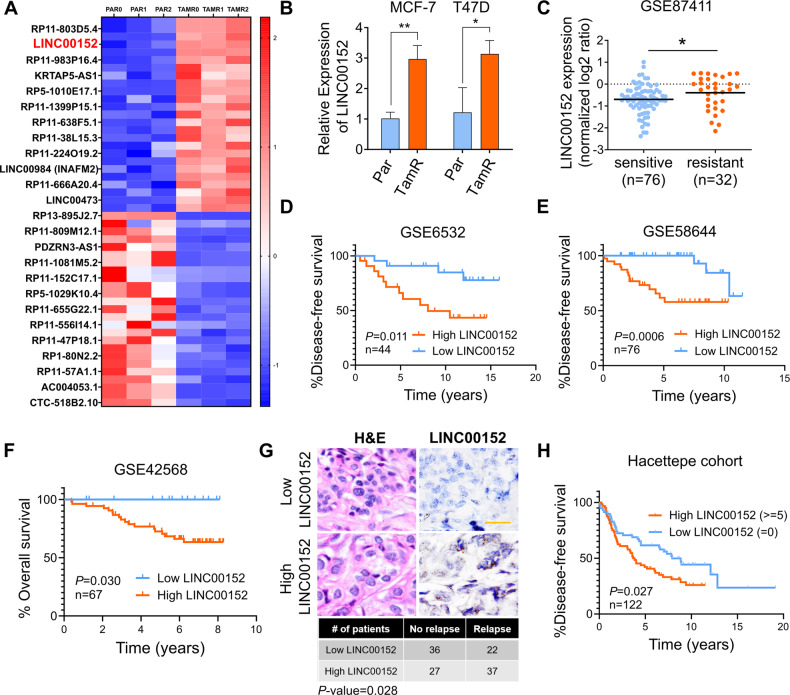
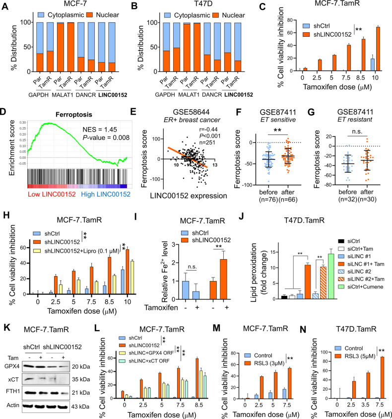
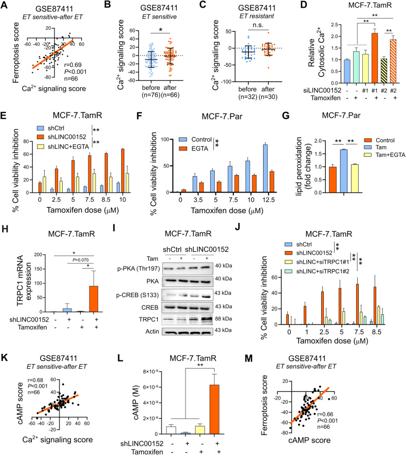
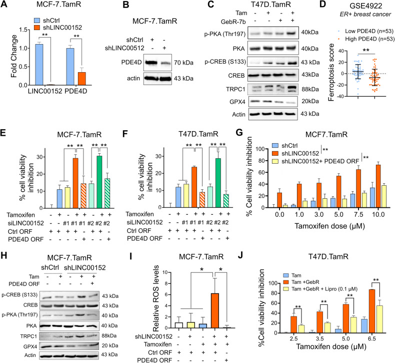
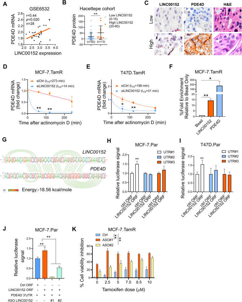
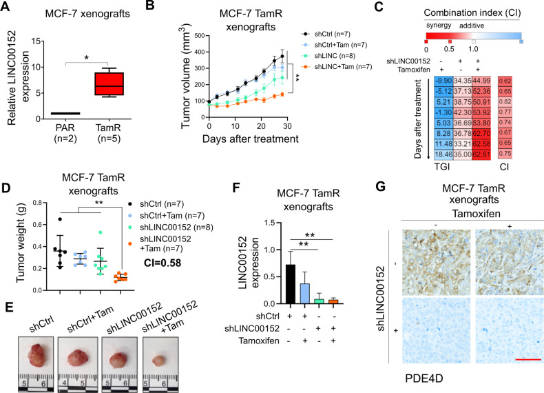
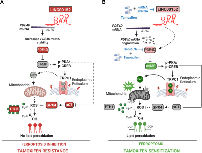
Tamoxifen has been the mainstay therapy to treat early, locally advanced, and metastatic estrogen receptor-positive (ER + ) breast cancer, constituting around 75% of all cases. However, the emergence of resistance is common, necessitating the identification of novel therapeutic targets. Here, we demonstrated that long-noncoding RNA LINC00152 confers tamoxifen resistance by blocking tamoxifen-induced ferroptosis, an iron-mediated cell death. Mechanistically, inhibiting LINC00152 reduces the mRNA stability of phosphodiesterase 4D (PDE4D), leading to activation of the cAMP/PKA/CREB axis and increased expression of the TRPC1 Ca2+ channel. This causes cytosolic Ca2+ overload and generation of reactive oxygen species (ROS) that is, on the one hand, accompanied by downregulation of FTH1, a member of the iron sequestration unit, thus increasing intracellular Fe2+ levels; and on the other hand, inhibition of the peroxidase activity upon reduced GPX4 and xCT levels, in part by cAMP/CREB. These ultimately restore tamoxifen-dependent lipid peroxidation and ferroptotic cell death which are reversed upon chelating Ca2+ or overexpressing GPX4 or xCT. Overexpressing PDE4D reverses LINC00152 inhibition-mediated tamoxifen sensitization by de-activating the cAMP/Ca2+/ferroptosis axis. Importantly, high LINC00152 expression is significantly correlated with high PDE4D/low ferroptosis and worse survival in multiple cohorts of tamoxifen- or tamoxifen-containing endocrine therapy-treated ER+ breast cancer patients. Overall, we identified LINC00152 inhibition as a novel mechanism of tamoxifen sensitization via restoring tamoxifen-dependent ferroptosis upon destabilizing PDE4D, increasing cAMP and Ca2+ levels, thus leading to ROS generation and lipid peroxidation. Our findings reveal LINC00152 and its effectors as actionable therapeutic targets to improve clinical outcome in refractory ER+ breast cancer.


Conflict of interest statement

O.S. is the co-founder of OncoCube Therapeutics LLC, the founder and president of LoxiGen, Inc., and the scientific advisory board member of A2A Pharmaceuticals. The other authors declare no potential competing interests.

Figures

Fig. 1
Fig. 1. LINC00152 is upregulated in tamoxifen resistance and its higher expression correlates with poor prognosis.
A Heatmap of the z-scores of the FPKM values of the top 25 most abundant, upregulated, and top 25, least abundant, downregulated lncRNAs in MCF-7 tamoxifen-resistant (TAMR) cells compared to parental (Par) cells in three biological replicates (0, 1, 2). The names of 25 genes are depicted on the heatmap. Red indicates upregulation and blue indicates downregulation in TamR cells. B qRT-PCR analysis of LINC00152 in both MCF-7 and T47D.Par and TamR cells. C LINC00152 levels in tamoxifen sensitive vs. resistant tumors at baseline from GSE87411. D, E The disease-free survival in tamoxifen-treated ER+ breast cancer patients with tumors expressing low vs. high LINC00152 expression from GSE6532 (D) and GSE58644 (E). F The overall survival in patients with tumors expressing low vs. high LINC00152 expression from GSE42568. G Representative in situ hybridization (ISH) staining of LINC00152 in low vs. high expressers among endocrine therapy-treated ER+ breast carcinoma specimens from the Hacettepe cohort. Scale bar = 20 µm. The numbers of Hacettepe patients having low vs high LINC00152 and with recurrence vs. no recurrence are provided at the bottom of the panel. H The disease-free survival in Hacettepe patients with tumors expressing low vs. high LINC00152 expression as quantified by ISH staining. Data are presented as mean values ± standard deviation (SD). P-values for the bar graphs and boxplots were calculated with the unpaired, two-tailed Student’s t-test. The significance for the Kaplan–Meier survival graphs was calculated with the Log-rank test. The chi-square test was used for (G). *P < 0.05, **P < 0.01.
Fig. 2
Fig. 2. Inhibiting LINC00152 overcomes tamoxifen resistance by inducing tamoxifen-dependent ferroptosis.
A, B The nuclear vs. cytoplasmic localization of LINC00152 in MCF-7 (A) and T47D (B) Par and TamR cells. MALAT1 and DANCR were used as controls for nuclear and cytoplasmic RNAs, respectively. C Percent cell viability inhibition in MCF-7.TamR cells expressing shLINC00152 upon treatment with tamoxifen. D GSEA analysis showing the ferroptosis-related genes enriched in low LINC00152-expressing ER+ breast cancer patients treated with endocrine therapy from GSE87411. E Pearson correlation analysis of LINC00152 expression with ferroptosis score in ER+ patients from GSE58644. F, G The levels of ferroptosis score before and after endocrine therapy in sensitive (F) and resistant (G) ER+ patients from GSE87411. H Percentage cell viability inhibition in shLINC00152-expressing MCF-7.TamR cells treated with tamoxifen with or without 0.1 µM of liproxstatin-1 (Lipro). I Relative intracellular Fe2+ levels in shCtrl or shLINC00152-expressing MCF-7.TamR cells that were treated with 7.5 µM tamoxifen for 1.5 h. J Lipid peroxidation assay in T47D.TamR cells transfected with siLINC00152 (siLINC) in combination with tamoxifen, showing fold change of lipid peroxidation. Cumene was used as a positive control. K Western blot analysis of the negative regulators of ferroptosis, xCT, GPX4, and FTH1 in MCF-7.TamR cells expressing shLINC00152 and treated with tamoxifen. Actin is used as a loading control in all Western blots unless stated otherwise. L Percentage cell viability inhibition in shLINC00152-expressing MCF-7.TamR cells transfected with GPX4 or xCT open reading frame (ORF). M, N Percentage cell viability inhibition in MCF-7.TamR (M) and T47D.TamR (N) cells treated with tamoxifen alone or in combination with 3 or 5 µM RSL3. Data are presented as mean values ± standard deviation (SD). P-values for (D) were calculated using the GSEA software (Broad Institute) while others were calculated with the paired (cell viability) or unpaired (others), two-tailed Student’s t-test. *P < 0.05, **P < 0.01, n.s. not significant.
Fig. 3
Fig. 3. LINC00152 inhibition increases TRPC1 expression and cytosolic Ca2+ downstream of cAMP signaling.
A The Pearson correlation analysis of Ca2+ signaling with ferroptosis score in endocrine therapy sensitive ER+ patients after therapy from GE87411. B, C The levels of Ca2+ signaling score before and after endocrine therapy in sensitive (B) and resistant (C) ER+ patients from GSE87411. D The levels of cytosolic Ca2+ in MCF-7.TamR cells that were transfected with two different siRNAs against LINC00152 and treated with tamoxifen. E Percentage cell viability in shLINC00152-expressing MCF-7.TamR cells treated with tamoxifen with or without EGTA. F Percentage cell viability inhibition in MCF-7.Par cells treated with tamoxifen with or without EGTA. G Relative lipid peroxidation in cells from (F). H qRT-PCR analysis of TRPC1 mRNA in shCtrl or shLINC00152-expressing MCF-7.TamR cells treated with tamoxifen. I Western Blot analysis of PDE4D, p-CREB (S133), p-PKA (Thr197) and TRPC1 in shCtrl or shLINC00152-expressing MCF-7.TamR cells treated with tamoxifen. J Percentage cell viability in shLINC00152-expressing MCF-7.TamR cells transfected with 2 different siRNAs against TRPC1. K The Pearson correlation analysis of Ca2+ signaling score with cAMP score in endocrine therapy sensitive ER+ patients after therapy from GSE124647. L cAMP levels in MCF-7.TamR shCtrl or shLINC00152 cells treated with tamoxifen. M The Pearson correlation analysis of cAMP score and ferroptosis score in endocrine therapy sensitive ER+ patients after therapy from GSE87411. Data are presented as mean values ±standard deviation (SD). P-values were calculated with the paired (cell viability) or unpaired (others), two-tailed Student’s t-test. *P < 0.05, **P < 0.01, n.s. not significant.
Fig. 4
Fig. 4. LINC00152 inhibits tamoxifen-dependent ferroptosis by regulating PDE4D and downstream cAMP/Ca2+ signaling in tamoxifen-resistant cells.
A qRT-PCR analysis of LINC00152 and PDE4D in the shLINC00152-expressing MCF-7.TamR cells. B Western blot analysis of PDE4D in the shLINC00152-expressing MCF-7.TamR cells. C Western blot analysis of p-CREB (S133), p-PKA (Thr197), TRPC1 and ferroptosis inhibitor GPX4 in T47D.TamR cells that were treated with tamoxifen in combination with GebR-7b. D The levels of ferroptosis score in low vs. high PDE4D-expressing ER+ patients from GSE4922. E, F Percentage cell viability inhibition in MCF-7 and T47D.TamR cells co-transfected with siLINC00152 and PDE4D ORF and treated with tamoxifen. G Percentage of viability inhibition in shLINC00152-expressing MCF-7.TamR cells stably overexpressing PDE4D and that were treated with tamoxifen. H Western blot analysis of p-CREB (S133), p-PKA (Thr197), TRPC1 and GPX4 in shLINC00152-expressing MCF-7.TamR cells stably overexpressing PDE4D and that were treated with tamoxifen. I Relative ROS levels in MCF-7.TamR cells expressing shLINC00152 and treated with tamoxifen. J Percentage viability inhibition in T47D.TamR cells treated with tamoxifen and GebR-7b with or without 0.1 µM of liproxstatin-1 (Lipro). Data are presented as mean values ± standard deviation (SD). P-values were calculated with the paired (cell viability) or unpaired (others), two-tailed Student’s t-test. *P < 0.05, **P < 0.01.
Fig. 5
Fig. 5. LINC00152 stabilizes PDE4D mRNA by interacting with its 3’UTR.
A The Pearson correlation analysis of LINC00152 and PDE4D mRNA in patients from GSE6532. B PDE4D protein expression determined by IHC in low vs. high LINC00152-expressing patients from the Hacettepe cohort. C Representative images of LINC00152 ISH, PDE4D IHC, and H&E staining in patients from the Hacettepe cohort. Scale bar = 20 µm. D, E qRT-PCR analysis of PDE4D in MCF-7.TamR (D) and T47D.TamR (E) cells transfected with siLINC00152 for 48 h, followed by treatment with 5 µg/ml of actinomycin (D). F Percentage fold enrichment of PDE4D in LINC00152 pull down in MCF-7.TamR cells. G Interaction between LINC00152 and PDE4D 3’UTR predicted by the IntaRNA tool. The visualization of the interaction was obtained using RILogo. H, I Luciferase assay in parental MCF7 (H) and T47D (I) cells co-transfected with LINC00152 overexpression vector and PDE4D 3’UTR, demonstrating binding to PDE4D 3’UTR. J Luciferase assay in MCF-7.Par cells co-transfected with LINC00152 overexpression vector and PDE4D 3’UTR with or without ASOs targeting the interaction site between LINC00152 and PDE4D 3’UTR. K Percent cell viability inhibition in ASO-transfected MCF-7.TamR cells under tamoxifen treatment. Data are presented as mean values ± standard deviation (SD). P-values were calculated with the paired (cell viability) or unpaired (others), two-tailed Student’s t-test. *P < 0.05, **P < 0.01.
Fig. 6
Fig. 6. LINC00152 inhibition overcomes tamoxifen resistance in vivo.
A PDE4D mRNA expression in MCF-7.Par (PAR) vs. TamR xenograft tumors. B Tumor volumes of MCF-7.TamR xenografts expressing shLINC00152 and treated with tamoxifen (3 mg/kg, daily). C Combination indices showing synergy between LINC00152 knockdown and tamoxifen in terms of tumor growth inhibition. D Tumor weights from mice in B. Combination index showing synergy between LINC00152 knockdown and tamoxifen in terms of tumor weight reduction is provided. E Tumor images from mice in (B). F qRT-PCR analysis of LINC00152 in tumors from (B). G IHC staining of PDE4D in tumors from (B). Scale bar = 100 µm. Data for the bar graphs and box plots are represented as mean values ± SD, while data for the tumor volume graph are represented as mean values ± standard error of the mean (SEM). P-values for the bar graphs and box plots were calculated with the unpaired, two-tailed Student’s t-test. The significance of the tumor volume graph was calculated with two-way ANOVA. *P < 0.05, **P < 0.01.
Fig. 7
Fig. 7. Schematic summary of LINC00152/PDE4D-driven tamoxifen resistance.
A LINC00152 is overexpressed in tamoxifen resistance, binds to PDE4D 3’UTR, and increases its mRNA stability. This results in reduced cAMP levels and de-activation of PKA/CREB, leading to decreased expression of TRPC1, reduction of cytosolic Ca2+, and blockage of reactive oxygen species (ROS) generation. Furthermore, the negative regulators of ferroptosis, xCT, GPX4, and FTH1, are upregulated, leading to increased intracellular Fe2+ and inhibition of lipid peroxidation that ultimately blocks tamoxifen-dependent ferroptosis and mediates tamoxifen resistance. B Targeting LINC00152 in combination with tamoxifen leads to reduced mRNA stability of PDE4D due to loss of binding to its 3’UTR, thereby activating cAMP/PKA/CREB, similar to targeting PDE4D with GebR-7b in combination with tamoxifen. This further increases TRPC1 levels, elevates cytosolic Ca2+, generates reactive oxygen species (ROS), and increases Fe2+ upon downregulation of xCT, FTH1, and GPX4, leading to lipid peroxidation and tamoxifen-dependent ferroptosis. Dashed lines indicate that the connection is not active.

Update of

Similar articles

Cited by

References

    1. Early Breast Cancer Trialists’ Collaborative G. Davies C, Godwin J, Gray R, Clarke M, Cutter D, et al. Relevance of breast cancer hormone receptors and other factors to the efficacy of adjuvant tamoxifen: patient-level meta-analysis of randomised trials. Lancet. 2011;378:771–84. doi: 10.1016/S0140-6736(11)60993-8. - DOI - PMC - PubMed
    1. Howell A, Howell SJ. Tamoxifen evolution. Br J Cancer. 2023;128:421–5. doi: 10.1038/s41416-023-02158-5. - DOI - PMC - PubMed
    1. Clemons M, Danson S, Howell A. Tamoxifen (“Nolvadex”): a review. Cancer Treat Rev. 2002;28:165–80. doi: 10.1016/S0305-7372(02)00036-1. - DOI - PubMed
    1. Ekholm M, Bendahl PO, Fernö M, Nordenskjöld B, Stål O, Rydén L. Two Years of Adjuvant Tamoxifen Provides a Survival Benefit Compared With No Systemic Treatment in Premenopausal Patients With Primary Breast Cancer: Long-Term Follow-Up (> 25 years) of the Phase III SBII:2pre Trial. J Clin Oncol 34(19):2232-2238. 10.1200/JCO.2015.65.6272. - PubMed
    1. Osborne CK, Fuqua SA. Mechanisms of tamoxifen resistance. Breast Cancer Res Treat. 1994;32:49–55. doi: 10.1007/BF00666205. - DOI - PubMed

Publication types

MeSH terms