. 2024 Jun 15;22(1):330.
doi: 10.1186/s12964-024-01704-9.

Inhibition of WNT/β-catenin signalling during sex-specific gonadal differentiation is essential for normal human fetal testis development

Affiliations

Inhibition of WNT/β-catenin signalling during sex-specific gonadal differentiation is essential for normal human fetal testis development

Malene Lundgaard Riis et al. Cell Commun Signal. .

Abstract

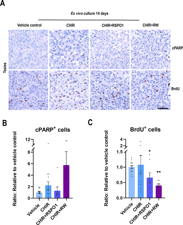
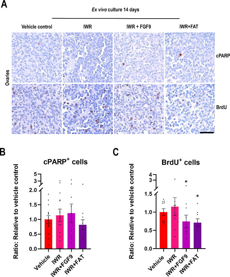
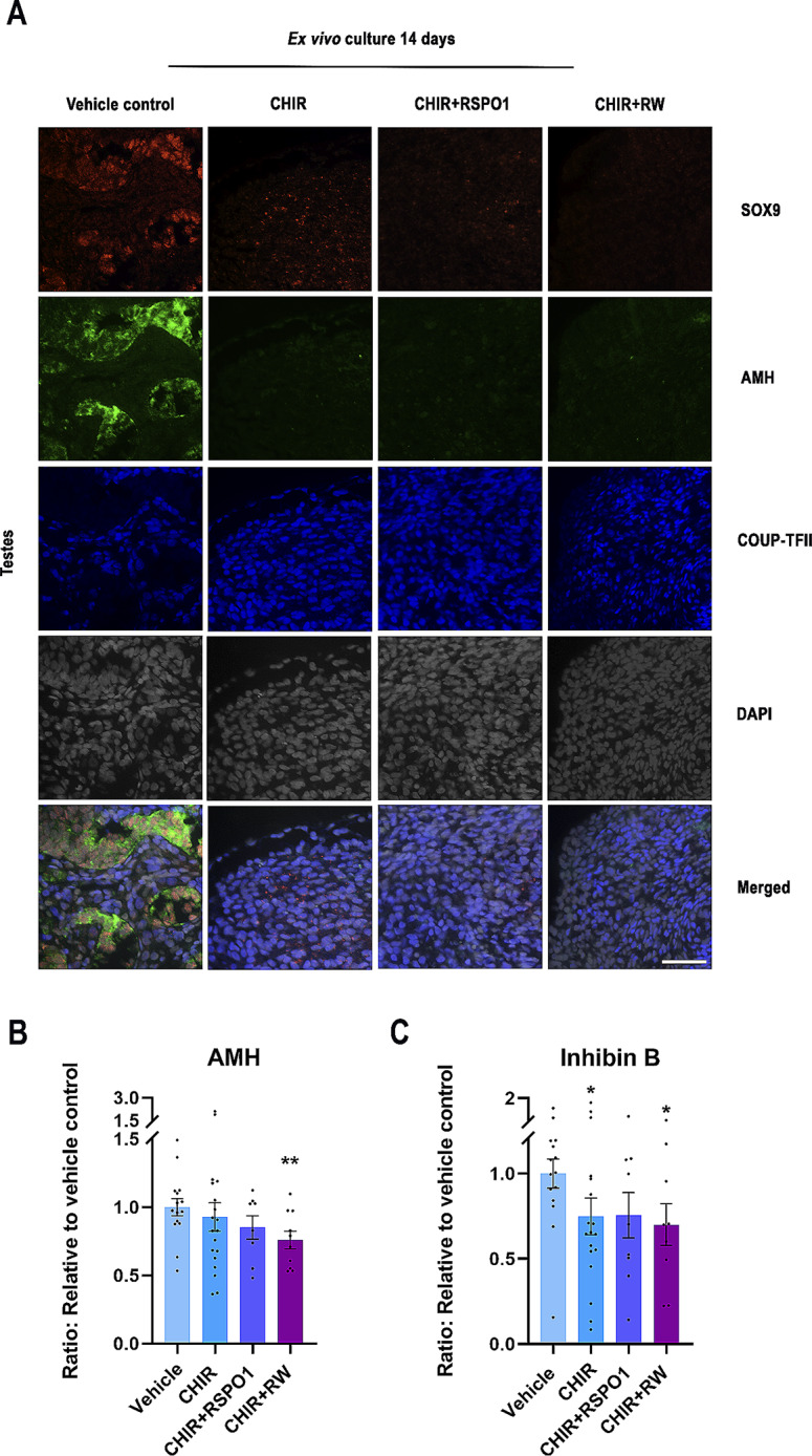
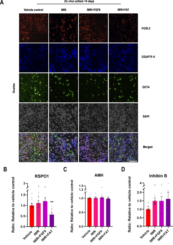
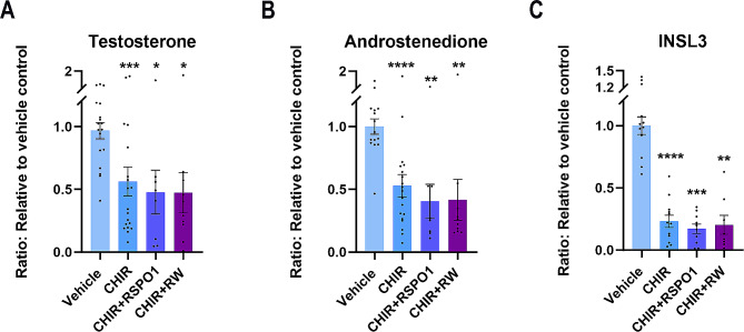
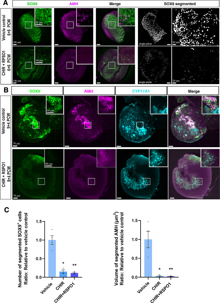
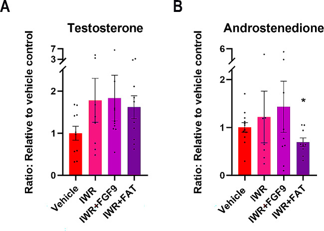
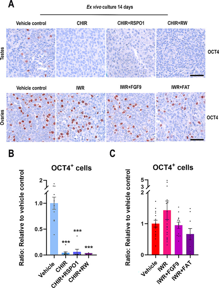
Sex-specific gonadal differentiation is directed by complex signalling promoting development in either male or female direction, while simultaneously inhibiting the opposite pathway. In mice, the WNT/β-catenin pathway promotes ovarian development and the importance of actively inhibiting this pathway to ensure normal testis development has been recognised. However, the implications of alterations in the tightly regulated WNT/β-catenin signalling during human fetal gonad development has not yet been examined in detail. Thus, the aim of this study was to examine the consequences of dysregulating the WNT/β-catenin signalling pathway in the supporting cell lineage during sex-specific human fetal gonad development using an established and extensively validated ex vivo culture model. Inhibition of WNT/β-catenin signalling in human fetal ovary cultures resulted in only minor effects, including reduced secretion of RSPO1 and reduced cell proliferation although this was not consistently found in all treatment groups. In contrast, promotion of WNT/β-catenin signalling in testes severely affected development and function. This included disrupted seminiferous cord structures, reduced cell proliferation, reduced expression of SOX9/AMH, reduced secretion of Inhibin B and AMH as well as loss of the germ cell population. Additionally, Leydig cell function was markedly impaired with reduced secretion of testosterone, androstenedione and INSL3. Together, this study suggests that dysregulated WNT/β-catenin signalling during human fetal gonad development severely impairs testicular development and function. Importantly, our study highlights the notion that sufficient inhibition of the opposite pathway during sex-specific gonadal differentiation is essential to ensure normal development and function also applies to human fetal gonads.

Keywords: Ex vivo culture; Germ cell development; Human fetal gonads; Ovarian and testicular differentiation; Sex-specific development; Supporting cell lineages; WNT/β-catenin signalling.


Conflict of interest statement

The authors declare no competing interests.

Figures

Fig. 1
Fig. 1
Stimulation of WNT/β-catenin signalling in ex vivo cultures of human fetal testes did not affect apoptosis but resulted in reduced proliferation. (A) Representative images of cPARP (apoptosis marker) and BrdU (proliferation marker) immunostaining in ex vivo cultured fetal testes treated with CHIR (3 µM), CHIR + RSPO1: CHIR (3 µM) + RSPO1 (100 ng/ml) or CHIR + RW: CHIR (3 µM) + RSPO1 (100 ng/ml) + WNT4 (100 ng/ml). Counterstaining was performed with Mayer’s haematoxylin; scale bar 50 μm. Age of fetal samples shown (at start of experiment): Vehicle control 8 + 2 PCW; CHIR 9 + 2 PCW; CHIR + RSPO1 8 + 1 PCW; CHIR + RW 7 + 4 PCW. (B) Quantification of cPARP-positive cells/mm2 in ex vivo cultured fetal testes treated with CHIR (n = 14), CHIR + RSPO1 (n = 7) or CHIR + RW (n = 5). (C) Quantification of BrdU-positive cells/mm2 in ex vivo cultured fetal testes treated with CHIR (n = 10), CHIR + RSPO1 (n = 8) or CHIR + RW (n = 7). Results are shown as fold change compared to internal vehicle control with data presented as mean ± SEM with individual datapoints included. Asterisk indicates statistical significance compared to vehicle control * P < 0.05 and ** P < 0.01
Fig. 2
Fig. 2
Inhibition of WNT/β-catenin signalling in ex vivo cultures of human fetal ovaries did not affect apoptosis but resulted in reduced proliferation. (A) Representative images of cPARP (apoptosis marker) and BrdU (proliferation marker) immunostaining in ex vivo cultured fetal ovaries treated with IWR (1µM), IWR + FGF9: IWR-1 (1µM) + FGF9 (50 ng/ml) or IWR + FAT: IWR-1 (1µM) + FGF9 (50 ng/ml) + Activin A (25 ng/ml) + Activin B (25 ng/ml) + TGFβ (25 ng/ml). Counterstaining was performed with Mayer’s haematoxylin; scale bar 50 μm. Age of fetal samples shown (at start of experiment): Vehicle control 8 + 2 PCW; IWR 9 + 3 PCW; IWR + FGF9 8 + 2 PCW; IWR + FAT 8 + 0 PCW. (B) Quantification of cPARP-positive cells/mm2in ex vivo cultured fetal ovaries treated with vehicle control, IWR (n = 16), IWR + FGF9 (n = 10) or IWR + FAT (n = 10). (C) Quantification of BrdU-positive cells/mm2 in ex vivo cultured fetal ovaries treated with vehicle control, IWR (n = 10), IWR + FGF9 (n = 10) or IWR + FAT (n = 8). Results are shown as fold change compared to internal vehicle control with data presented as mean ± SEM with individual datapoints included. Asterisk indicates statistical significance compared to vehicle control * P < 0.05
Fig. 3
Fig. 3
Stimulation of WNT/β-catenin signalling in ex vivo cultures of human fetal testes affected Sertoli cell identity and function. (A) Representative images of triple immunofluorescence staining of SOX9 (Sertoli cell marker, red), AMH (Sertoli cell marker, green), COUPTF-II (Interstitial cell marker, blue) and DAPI (grey) in ex vivo cultured fetal testes treated with CHIR (3 µM), CHIR + RSPO1: CHIR (3 µM) + RSPO1 (100 ng/ml) or CHIR + RW: CHIR (3 µM) + RSPO1 (100 ng/ml) + WNT4 (100 ng/ml). Age of fetal samples shown (at start of experiment): Vehicle control 9 + 0 PCW; CHIR 9 + 0 PCW; CHIR + RSPO1 9 + 4 PCW; CHIR + RW 9 + 0 PCW. Scale bar corresponds to 50 μm. (B) Secretion of AMH measured in media from ex vivo cultured fetal testes treated with CHIR (n = 19), CHIR + RSPO1 (n = 9) or CHIR + RW (n = 10). (C) Inhibin B measured in media from ex vivo cultured fetal testes treated with CHIR (n = 18), CHIR + RSPO1 (n = 9) or CHIR + RW (n = 9). Results are shown as fold change compared to internal vehicle control with data presented as mean ± SEM with individual datapoints included. Asterisk indicates statistical significance compared to vehicle control with * P < 0.05 and ** P < 0.01
Fig. 4
Fig. 4
Inhibiting WNT/β-catenin signalling in ex vivo cultures of human fetal ovaries had only minor effects on granulosa cell identity and function. (A) Representative images of triple immunofluorescence staining of FOXL2 (Granulosa cell marker, red), (COUPTF-II (Interstitial cell marker, blue), OCT4 (oogonia marker, green) and DAPI (grey) in ex vivo cultured fetal ovaries treated with IWR (1µM), IWR + FGF9: IWR-1 (1µM) + FGF9 (50 ng/ml) or IWR + FAT: IWR-1 (1µM) + FGF9 (50 ng/ml) + Activin A (25 ng/ml) + Activin B (25 ng/ml) + TGFβ (25 ng/ml). Age of fetal samples shown (at start of experiment): Vehicle control 8 + 0 PCW; IWR 7 + 6 PCW; IWR + FGF9 7 + 6 PCW; IWR + FAT 7 + 6 PCW. Scale bar corresponds to 50 μm. (B) Secretion of RSPO1 measured in media from ex vivo cultured fetal ovaries treated with IWR (n = 19), IWR + FGF9 (n = 11) or IWR + FAT (n = 10). (C) AMH measured in media from ex vivo cultured fetal ovaries treated with IWR (n = 13), IWR + FGF9 (n = 8) or IWR + FAT (n = 7). (D) Inhibin B measured in media from ex vivo cultured fetal ovaries treated with IWR (n = 13), IWR + FGF9 (n = 12) or IWR + FAT (n = 11). Results are shown as fold change compared to internal vehicle control with data presented as mean ± SEM with individual datapoints included. Asterisk indicates statistical significance compared to vehicle control with ** P < 0.01
Fig. 5
Fig. 5
Stimulation of WNT/β-catenin signalling in ex vivo cultures of human fetal testes reduced the production of androgens and INSL3. (A) Secretion of testosterone measured in media from ex vivo cultured fetal testes treated with CHIR (n = 19), CHIR + RSPO1 (n = 8) or CHIR + RW (n = 10) (B) androstenedione measured in media from ex vivo cultured fetal testes treated with CHIR (n = 19), CHIR + RSPO1 (n = 8) or CHIR + RW (n = 10). (C) INSL3 measured in media from ex vivo cultured fetal testes treated with CHIR (n = 12), CHIR + RSPO1 (n = 10) or CHIR + RW (n = 8). Results are shown as fold change compared to internal vehicle control with data presented as mean ± SEM with individual datapoints included. Asterisk indicates statistical significance compared to vehicle control with * P < 0.05, ** P < 0.01, *** P < 0.001 and **** P < 0.0001
Fig. 6
Fig. 6
Stimulation of WNT/β-catenin signalling in ex vivo cultures of human fetal testes reduced Sertoli cell number and impaired seminiferous cord structures in 3D imaging analyses. A) Representative images of SOX9 and AMH in toto-immunostaining of ex vivo cultured fetal testes (6 + 6 PCW) treated or not with CHIR (3 µM) + RSPO1 (100 ng/ml). Right-hand panels are images of single-plane SOX9 segmented areas. B) Representative images of SOX9, AMH and CYP17A1 in toto-immunostaining of ex vivo cultured fetal testes (9 + 4 PCW) treated or not with CHIR (3 µM) + RSPO1 (100 ng/ml). C) Automated quantitative analysis was performed using a machine-learning based software to assess, respectively, the total number of SOX9 nuclei and the volume of AMH-positive segmented areas in control testes, CHIR-treated testes and CHIR + RSPO1-treated testes with n = 3 (fetal testes age: 6 + 6, 9 + 4 and 9 + 6 PCW). Results are shown as fold change compared to internal vehicle control with data presented as mean ± SEM with individual datapoints included. Asterisk indicates statistical significance compared to vehicle control with * P < 0.05 and ** P < 0.01
Fig. 7
Fig. 7
Inhibition of WNT/β-catenin signalling in ex vivo cultures of human fetal ovaries only slightly affect steroid production. (A) Secretion of testosterone measured in media from ex vivo cultured fetal ovaries treated with IWR (n = 8), IWR + FGF9 (n = 10) or IWR + FAT (n = 10) and (B) androstenedione measured in media from ex vivo cultured fetal ovaries treated with IWR (n = 7), IWR + FGF9 (n = 9) or IWR + FAT (n = 9). Results are shown as fold change compared to internal vehicle control with data presented as mean ± SEM with individual datapoints included. Asterisk indicates statistical significance compared to vehicle control with * P < 0.05
Fig. 8
Fig. 8
Manipulation of WNT/β-catenin signalling in ex vivo cultures of human fetal gonads severely reduce the number of germ cells in testes but not ovaries. (A) Representative images of OCT4 immunostaining in ex vivo cultured fetal testes treated with CHIR (3 µM) or CHIR + RW: CHIR (3 µM) + RSPO1 (100 ng/ml) + WNT4 (100 ng/ml) and ex vivo cultured fetal ovaries treated with IWR (1µM), IWR + FGF9: IWR-1 (1µM) + FGF9 (50 ng/ml) or IWR + FAT: IWR-1 (1µM) + FGF9 (50 ng/ml) + Activin A (25 ng/ml) + Activin B (25 ng/ml) + TGFβ (25 ng/ml). Counterstaining was performed with Mayer’s haematoxylin; scale bar 50 μm. Age of fetal samples shown (at start of experiment): Vehicle control 9 + 0 PCW; CHIR 8 + 2 PCW; CHIR + RSPO1 7 + 2 PCW; CHIR + RW 9 + 0 PCW for testes samples and vehicle control 7 + 0 PCW; IWR 7 + 2 PCW; IWR + FGF9 7 + 1 PCW; IWR + FAT 7 + 6 PCW for ovarian samples. (B) Quantification of the number of OCT4+ cells/mm2 in ex vivo cultured fetal testes treated with CHIR (n = 8), CHIR + RSPO1 (n = 7) or CHIR + RW (n = 5). Results are shown as fold change compared to internal vehicle control with data presented as mean ± SEM with individual datapoints included. Asterisk indicates statistical significance compared to vehicle control with *** P < 0.001. (C) Quantification of the number of OCT4+ cells/mm2 in ex vivo cultured fetal ovaries treated with IWR (n = 19), IWR + FGF9 (n = 12) or IWR + FAT (n = 9). Results are shown as fold change compared to internal vehicle control with data presented as mean ± SEM with individual datapoints included

Similar articles

Cited by

References

    1. Byskov AG. Differentiation of mammalian embryonic gonad. Physiol Rev. 1986;66:71–117. doi: 10.1152/physrev.1986.66.1.71. - DOI - PubMed
    1. Lin Y-T, Capel B. Cell fate commitment during mammalian sex determination. Curr Opin Genet Dev. 2015;32:144–52. doi: 10.1016/j.gde.2015.03.003. - DOI - PMC - PubMed
    1. Capel B. WOMEN IN REPRODUCTIVE SCIENCE: to be or not to be a testis. Reproduction. 2019;158:F101–11. doi: 10.1530/REP-19-0151. - DOI - PMC - PubMed
    1. Rotgers E, Jørgensen A, Yao HH-C. At the crossroads of fate-somatic cell lineage specification in the fetal gonad. Endocr Rev. 2018;39:739–59. doi: 10.1210/er.2018-00010. - DOI - PMC - PubMed
    1. Lundgaard Riis M, Jørgensen A. Deciphering sex-specific differentiation of human fetal gonads: insight from experimental models. Front Cell Dev Biol. 2022. - PMC - PubMed

Publication types