. 2024 Jun 15;15(1):5133.
doi: 10.1038/s41467-024-49402-x.

Disease progression modelling reveals heterogeneity in trajectories of Lewy-type α-synuclein pathology

Affiliations

Disease progression modelling reveals heterogeneity in trajectories of Lewy-type α-synuclein pathology

Sophie E Mastenbroek et al. Nat Commun. .

Abstract

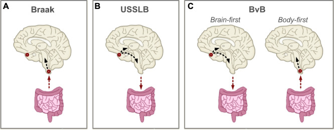
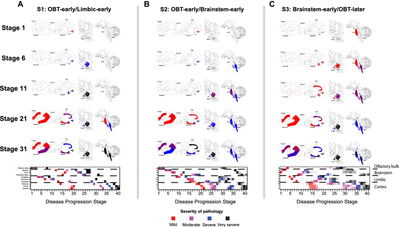
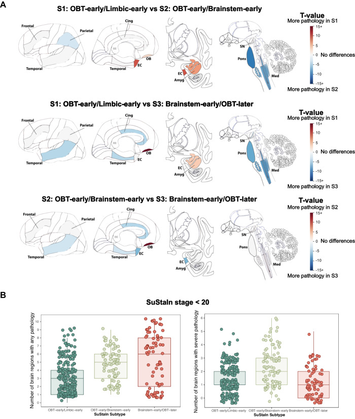
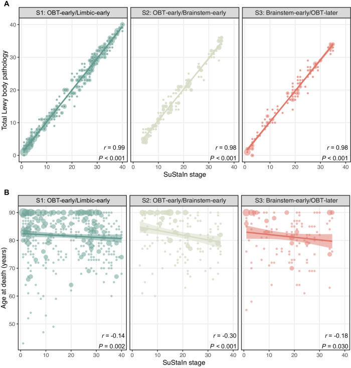
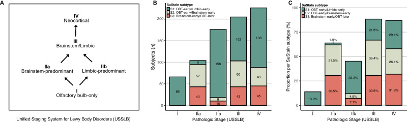
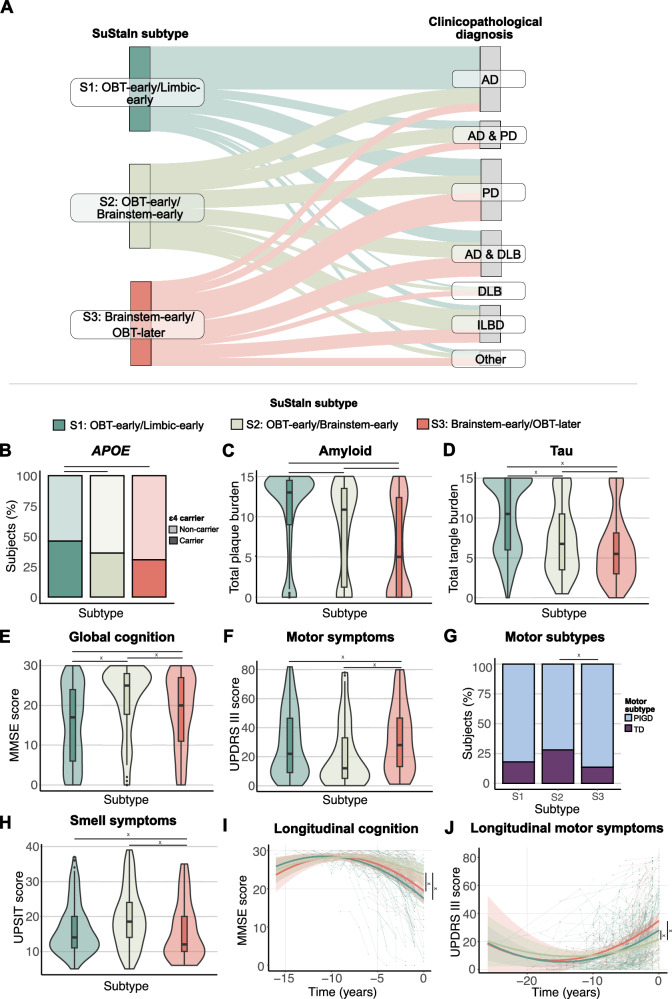
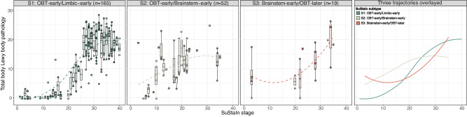
Lewy body (LB) diseases, characterized by the aggregation of misfolded α-synuclein proteins, exhibit notable clinical heterogeneity. This may be due to variations in accumulation patterns of LB neuropathology. Here we apply a data-driven disease progression model to regional neuropathological LB density scores from 814 brain donors with Lewy pathology. We describe three inferred trajectories of LB pathology that are characterized by differing clinicopathological presentation and longitudinal antemortem clinical progression. Most donors (81.9%) show earliest pathology in the olfactory bulb, followed by accumulation in either limbic (60.8%) or brainstem (21.1%) regions. The remaining donors (18.1%) initially exhibit abnormalities in brainstem regions. Early limbic pathology is associated with Alzheimer's disease-associated characteristics while early brainstem pathology is associated with progressive motor impairment and substantial LB pathology outside of the brain. Our data provides evidence for heterogeneity in the temporal spread of LB pathology, possibly explaining some of the clinical disparities observed in Lewy body disease.


Conflict of interest statement

L.E.C. has received research support from GE Healthcare (paid to institution). F.B. acts as a consultant for Biogen-Idec, IXICO, Merck-Serono, Novartis, Combinostics, and Roche. He has received grants, or grants are pending, from the Amyloid Imaging to Prevent Alzheimer’s Disease (AMYPAD) initiative, the Biomedical Research Centre at University College London Hospitals, the Dutch MS Society, ECTRIMS–MAGNIMS, EU-H2020, the Dutch Research Council (NWO), the UK MS Society, and the National Institute for Health Research, University College London. He has received payments for the development of educational presentations from Ixico and his institution from Biogen-Idec and Merck. He is on the editorial board of Radiology, European Neuroradiology, Multiple Sclerosis Journal, and Neurology. Is on the board of directors of Queen Square Analytics. R.O. has received research support from Avid Radiopharmaceuticals, has given lectures in symposia sponsored by GE Healthcare and is an editorial board member of Alzheimer’s Research & Therapy and the European Journal of Nuclear Medicine and Molecular Imaging. O.H. has acquired research support (for the institution) from ADx, AVID Radiopharmaceuticals, Biogen, Eli Lilly, Eisai, Fujirebio, GE Healthcare, Pfizer, and Roche. In the past 2 years, he has received consultancy/speaker fees from AC Immune, Amylyx, Alzpath, BioArctic, Biogen, Cerveau, Eisai, Eli Lilly, Fujirebio, Merck, Novartis, Novo Nordisk, Roche, Sanofi and Siemens. T.G.B. is a consultant for Aprinoia Therapeutics, Biogen and Avid Radiopharmaceuticals. A.A. has received, over the last 10 years, honoraria or support for consulting; participating in independent data safety monitoring boards; providing educational lectures, programs, and materials; or serving on advisory or oversight boards for AbbVie, Acadia, Allergan, the Alzheimer’s Association, Alzheimer’s Disease International, Axovant, AZ Therapies, Biogen, Eisai, Grifols, Harvard Medical School Graduate Continuing Education, JOMDD, Lundbeck, Merck, Prothena, Roche/Genentech, Novo Nordisk, Qynapse, Sunovion, Suven, and Synexus. Dr. Atri receives book royalties from Oxford University Press for a medical book on dementia. Dr. Atri receives institutional research grant/contract funding from NIA/NIH 1P30AG072980, NIA/NIH U22AG057437, AZ DHS CTR040636, Washington University St Louis, and Gates Ventures. Dr. Atri’s institution receives/received funding for clinical trial grants, contracts, and projects from government, consortia, foundations and companies for which he serves/served as contracted site-PI. S.H.M. serves as a site co-investigator and receives funding for the PPMI study from the Michael J Fox Foundation. S.H.M. has received research funding from the Arizona Biomedical Research Consortium (ABRC) and International Essential Tremor Foundation (IETF). S.H.M. also has received funding for clinical trial grants, contracts, and projects from government and companies for which S.H.M. serves or has served as contracted site-PI. P.C. has received research support from Lewy Body Dementia association and Arizona Alzheimer’s Consortium (both paid to institution). The remaining authors declare no competing interests.

Figures

Fig. 1
Fig. 1. Hypothetical disease progression models of Lewy body pathology.
Summary of theoretical disease models describing the progression of Lewy body (LB) pathology. Origins of LB pathology are designated with red circles. Arrows denote direction of spreading. A The Braak staging scheme postulates that LB pathology enters the brain through the gut and/or the nasal epithelium, resulting in simultaneous deposition in brainstem regions and the olfactory bulb,. Only brainstem pathology subsequently propagates throughout the brain. B The Unified Staging System for Lewy Body Disorders (USSLB) posits that LB pathology starts in the olfactory bulb in most cases, followed by either brainstem or limbic regions. It states that pathology always starts in the brain and propagates to the body. C The brain-first vs. body-first (BvB) model hypothesizes the existence of two subtypes of LB diseases, with one originating from the brain (i.e., brain-first) and the other from the body (i.e., body-first),. In the former, pathology starts in olfactory regions, followed by either brainstem or limbic regions. In the latter, pathology enters the brain through brainstem regions.
Fig. 2
Fig. 2. Three spatiotemporal trajectories of LB pathology.
SuStaIn-inferred disease progression patterns of regional Lewy-type α-synuclein pathology. Three distinct trajectories were identified and termed (A) “S1: OBT-early/Limbic-early”, (B) “S2: OBT-early/Brainstem-early”, and (C) “S3: Brainstem-early/OBT-later”. Brain maps of α-synuclein deposition are shown for SuStaIn stage 1, 6, 11, 21, and 31, with colors indicating severity of pathology (red = mild, purple = moderate, blue = severe, black = very severe). Below, positional variance diagrams are shown. Each box represents the certainty that a brain region has reached a certain level of pathology (mild, moderate, severe, and very severe) at a given SuStaIn (i.e., disease progression) stage, with darker colors representing more confidence. Brain schematics were generated using: https://github.com/AllenInstitute/hba_brain_schematic (Copyright © 2023. Allen Institute. All rights reserved.). Amyg amygdala, Cing anterior cingulate, EC entorhinal cortex, Med medulla, SN substantia nigra, SuStaIn Subtype and Stage Inference.
Fig. 3
Fig. 3. Regional burden of LB pathology differs between subtypes.
A T-maps adjusted for SuStaIn stage and multiple comparisons, showing regions that are significantly different between LB subtypes. For visibility, t-values are shown on a scale from 15 to −15, but can represent values below and above as indicated by the 15+. Non-significant regions are shown in white. Brain schematics were generated using: https://github.com/AllenInstitute/hba_brain_schematic (Copyright © 2023. Allen Institute. All rights reserved.). B Number of brain regions affected by any or severe pathology in SuStaIn stages <20 (n = 420). The left panel indicates the number of brain regions displaying any Lewy body pathology (density score > 0) and the right panel indicates the number of brain regions with severe or very severe Lewy body pathology (density score > 2). Boxplots show the median, lower, and upper quartiles with whiskers representing minimum and maximum values. Amyg amygdala, Cing anterior cingulate, EC entorhinal cortex, Med medulla, OBT olfactory bulb and tract, SN substantia nigra, SuStaIn Subtype and Stage Inference.
Fig. 4
Fig. 4. SuStaIn stage is inversely correlated to age at death.
Two-sided spearman correlations between SustaIn stage and (A) total Lewy body pathology and (B) age at death. Total Lewy body pathology was defined by the sum of the α-synuclein density scores across 10 brain regions and was available for 673 (86.2%) cases. SuStaIn stage was strongly positively correlated to total Lewy body pathology and negatively associated with age in all subtypes. Error bands represent the standard error. Dot size represents the number of cases, with larger dots indicating larger sample size. OBT olfactory bulb and tract, SuStaIn Subtype and Stage Inference.
Fig. 5
Fig. 5. Data-driven LB subtypes resemble the previous Unified Staging Scheme for Lewy Body Disorders.
Staging of subjects (n = 777) according to the Unified Staging Scheme for Lewy Body Disorders (A) across subtypes, shown as number of subjects (B) or proportion within each SuStaIn subtype (C).
Fig. 6
Fig. 6. LB subtypes are characterized by distinct clinicopathological, genetic, and clinical characteristics.
Results of SuStaIn subtype comparisons. A Clinicopathological diagnosis in relation to SuStaIn subtype. The Sankey diagram shows the proportion (%) of SuStaIn subtypes across clinicopathological diagnoses. Subtype differences assessed with two-sided linear and logistic regression models, for continuous and categorical variables respectively, are shown for (B) APOE ε4 carriership (n = 774); C total postmortem plaque burden (n = 772); D total postmortem neurofibrillary burden (n = 769); E MMSE measuring global cognition (n = 646); F UPDRS part III measuring motor symptoms (assessed off medication) (n = 385); G motor subtypes (n = 385); and (H) UPSIT measuring smell ability (n = 255). Clinical variables were measured closest to time of death. Horizontal lines reflect significant differences. Longitudinal trajectories of (I) MMSE and (J) UPDRS part III in the different subtypes. Model-predicted associations are plotted for each subtype from linear mixed-effect models including a polynomial (non-linear) term for time. Covariates were age, sex, education and SuStaIn stage. Time 0 indicates baseline anchored to the date of autopsy. Individual trajectories are shown in the background. Vertical lines represent significant differences (all p < 0.05). x indicates differences that persist after adjusting for plaque burden. Boxplots show the median, lower, and upper quartiles with whiskers representing minimum and maximum values. AD Alzheimer’s disease, DLB dementia with Lewy bodies, ILBD incidental Lewy body disease, MMSE Mini-Mental State Examination, OBT olfactory bulb and tract, PD Parkinson’s disease, PIGD Postural Instability and Gait difficulties, SuStaIn Subtype and Stage Inference, TD tremor dominant, UPDRS Unified Parkinson’s Disease Rating Scale, UPSIT Smell Identification Test.
Fig. 7
Fig. 7. Early LB pathology in brainstem regions is associated with substantial non-brain LB pathology in early SuStaIn stages.
Total peripheral Lewy body pathology across SuStaIn stages, shown by SuStaIn subtype. Total pathology scores were computed as the sum of the α-synuclein density scores (0–4) assessed in the cervical, thoracic, lumbar, and sacral spinal cord gray matter, vagus nerve, submandibular gland, and esophagus, with higher scores indicating more severe pathology. Dashed lines represent modeled trajectories computed with LOESS regressions. The right-most panel shows the 4 inferred trajectories overlayed. Boxplots show the median, lower, and upper quartiles with whiskers representing minimum and maximum values. OBT olfactory bulb and tract, SuStaIn Subtype and Stage Inference.

Update of

Similar articles

Cited by

References

    1. Spillantini MG, et al. α-Synuclein in Lewy bodies. Nature. 1997;388:839–840. doi: 10.1038/42166. - DOI - PubMed
    1. Galaz, Z. et al. in Intertwining Graphonomics with Human Movements (eds Cristina Carmona-Duarte, Moises Diaz, Miguel A. Ferrer, & Aythami Morales) 255–268 (Springer International Publishing, 2022).
    1. Postuma RB, et al. MDS clinical diagnostic criteria for Parkinson’s disease. Mov. Disord. 2015;30:1591–1601. doi: 10.1002/mds.26424. - DOI - PubMed
    1. McKeith IG, et al. Diagnosis and management of dementia with Lewy bodies: fourth consensus report of the DLB Consortium. Neurology. 2017;89:88–100. doi: 10.1212/WNL.0000000000004058. - DOI - PubMed
    1. Albrecht F, et al. Unraveling Parkinson’s disease heterogeneity using subtypes based on multimodal data. Parkinsonism Relat. Disord. 2022;102:19–29. doi: 10.1016/j.parkreldis.2022.07.014. - DOI - PubMed

MeSH terms