

The roles of Th cells in myocardial infarction
- PMID: 38879568
- PMCID: PMC11180143
- DOI: 10.1038/s41420-024-02064-6
The roles of Th cells in myocardial infarction
Abstract
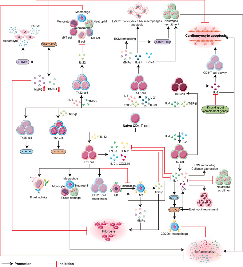
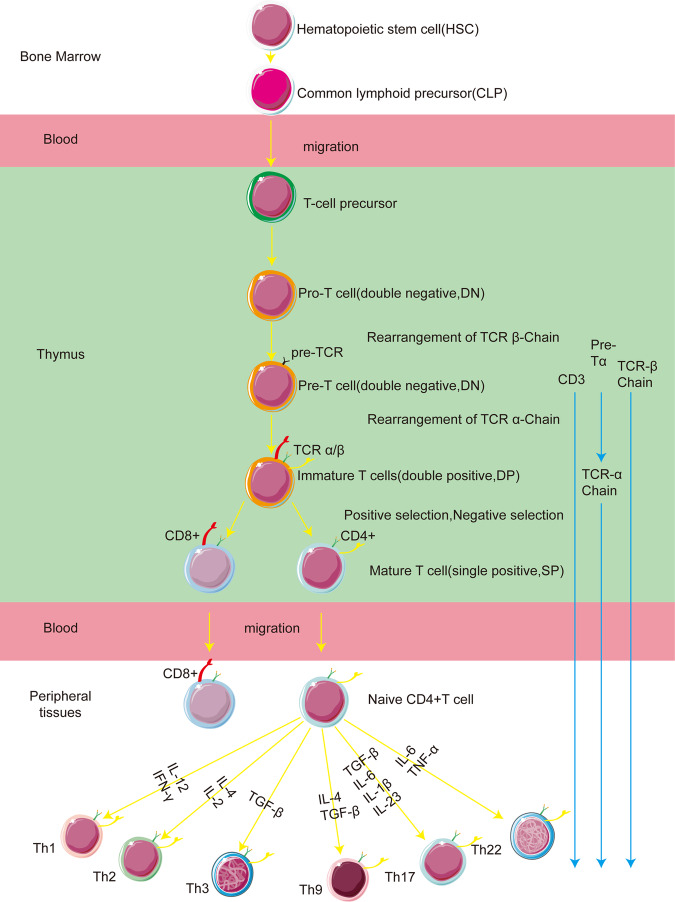
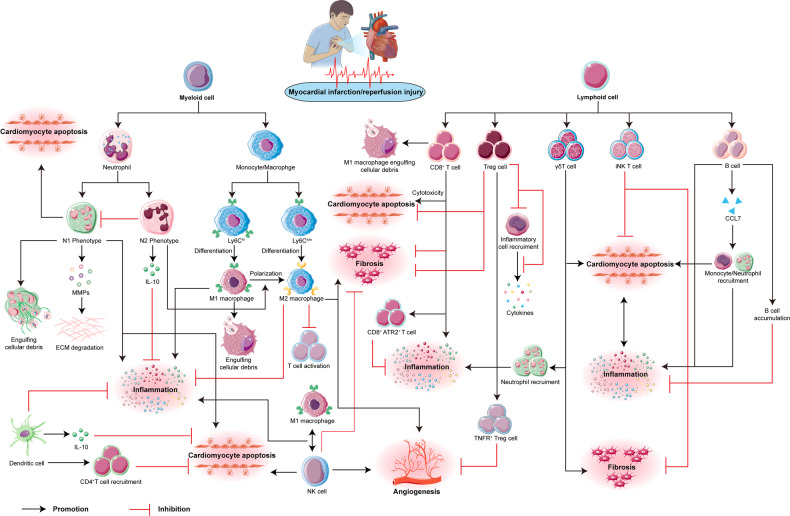
Myocardial infarction, commonly known as a heart attack, is a serious condition caused by the abrupt stoppage of blood flow to a part of the heart, leading to tissue damage. A significant aspect of this condition is reperfusion injury, which occurs when blood flow is restored but exacerbates the damage. This review first addresses the role of the innate immune system, including neutrophils and macrophages, in the cascade of events leading to myocardial infarction and reperfusion injury. It then shifts focus to the critical involvement of CD4+ T helper cells in these processes. These cells, pivotal in regulating the immune response and tissue recovery, include various subpopulations such as Th1, Th2, Th9, Th17, and Th22, each playing a unique role in the pathophysiology of myocardial infarction and reperfusion injury. These subpopulations contribute to the injury process through diverse mechanisms, with cytokines such as IFN-γ and IL-4 influencing the balance between tissue repair and injury exacerbation. Understanding the interplay between the innate immune system and CD4+ T helper cells, along with their cytokines, is crucial for developing targeted therapies to mitigate myocardial infarction and reperfusion injury, ultimately improving outcomes for cardiac patients.
© 2024. The Author(s).
Conflict of interest statement
The authors declare no competing interests.
Figures
Similar articles
-
T Helper (Th) Cell Profiles in Pregnancy and Recurrent Pregnancy Losses: Th1/Th2/Th9/Th17/Th22/Tfh Cells.Front Immunol. 2020 Aug 18;11:2025. doi: 10.3389/fimmu.2020.02025. eCollection 2020. Front Immunol. 2020. PMID: 32973809 Free PMC article. Review.
-
Circulating Th1, Th2, Th9, Th17, Th22, and Treg Levels in Aortic Dissection Patients.Mediators Inflamm. 2018 Sep 6;2018:5697149. doi: 10.1155/2018/5697149. eCollection 2018. Mediators Inflamm. 2018. PMID: 30258282 Free PMC article.
-
T helper (Th) cell profiles and cytokines/chemokines in characterization, treatment, and monitoring of autoimmune diseases.Methods. 2023 Dec;220:115-125. doi: 10.1016/j.ymeth.2023.11.003. Epub 2023 Nov 13. Methods. 2023. PMID: 37967756
-
T-Helper 22 Cell Type Responses to Nickel in Contact Allergic Subjects Are Associated with T-Helper 1, T-Helper 2, and T-Helper 17 Cell Cytokine Profile Responses and Patch Test Reactivity.Int Arch Allergy Immunol. 2023;184(8):832-840. doi: 10.1159/000530105. Epub 2023 Apr 27. Int Arch Allergy Immunol. 2023. PMID: 37105142 Free PMC article.
-
Recent advances in potential targets for myocardial ischemia reperfusion injury: Role of macrophages.Mol Immunol. 2024 May;169:1-9. doi: 10.1016/j.molimm.2024.02.007. Epub 2024 Mar 5. Mol Immunol. 2024. PMID: 38447462 Review.
Cited by
-
Inflammatory Cell-Targeted Delivery Systems for Myocardial Infarction Treatment.Bioengineering (Basel). 2025 Feb 19;12(2):205. doi: 10.3390/bioengineering12020205. Bioengineering (Basel). 2025. PMID: 40001724 Free PMC article. Review.
-
Gut microbiome and inflammation in cardiovascular drug response: trends in therapeutic success and commercial focus.Inflammopharmacology. 2025 Jan;33(1):49-68. doi: 10.1007/s10787-024-01593-x. Epub 2024 Nov 2. Inflammopharmacology. 2025. PMID: 39488611 Review.
-
Immune in myocardial ischemia/reperfusion injury: potential mechanisms and therapeutic strategies.Front Immunol. 2025 May 8;16:1558484. doi: 10.3389/fimmu.2025.1558484. eCollection 2025. Front Immunol. 2025. PMID: 40406107 Free PMC article. Review.
-
Pathobiology of HIV-related metabolic and cardiovascular comorbidities: Towards a unifying mechanism.Afr J Lab Med. 2024 Oct 25;13(1):2582. doi: 10.4102/ajlm.v13i1.2582. eCollection 2024. Afr J Lab Med. 2024. PMID: 39507476 Free PMC article. No abstract available.
-
The Role of Inflammation in the Pathogenesis of Cardiogenic Shock Secondary to Acute Myocardial Infarction: A Narrative Review.Biomedicines. 2024 Sep 11;12(9):2073. doi: 10.3390/biomedicines12092073. Biomedicines. 2024. PMID: 39335587 Free PMC article. Review.
References
-
- Clay S, Blankenship JC. Left ventricular thrombus after myocardial infarction: opinions and equipoise. cardiovasc drugs therapy. 2024;5:online ahead of print. - PubMed
Publication types
Grants and funding
LinkOut - more resources
Full Text Sources
Research Materials
