Interspecies co-feeding transmission of Powassan virus between a native tick, Ixodes scapularis, and the invasive East Asian tick, Haemaphysalis longicornis
- PMID: 38879603
- PMCID: PMC11180395
- DOI: 10.1186/s13071-024-06335-0
Interspecies co-feeding transmission of Powassan virus between a native tick, Ixodes scapularis, and the invasive East Asian tick, Haemaphysalis longicornis
Abstract
Background: Powassan virus, a North American tick-borne flavivirus, can cause severe neuroinvasive disease in humans. While Ixodes scapularis are the primary vectors of Powassan virus lineage II (POWV II), also known as deer tick virus, recent laboratory vector competence studies showed that other genera of ticks can horizontally and vertically transmit POWV II. One such tick is the Haemaphysalis longicornis, an invasive species from East Asia that recently established populations in the eastern USA and already shares overlapping geographic range with native vector species such as I. scapularis. Reports of invasive H. longicornis feeding concurrently with native I. scapularis on multiple sampled hosts highlight the potential for interspecies co-feeding transmission of POWV II. Given the absence of a clearly defined vertebrate reservoir host for POWV II, it is possible that this virus is sustained in transmission foci via nonviremic transmission between ticks co-feeding on the same vertebrate host. The objective of this study was to evaluate whether uninfected H. longicornis co-feeding in close proximity to POWV II-infected I. scapularis can acquire POWV independent of host viremia.
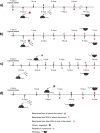
Methods: Using an in vivo tick transmission model, I. scapularis females infected with POWV II ("donors") were co-fed on mice with uninfected H. longicornis larvae and nymphs ("recipients"). The donor and recipient ticks were infested on mice in various sequences, and mouse infection status was monitored by temporal screening of blood for POWV II RNA via quantitative reverse transcription polymerase chain reaction (q-RT-PCR).
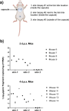
Results: The prevalence of POWV II RNA was highest in recipient H. longicornis that fed on viremic mice. However, nonviremic mice were also able to support co-feeding transmission of POWV, as demonstrated by the detection of viral RNA in multiple H. longicornis dispersed across different mice. Detection of viral RNA at the skin site of tick feeding but not at distal skin sites indicates that a localized skin infection facilitates transmission of POWV between donor and recipient ticks co-feeding in close proximity.
Conclusions: This is the first report examining transmission of POWV between co-feeding ticks. Against the backdrop of multiple unknowns related to POWV ecology, findings from this study provide insight on possible mechanisms by which POWV could be maintained in nature.
Keywords: Haemaphysalis longicornis; Co-feeding transmission; Nonviremic transmission; Powassan virus.
© 2024. The Author(s).
Conflict of interest statement
The authors declare that they have no competing interest.
Figures


Similar articles
-
Horizontal and Vertical Transmission of Powassan Virus by the Invasive Asian Longhorned Tick, Haemaphysalis longicornis, Under Laboratory Conditions.Front Cell Infect Microbiol. 2022 Jul 1;12:923914. doi: 10.3389/fcimb.2022.923914. eCollection 2022. Front Cell Infect Microbiol. 2022. PMID: 35846754 Free PMC article.
-
Vector competence of human-biting ticks Ixodes scapularis, Amblyomma americanum and Dermacentor variabilis for Powassan virus.Parasit Vectors. 2021 Sep 9;14(1):466. doi: 10.1186/s13071-021-04974-1. Parasit Vectors. 2021. PMID: 34503550 Free PMC article.
-
Smelly communication between haemaphysalis longicornis and infected hosts with indolic odorants: A case from severe fever with thrombocytopenia syndrome virus.PLoS Negl Trop Dis. 2025 Jun 5;19(6):e0013139. doi: 10.1371/journal.pntd.0013139. eCollection 2025 Jun. PLoS Negl Trop Dis. 2025. PMID: 40472053 Free PMC article.
-
Pathogenicity and virulence of Powassan virus.Virulence. 2025 Dec;16(1):2523887. doi: 10.1080/21505594.2025.2523887. Epub 2025 Jun 26. Virulence. 2025. PMID: 40545598 Free PMC article. Review.
-
Human pathogens associated with the blacklegged tick Ixodes scapularis: a systematic review.Parasit Vectors. 2016 May 5;9:265. doi: 10.1186/s13071-016-1529-y. Parasit Vectors. 2016. PMID: 27151067 Free PMC article.
Cited by
-
Strange relatives: the enigmatic arbo-jingmenviruses and orthoflaviviruses.Npj Viruses. 2025 Apr 4;3(1):24. doi: 10.1038/s44298-025-00106-z. Npj Viruses. 2025. PMID: 40295693 Free PMC article. Review.
-
Public health significance of the white-tailed deer (Odocoileus virginianus) and its role in the eco-epidemiology of tick- and mosquito-borne diseases in North America.Parasit Vectors. 2025 Feb 6;18(1):43. doi: 10.1186/s13071-025-06674-6. Parasit Vectors. 2025. PMID: 39915849 Free PMC article. Review.
-
TITAN-RNA: A hybrid-capture sequencing panel detects known and unknown Flaviviridae for diagnostics and vector surveillance.bioRxiv [Preprint]. 2025 Jun 8:2025.06.08.658352. doi: 10.1101/2025.06.08.658352. bioRxiv. 2025. PMID: 40501929 Free PMC article. Preprint.
-
Protocol for encapsulating ticks to study tick hematophagy and tick-virus-host interactions.STAR Protoc. 2025 Jun 30;6(3):103925. doi: 10.1016/j.xpro.2025.103925. Online ahead of print. STAR Protoc. 2025. PMID: 40591457 Free PMC article.
-
Atomic-resolution structure of a chimeric Powassan tick-borne flavivirus.Sci Adv. 2025 Jul 11;11(28):eadw7700. doi: 10.1126/sciadv.adw7700. Epub 2025 Jul 9. Sci Adv. 2025. PMID: 40632869 Free PMC article.
References
MeSH terms
Grants and funding
LinkOut - more resources
Full Text Sources

