. 2024 Jun 15;15(1):5121.
doi: 10.1038/s41467-024-49520-6.

Light chain mutations contribute to defining the fibril morphology in systemic AL amyloidosis

Affiliations

Light chain mutations contribute to defining the fibril morphology in systemic AL amyloidosis

Sara Karimi-Farsijani et al. Nat Commun. .

Abstract

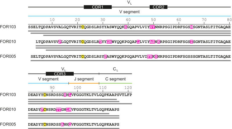
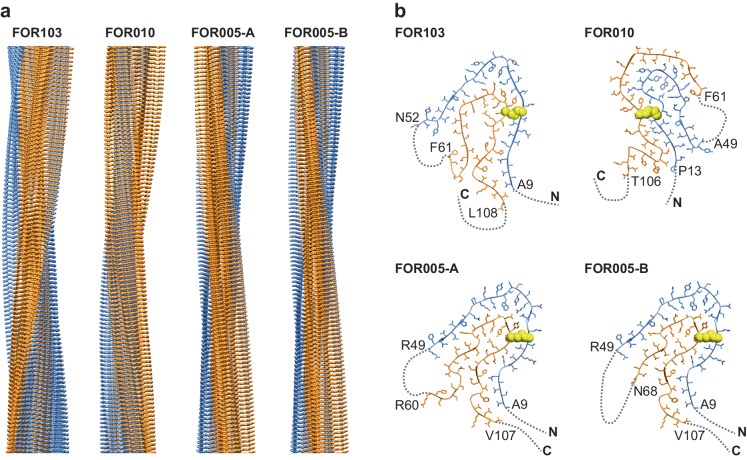
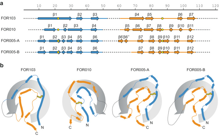
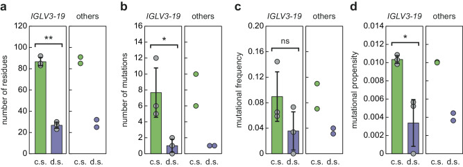
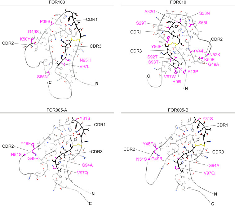
Systemic AL amyloidosis is one of the most frequently diagnosed forms of systemic amyloidosis. It arises from mutational changes in immunoglobulin light chains. To explore whether these mutations may affect the structure of the formed fibrils, we determine and compare the fibril structures from several patients with cardiac AL amyloidosis. All patients are affected by light chains that contain an IGLV3-19 gene segment, and the deposited fibrils differ by the mutations within this common germ line background. Using cryo-electron microscopy, we here find different fibril structures in each patient. These data establish that the mutations of amyloidogenic light chains contribute to defining the fibril architecture and hence the structure of the pathogenic agent.


Conflict of interest statement

The authors declare no competing interests.

Figures

Fig. 1
Fig. 1. Primary structures of the analysed fibril proteins.
Alignment of the amino acid sequences of the FOR103, FOR010 and FOR005 fibril proteins. The bars below the sequence refer to the experimentally observed fibril proteins. The FOR010 data were taken from SI Table 1. The FOR103 and FOR005 data were taken from previous studies,. Magenta: amino acid changes compared to GL sequences of IGLV3-19 and IGLJ2 segments; yellow: cysteine residues involved in disulphide bond formation.
Fig. 2
Fig. 2. Cryo-EM structures of IGLV3-19-derived AL amyloid fibrils.
a Side views of the molecular models of FOR103, FOR010 and FOR005 (A and B) fibrils. The FOR103 structure is right-hand twisted. The other fibrils possess a left-hand twist. b Cross-sectional views of one molecular layer of the four fibrils. The first and last residues of the ordered regions (ribbon diagrams with side chains) are highlighted. Structurally disordered regions are represented as dotted lines. The disulphide bond is shown in a space-filled representation (yellow). The N-terminal ordered region is coloured in blue in all panels, while the C-terminal region is consistently coloured orange.
Fig. 3
Fig. 3. Secondary structural composition of the fibrils.
a Location of the fibril cross-β structure in the sequence of the four fibril proteins. The numbering reflects the residue numbers of the respective precursor LCs after the removal of their signal sequences. Continuous lines refer to the ordered regions observed in the 3D map. Structurally disordered regions are shown as dotted lines. Cysteine residues participating in disulphide bonds are indicated in yellow. b Cross-sectional views of the four fibril proteins. The swirl of the amyloid key is indicated by the grey rotational arrow.
Fig. 4
Fig. 4. Mutational propensities of the analysed fibril proteins.
a Length of the disordered segments (d.s.) and core segments (c.s.) of the presently analysed IGLV3-19-derived AL amyloid fibrils (left, n = 3, biological replicates) and of two other fibril structures (right, n = 2, biological replicates): FOR001 (IGLV1-51) and FOR006 (IGLV1-44). b Number of the mutations in the two types of segments for the fibrils described in (a). c Mutational frequencies of these fibrils. d Mutational propensities of these fibrils. The colour coding is consistent in all panels. Grey data points: individual values of the IGLV3-19-derived AL amyloid fibrils. Bars, error bars: average values of these fibrils and standard deviation. Significant differences between the core and disordered segments are indicated by asterisks: p-value of 0.1 (*) and p-value of 0.01 (**). Non-significant (ns). Statistical analysis was performed using a two-sided t test. Source data are provided as a Source Data file.
Fig. 5
Fig. 5. Mutations of the fibril proteins.
Cross-sectional views of one molecular layer of the fibrils. Magenta: mutations compared to GL sequences of IGLV3-19 and IGLJ2 segments; yellow: cysteine residues involved in disulphide bond formation. Black: CDRs as described previously.

Similar articles

Cited by

References

    1. Merlini G, et al. Systemic immunoglobulin light chain amyloidosis. Nat. Rev. Dis. Prim. 2018;4:1–19. - PubMed
    1. Buxbaum JN, Chuba JV, Hellman GC, Solomon A, Gallo GR. Monoclonal immunoglobulin deposition disease: light chain and light and heavy chain deposition diseases and their relation to light chain amyloidosis. Ann. Intern. Med. 1990;112:455–464. doi: 10.7326/0003-4819-76-3-112-6-455. - DOI - PubMed
    1. Blancas-Mejía LM, Ramirez-Alvarado M. Systemic amyloidoses. Annu. Rev. Biochem. 2013;82:745–774. doi: 10.1146/annurev-biochem-072611-130030. - DOI - PMC - PubMed
    1. Del Pozo-Yauner L, et al. Role of the mechanisms for antibody repertoire diversification in monoclonal light chain deposition disorders: when a friend becomes foe. Front. Immunol. 2023;14:1203425. doi: 10.3389/fimmu.2023.1203425. - DOI - PMC - PubMed
    1. Bellotti V, Mangione P, Merlini G. Review: immunoglobulin light chain amyloidosis—the archetype of structural and pathogenic variability. J. Struct. Biol. 2000;130:280–289. doi: 10.1006/jsbi.2000.4248. - DOI - PubMed