. 2024 Jun 15;15(6):419.
doi: 10.1038/s41419-024-06809-0.

Trpv6 channel targeting using monoclonal antibody induces prostate cancer cell apoptosis and tumor regression

Affiliations

Trpv6 channel targeting using monoclonal antibody induces prostate cancer cell apoptosis and tumor regression

Aurélien Haustrate et al. Cell Death Dis. .

Abstract

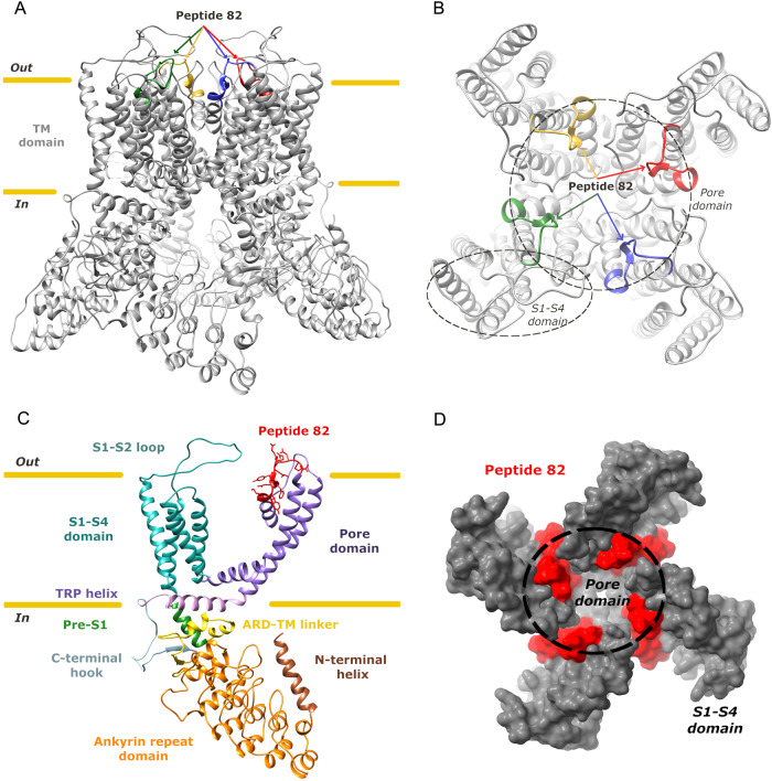
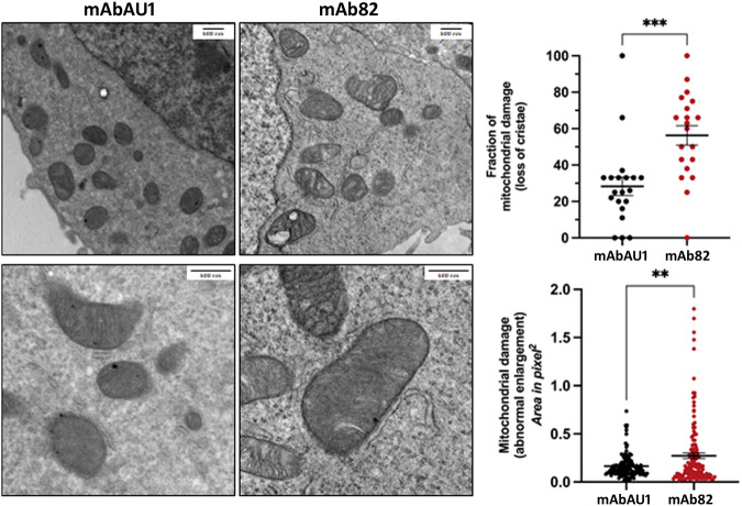
TRPV6 calcium channel is a prospective target in prostate cancer (PCa) since it is not expressed in healthy prostate while its expression increases during cancer progression. Despite the role of TRPV6 in PCa cell survival and apoptotic resistance has been already established, no reliable tool to target TRPV6 channel in vivo and thus to reduce tumor burden is known to date. Here we report the generation of mouse monoclonal antibody mAb82 raised against extracellular epitope of the pore region of the channel. mAb82 inhibited TRPV6 currents by 90% at 24 µg/ml in a dose-dependent manner while decreasing store-operated calcium entry to 56% at only 2.4 µg/ml. mAb82 decreased PCa survival rate in vitro by 71% at 12 µg/ml via inducing cell death through the apoptosis cascade via activation of the protease calpain, following bax activation, mitochondria enlargement, and loss of cristae, Cyt C release, pro-caspase 9 cleavage with the subsequent activation of caspases 3/7. In vivo, mice bearing either PC3Mtrpv6+/+ or PC3Mtrpv6-/-+pTRPV6 tumors were successfully treated with mAb82 at the dose as low as 100 µg/kg resulting in a significant reduction tumor growth by 31% and 90%, respectively. The survival rate was markedly improved by 3.5 times in mice treated with mAb82 in PC3Mtrpv6+/+ tumor group and completely restored in PC3Mtrpv6-/-+pTRPV6 tumor group. mAb82 showed a TRPV6-expression dependent organ distribution and virtually no toxicity in the same way as mAbAU1, a control antibody of the same Ig2a isotype. Overall, our data demonstrate for the first time the use of an anti-TRPV6 monoclonal antibody in vitro and in vivo in the treatment of the TRPV6-expressing PCa tumors.


Conflict of interest statement

The authors declare no competing interests.

Figures

Fig. 1
Fig. 1. Localization of a targeted peptide 82 within the structure of TRPV6.
Side (A) and top (B) views of the human TRPV6 tetramer (PDB https://www.rcsb.org/ ID: 6BO8), with the position of peptide 82 of each subunit shown in a different color. C single human TRPV6 monomer with each domain in a different color and the peptide 82 in red. D surface representation of the top view of the human TRPV6 tetramer with the peptide 82 in red. The base figure was adapted from [48].
Fig. 2
Fig. 2. Validation of the mouse anti human TRPV6 mAb82 antibody.
A immunoblotting of the PC3Mtrpv6+/+, PC3Mtrpv6-/-, and LNCaP cell lines using mAb82 antibody. B immunohistochemical staining of the PC3Mtrpv6-/- and PC3Mtrpv6-/-+pTRPV6 stable clones bearing tumors grown in immunodeficient Swiss nude mice using mAb82 followed by HRP-conjugated goat anti-mouse antibody (magnification x200). C immunofluorescent staining of TRPV6 channels in non-permeabilized PC3Mtrpv6-/- versus PC3Mtrpv6+/+ -mCherry expressing cell lines using mAb82 antibody followed by Alexa Fluor 488 goat anti-mouse antibody. D Fluorescence Activated Cell Sorting using mAb82 antibody followed by Alexa Fluor 488 goat anti-mouse antibody in non-permeabilized PC3Mtrpv6-/-, PC3Mtrpv6+/+, PC3Mtrpv6-/-+pTRPV6, and LNCaP cell lines. E representative image of immunohistochemical staining of the human patient samples of normal versus cancerous prostates using mAb82 followed by HRP-conjugated goat anti-mouse antibody (magnification x100). F Binding of mAb82 anti-TRPV6 antibody to the hTRPV6 peptide 82 as compared to control irrelevant mAb, anti-beta galactosidase from E.coli (mAbCT) using ELISA, and the corresponding EC50 value.
Fig. 3
Fig. 3. Effects of mAb82 on Ca2+ currents via the TRPV6 channel and Store-Operated Calcium Entry (SOCE) in LNCaP cells.
A representative IV curves induced by the −100 to +100 mV voltage ramp recorded in HEK cells transfected with the vEF1ap-5’UTR-TRPV6wt_CMVp-GFP vector. Curves show whole-cell TRPV6 currents in either base HBSS/basal medium (black), DVF medium alone (red), or DVF medium containing either 12 µg/ml of mAb82 anti-TRPV6 antibody (pink) or 24 µg/ml (yellow). B representative trace of the whole-cell currents during the application of the DVF solution as well as different doses of mAb82 antibody, as indicated by arrows. C bar plots summarizing average whole-cell currents under conditions indicated above for mAb82, (n = 15 for 1.2 µg/ml; n = 20 for 2.4 µg/ml; n = 12 for 6 µg/ml; n = 11 for 12 µg/ml; and n = 11 for 24 µg/ml). D SOCE in the LNCaP cells pretreated 5 min with either mouse monoclonal mAbAU1 as a control antibody or a mouse anti-human TRPV6 mAb82 antibody, both at 2.4 µg/ml. E corresponding quantitative representation of the ER content (calculated as a maximum amplitude), n = 3, *p < 0.05; the SOCE affected by antibody-induced treatments shown in (D); n = 3, ***p < 0.001; and the slope of SOCE calculated as a ratio delta/sec for each condition; n = 3, *p < 0.05.
Fig. 4
Fig. 4. TRPV6 inhibition by mAb82 decreases LNCaP cell survival.
A Cell survival assay (MTS) of LNCaP cells treated either with the different doses of a control mAbAU1 antibody of the same IgG2a isotype or with mAb82 anti-TRPV6 antibody during 3 days, n = 3, *p < 0.05; **p < 0.01, and ***p < 0.001. B apoptosis assay using TMR-red TUNEL assay of LNCaP cells treated with anti-TRPV6 antibody mAb82 at 2.4 µg/ml versus 1 µM thapsigargin (TG) treatment as a positive control; n = 3. C Statistical analysis of the number of apoptotic cells observed in (B) over three days, n = 3; ***p < 0.001. D Caspase-Glo 3/7 activity in LNCaP cells treated for 72 h with both mAbAU1 and mAb82 at the indicated dose as compared to 1 µM of Thapsigargin treatment. n = 3, ****p < 0.0001. E Immunoblotting of the key proteins involved in apoptosis under mAbAU1 and mAb82 treatment for 72 h. n = 3. F Video microscopy of LNCaP cells stained with both vital Hoechst at 2.5 µg/ml and PI 1 µg/ml and treated with 6 µg/ml of either mAbAU1 or mAb82 for 48 h.
Fig. 5
Fig. 5. Representative transmission electron microscopy.
(TEM) images (left) of LNCaP cells treated with either mAbAU1 and mAb82 antibodies, and their corresponding graphs (right) showing quantitative analysis of mitochondrial damage in the form of loss of cristae and abnormal enlargement. Significant difference at **P < 0.01 and ***P < 0.001 (n = 10 different sections; t test). Error bars represent mean ± s.e.m.
Fig. 6
Fig. 6. mAb82 distribution in vivo using immunodeficient Swiss nude mouse model.
A intraperitoneal injection of mAb82-cf790 at 100 µg/kg of body weight in Swiss nude mice without tumor burden and in vivo imaging over different time periods. B, C intraperitoneal injection of both mAb82-cf790 and mAbAU1-cf790 into Swiss nude mice at 100 µg/kg of body weight and organ distribution of both antibodies 4 h after injection. D mAb82-cf790 and mAbAU1-cf790 antibodies distributions at different time periods in Swiss nude mice bearing PC3Mtrpv6+/+ tumors. E relative toxicity of mAb82 and mAbAU1 after 24- and 72-h post-injection, measured using TMR-red TUNEL apoptosis assay on liver, intestine, and kidney tissue samples. F Weight of mice internal organs normalized to the body weight, n = 4. Treatment with mAbAU1 was normalized to 1. Mice were injected twice per week during two weeks via intraperitoneal injection of either mAbAU1 or mAb82, at 150 µg/kg of the body weight diluted in PBS.
Fig. 7
Fig. 7. Targeting TRPV6-expressing tumors in vivo using mAb82 decreases tumor burden and increases mice survival.
A tumor growth curves of Swiss nude mice bearing tumors generated by PC3Mtrpv6+/+, treated with 100 µg/kg body weight of mAb82 or mAbAU1 as a control antibody of the same IgG2 isotype. B mice survival curves from (A). C tumor growth curves of Swiss nude mice bearing tumors generated by PC3Mtrpv6-/-, treated with 100 µg/kg body weight of mAb82 or mAbAU1 as a control antibody of the same IgG2 isotype. D mice survival curves from (C). E tumor growth curves of Swiss nude mice bearing tumors generated by PC3Mtrpv6-/-+pTRPV6, treated with 100 µg/kg body weight of mAb82 or mAbAU1 as a control antibody of the same IgG2 isotype. F mice survival curves from (E). G in vivo imaging at different time intervals of mCherry-labeled tumors derived from Swiss nude mice bearing tumors generated by PC3Mtrpv6-/-+pTRPV6, treated with 100 µg/kg body weight of mAb82 or mAbAU1 as a control antibody of the same IgG2 isotype. H Visual representation of Swiss nude mice bearing tumors generated by PC3Mtrpv6-/-+pTRPV6, after 42 days of treatment with 100 µg/kg body weight of mAb82 or mAbAU1 as a control antibody of the same IgG2 isotype.

References

    1. Prevarskaya N, Skryma R, Shuba Y. Ion channels and the hallmarks of cancer. Trends Mol Med. 2010;16:107–21. doi: 10.1016/j.molmed.2010.01.005. - DOI - PubMed
    1. Audero MM, Prevarskaya N, Fiorio Pla A. Ca2+ signalling and hypoxia/acidic tumour microenvironment interplay in tumour progression. Int J Mol Sci. 2022;23:7377. doi: 10.3390/ijms23137377. - DOI - PMC - PubMed
    1. Prevarskaya N, Ouadid-Ahidouch H, Skryma R, Shuba Y. Remodelling of Ca2+ transport in cancer: how it contributes to cancer hallmarks? Philos Trans R Soc Lond B Biol Sci. 2014;369:20130097. doi: 10.1098/rstb.2013.0097. - DOI - PMC - PubMed
    1. Sanchez-Collado J, Jardin I, López JJ, Ronco V, Salido GM, Dubois C, et al. Role of Orai3 in the pathophysiology of cancer. Int J Mol Sci. 2021;22:11426. doi: 10.3390/ijms222111426. - DOI - PMC - PubMed
    1. Fiorio Pla A, Kondratska K, Prevarskaya N. STIM and ORAI proteins: crucial roles in hallmarks of cancer. Am J Physiol Cell Physiol. 2016;310:C509–19. doi: 10.1152/ajpcell.00364.2015. - DOI - PubMed

Publication types