. 2024 Oct;31(10):1362-1374.
doi: 10.1038/s41418-024-01327-0. Epub 2024 Jun 15.

Lactylation of NAT10 promotes N4-acetylcytidine modification on tRNASer-CGA-1-1 to boost oncogenic DNA virus KSHV reactivation

Affiliations

Lactylation of NAT10 promotes N4-acetylcytidine modification on tRNASer-CGA-1-1 to boost oncogenic DNA virus KSHV reactivation

Qin Yan et al. Cell Death Differ. 2024 Oct.

Abstract

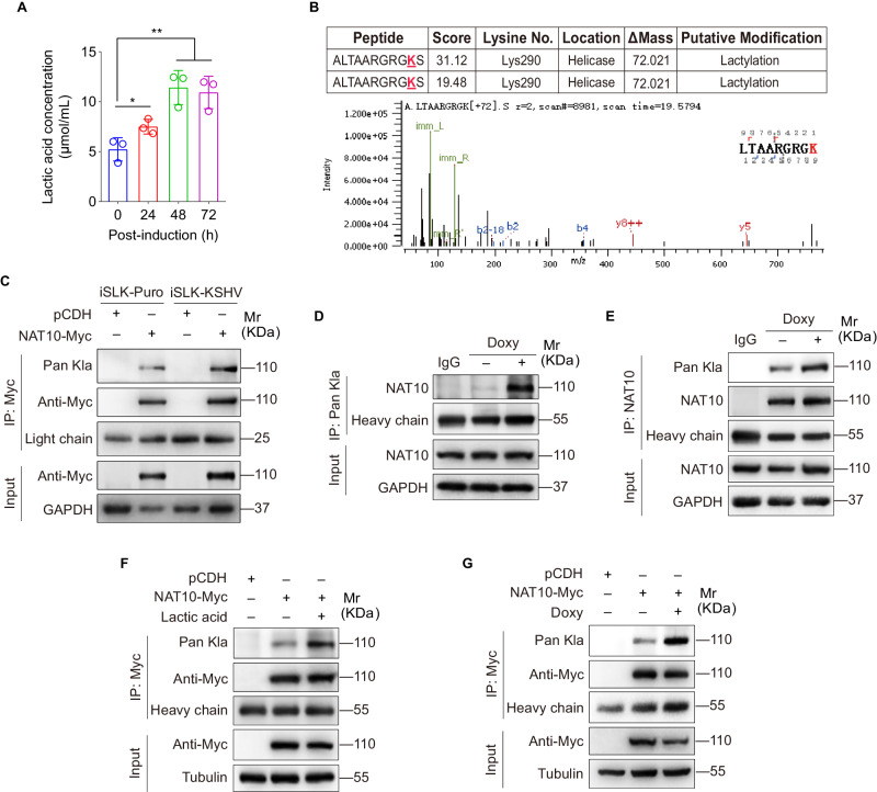
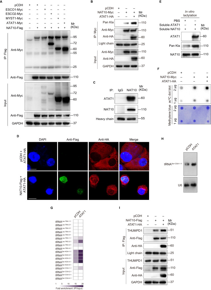
N4-acetylcytidine (ac4C), a conserved but recently rediscovered RNA modification on tRNAs, rRNAs and mRNAs, is catalyzed by N-acetyltransferase 10 (NAT10). Lysine acylation is a ubiquitous protein modification that controls protein functions. Our latest study demonstrates a NAT10-dependent ac4C modification, which occurs on the polyadenylated nuclear RNA (PAN) encoded by oncogenic DNA virus Kaposi's sarcoma-associated herpesvirus (KSHV), can induce KSHV reactivation from latency and activate inflammasome. However, it remains unclear whether a novel lysine acylation occurs in NAT10 during KSHV reactivation and how this acylation of NAT10 regulates tRNAs ac4C modification. Here, we showed that NAT10 was lactylated by α-tubulin acetyltransferase 1 (ATAT1), as a writer at the critical domain, to exert RNA acetyltransferase function and thus increase the ac4C level of tRNASer-CGA-1-1. Mutagenesis at the ac4C site in tRNASer-CGA-1-1 inhibited its ac4C modifications, translation efficiency of viral lytic genes, and virion production. Mechanistically, KSHV PAN orchestrated NAT10 and ATAT1 to enhance NAT10 lactylation, resulting in tRNASer-CGA-1-1 ac4C modification, eventually boosting KSHV reactivation. Our findings reveal a novel post-translational modification in NAT10, as well as expand the understanding about tRNA-related ac4C modification during KSHV replication, which may be exploited to design therapeutic strategies for KSHV-related diseases.


Conflict of interest statement

The authors declare no competing interests.

Figures

Fig. 1
Fig. 1. NAT10 catalyzes tRNASer-CGA-1-1 ac4C modification to promote viral mRNAs translation and virion production.
A The total RNAs from iSLK-KSHV NAT10+/- cells (NAT10+/-) and iSLK-KSHV NAT10+/+ cells (WT) were subjected to dot blot assay with anti-ac4C antibody. Methylene blue staining was used as the internal control. B The heatmap for differentially expressed tRNAs in cells shown as in (A) by tRNA acRIP-seq analysis. The pseudo-color represented as the fold enrichment (IP/Input) of WT and NAT10+/− group, respectively. Any tRNAs not detected in tRNA acRIP-seq were plotted as white in the heatmap. C The total RNAs from cells shown as in (A) were subjected to Northern blot with probes to tRNASer-CGA-1-1, tRNASer-CGA-4-1, tRNALeu-TAG-3-1, tRNASer-AGA, tRNALeu-TAA, and U6 as the control. D The RT-qPCR analysis for the ribosome-nascent chain-complex-bound mRNA (RNC-qPCR) of representative viral genes of KSHV (RTA, K5, K8, ORF45, ORF57, vIRF1, vIL-6, vBCL-2, ORF65, and K8.1) in cells shown as in (A). ***P < 0.001 by Student’s t-test. E Schematic representation of the acetylation site mutation of tRNASer-CGA-1-1 (Upper) and tRNALeu-TAG-3-1 (Lower). The C (marked in red) at position 12 was mutated to U (marked in dark blue). F The total RNAs from iSLK-KSHV cells with overexpression of the wild type tRNASer-CGA-1-1 or tRNALeu-TAG-3-1 (WT), mutant tRNASer-CGA-1-1 or tRNALeu-TAG-3-1 (Mut), and their control empty vector (EV) for 48 h were respectively subjected to Northern blot with probes to tRNASer-CGA-1-1, tRNALeu-TAG-3-1 and U6 as the control. G. The RT-qPCR analysis for the ribosome-nascent chain-complex-bound mRNA (RNC-qPCR) of representative viral genes of KSHV (RTA, K5, K8, vIRF1, vIL-6, vBCL-2, and ORF65) in iSLK-KSHV cells with overexpression of the wild type (tRNASer-CGA-1-1-WT) or its mutant (tRNASer-CGA-1-1-Mut) tRNASer-CGA-1-1 for 48 h, and their control (tRNA-EV). ***P < 0.001 by Student’s t-test. H The expression levels of vIRF1 and NAT10 in iSLK-KSHV cells with overexpression of the wild type (WT), mutant (Mut) tRNASer-CGA-1-1, and their control (EV) for 48 h were detected by Western blot. I The SDS gel analysis of the in vitro translation reaction supplemented with the wild type (tRNASer-CGA-1-1-WT) or the mutant (tRNASer-CGA-1-1-Mut) tRNASer-CGA-1-1, in comparison with reaction mixtures that not containing mRNA (No mRNA). J By the assessment of ORF26, real-time DNA-PCR for cells shown as in (H) was performed to detect viral copy number after doxycycline stimulation for 72 h. ***P < 0.001 by Student’s t-test.
Fig. 2
Fig. 2. NAT10 undergoes lactate-driven lactylation during KSHV reactivation.
A The culture medium of iSLK-KSHV cells with doxycycline induction for 0, 24, 48, and 72 h were harvested for detection of lactic acid concentration with the Lactate Assay Kit (Jiancheng BioEngineering, Nanjing, China). *P < 0.05 and **P < 0.01 by Student’s t-test versus the 0 h group. B Lactylation of NAT10 lysine residues in iSLK-KSHV cells transduced with lentiviral NAT10 was identified by LC–MS/MS analysis. The sequence of identified peptides, score of peptide segment for matching degree, the number and location of the identified lysines (K) in LC-MS/MS analysis were shown (Upper). The increased 72.021 Da on lysines was considered as lactylation. The b and y ions in the spectra of the peptide were marked in blue and red, respectively (Lower). C The iSLK-Puro or iSLK-KSHV cells were transduced with lentiviral NAT10 (NAT10-Myc) or control lentivirus (pCDH) for 48 h, and then subjected to anti-Myc immunoprecipitation assay (IP). Lysine lactylation in the immunoprecipitated NAT10 was examined by Western blot with anti-Pan-lactyl-lysine antibody (Pan Kla). D, E The iSLK-KSHV cells induced with (+) or without (−) doxycycline (Doxy) were subjected to the anti-Pan Kla (D) or anti-NAT10 (E) immunoprecipitation, and the pull-downed proteins were examined by Western blot with anti-NAT10 (D) or anti-Pan Kla (E) antibodies, respectively. Anti-IgG antibody was used as the control. F The iSLK-KSHV cells transduced with lentiviral NAT10 (NAT10-Myc) or control lentivirus (pCDH) for 48 h were treated with or without lactic acid (20 mM) for 24 h, and then subjected to anti-Myc IP assay and detected by Western blot with the anti-Pan Kla antibody. G The iSLK-KSHV cells transduced with lentiviral NAT10 (NAT10-Myc) or control lentivirus (pCDH) for 48 h were treated with or without doxycycline (Doxy) for 72 h, and then subjected to anti-Myc IP assay and detected by Western blot with the anti-Pan Kla antibody.
Fig. 3
Fig. 3. NAT10 lactylation at Lys290 promotes viral mRNAs translation and virion production of KSHV.
A Schematic representation of DUF1726 domain (marked in blue), RNA helicase domain (marked in yellow), N-acetyltransferase domain (marked in green) and RNA-binding domain (marked in purple) of NAT10. The identified lactylation lysine site (K) within the RNA helicase domain was marked in red, which was mutated to arginine (R) and marked in dark blue. B The iSLK-KSHV cells transduced with the wild type (NAT10-Flag), the mutant NAT10 (K290R-Flag) or its control (pCDH) for 48 h were subjected to the anti-Flag immunoprecipitation, and then examined by Western blot with the anti-Pan Kla antibody. C The sequences around NAT10 Lys290 from different species were aligned. Conserved Lys290 was marked in red. D The total RNAs from cells shown as in (B) were subjected to dot blot assay with anti-ac4C antibody. Methylene blue staining was used as the internal control. E The iSLK-KSHV cells with THUMPD1 overexpression (THUMPD1-Myc) were transduced with the wild type (NAT10-Flag), the mutant NAT10 (K290R-Flag) or its control (pCDH) for 48 h. Cells were subjected to the anti-Flag immunoprecipitation and analyzed by Western blot using anti-Myc antibody. F The total RNAs from iSLK-KSHV cells transduced with the wild type (NAT10) and the mutant (K290R) NAT10 for 48 h were subjected to Northern blot with probes to tRNASer-CGA-1-1, and U6 as the control. G The expression levels of vIRF1 and K-bZIP in cells treated as in (B) were examined by Western blot with the corresponding antibodies. H The RT-qPCR analysis for the ribosome-nascent chain-complex-bound mRNA (RNC-qPCR) of representative viral genes of KSHV (RTA, K5, K8, ORF45, ORF57, vIRF1, vIL-6, vBCL-2, ORF65, and K8.1) in cells shown as in (B). *P < 0.05, **P < 0.01 and ***P < 0.001 by Student’s t-test. I The RT-qPCR analysis for the ribosome-nascent chain-complex-bound mRNA (RNC-qPCR) of representative viral genes of KSHV (RTA, K5, K8, vIRF1, vIL-6, vBCL-2, and ORF65) in iSLK-KSHV cells with the wild type (NAT10-WT) or the mutant (NAT10-K290R) NAT10 overexpression, which were complemented with the wild type (tRNASer-CGA-1-1-WT) or the mutant (tRNASer-CGA-1-1-Mut) tRNASer-CGA-1-1 for 48 h. *P < 0.05, **, P < 0.01 and ***P < 0.001 by Student’s t-test. J By the assessment of ORF26, real-time DNA-PCR for cells treated as in (B) was performed to examine viral copy number after doxycycline stimulation for 72 h. ***P < 0.001 by Student’s t-test.
Fig. 4
Fig. 4. NAT10 is lactylated by ATAT1.
A The iSLK-KSHV cells transduced with lentiviral NAT10 (NAT10-Flag) or its control (pCDH) were infected by lentiviral ESCO1 (ESCO1-Myc), ESCO2 (ESCO2-Myc), MYST1 (MYST1-Myc), and ATAT1 (ATAT1-Myc) for 48 h, and then subjected to anti-Flag immunoprecipitation. The interaction between NAT10 and acyltransferases was examined by Western blot with anti-Myc antibody. B The iSLK-KSHV cells transduced with lentiviral NAT10 (NAT10-Myc) or its control (pCDH) were infected by lentiviral ATAT1 (ATAT1-HA) for 48 h, and then subjected to anti-Myc immunoprecipitation to examine the lactylation level of NAT10 with anti-Pan Kla antibody. C Immunoprecipitation assay was performed to examine the interaction between endogenous NAT10 and ATAT1. D The iSLK-KSHV cells with ATAT1 (ATAT1-HA) overexpression were infected with lentiviral NAT10 (NAT10-Flag) or its control (pCDH) for 48 h, and then were employed to examine the co-localization of NAT10 and ATAT1 by immunofluorescence staining. Scar bars, 40 μm. E ATAT1-catalyzed NAT10 lactylation was determined by mixing soluble ATAT1, NAT10, and lactyl CoA (20 μΜ) in the in vitro lactylation assay. Western blot analysis was performed with the indicated antibodies. F The total RNAs from cells treated as in (B) were subjected to dot blot assay with anti-ac4C antibody. Methylene blue staining was used as the internal control. G The heatmap for differentially expressed tRNAs in iSLK-KSHV cells transduced with ATAT1 (ATAT1) or its control (pCDH) for 48 h by tRNA acRIP-seq analysis. The pseudo-color represented as the fold enrichment (IP/Input) of pCDH and ATAT1 group, respectively. Any tRNAs not detected in tRNA acRIP-seq were plotted as white in the heatmap. H The total RNAs from cells shown as in (G) were subjected to Northern blot with probes to tRNASer-CGA-1-1 and U6 as the control. I The cells treated as in (B) were subjected to the anti-Flag immunoprecipitation and analyzed by Western blot using anti-THUMPD1 antibody.
Fig. 5
Fig. 5. ATAT1, as the lactylation writer of NAT10 promotes viral lytic transcripts translation and virus reactivation.
A The expression levels of vIRF1 and K-bZIP in iSLK-KSHV cells transduced with lentiviral sgATAT1 (sgATAT1 #2, sgATAT1 #3) or control lentivirus (Lenti-V2) for 72 h were examined by Western blot with the corresponding antibodies. B The RT-qPCR analysis for the ribosome-nascent chain-complex-bound mRNA (RNC-qPCR) of representative viral genes of KSHV (K5, K8, vIRF1, vIL-6, vBCL-2, and ORF65) in cells treated as in (A). *P < 0.05, **P < 0.01 and ***P < 0.001 by Student’s t-test. C By the assessment of ORF26, real-time DNA-PCR for cells treated as in (A) was performed to examine viral copy number after doxycycline stimulation for 72 h. ***, P < 0.001 by Student’s t-test. D The RT-qPCR analysis for the ribosome-nascent chain-complex-bound mRNA (RNC-qPCR) of representative viral genes of KSHV (RTA, K5, K8, ORF45, ORF57, vIRF1, vIL-6, vBCL-2, ORF65, and K8.1) in iSLK-KSHV cells transduced with lentiviral ATAT1 (ATAT1) or its control (pCDH) for 48 h. ***, P < 0.001 by Student’s t-test. E By the assessment of ORF26, real-time DNA-PCR for cells treated as in (D) was performed to detect viral copy number after doxycycline stimulation for 72 h. **P < 0.01 by Student’s t-test.
Fig. 6
Fig. 6. KSHV PAN RNA assists in the recruitment of ATAT1 to NAT10.
A The iSLK-KSHV cells transduced with lentiviral NAT10 (NAT10-Myc) or control (pCDH) for 48 h were treated with or without doxycycline (Doxy) for 72 h, and then subjected to anti-Myc IP assay and analyzed by Western blot with the anti-ATAT1 antibody. B, C The iSLK-KSHV cells with doxycycline induction for 72 h were subjected to the anti-ATAT1 (B), anti-NAT10 (C), or immunoglobulin G (Anti-IgG) RNA immunoprecipitation, and the precipitated PAN RNA were examined by RT-qPCR. ***, P < 0.001 by Student’s t-test versus the IgG group. D HEK293T cells with NAT10 overexpression (NAT10-Flag) were transduced with lentiviral PAN (pHAGE-PAN) or control (pHAGE) for 48 h. Cells were subjected to the anti-Flag immunoprecipitation and analyzed by Western blot using anti-ATAT1 and anti-Pan Kla antibodies. E The iSLK-KSHV cells with NAT10 overexpression (NAT10-Myc) were transduced with lentiviral PAN shRNAs (shPAN-1, shPAN-2, shPAN-3, and shPAN-4) or control (shCtrl) for 48 h. Cells were subjected to the anti-Myc immunoprecipitation and analyzed by Western blot using anti-ATAT1 and anti-Pan Kla antibodies. F The iSLK-KSHV cells transduced with lentiviral NAT10 (NAT10-Myc) or control (pCDH) for 48 h were treated with or without doxycycline (Doxy) for 72 h, while the induced NAT10-overexpressing cells were further treated with RNase. Cell proteins were subjected to anti-Myc IP assay and detected by Western blot with anti-ATAT1 and anti-Pan Kla antibodies. G Schematic illustration for the mechanism of lactylated NAT10-dependent tRNASer-CGA-1-1 ac4C modification during KSHV reactivation. As for NAT10 lactylation, ATAT1 serves as a writer. During KSHV reactivation, PAN RNA assists in the interaction between ATAT1 and NAT10 to promote the lactylation level of NAT10. Lactylated NAT10 occurring on Lys290 mediates tRNASer-CGA-1-1 ac4C modification, and eventually increases the translation efficiency of a series viral lytic transcripts, resulting in progeny virion production and virus reactivation.

Similar articles

Cited by

References

    1. Roundtree IA, Evans ME, Pan T, He C. Dynamic RNA modifications in gene expression regulation. Cell. 2017;169:1187–1200. - PMC - PubMed
    1. Bartee D, Nance KD, Meier JL. Site-specific synthesis of N(4)-acetylcytidine in RNA reveals physiological duplex stabilization. J Am Chem Soc. 2022;144:3487–96. - PMC - PubMed
    1. Ito S, Horikawa S, Suzuki T, Kawauchi H, Tanaka Y, Suzuki T, et al. Human NAT10 is an ATP-dependent RNA acetyltransferase responsible for N4-acetylcytidine formation in 18 S ribosomal RNA (rRNA). J Biol Chem. 2014;289:35724–30. - PMC - PubMed
    1. Sas-Chen A, Thomas JM, Matzov D, Taoka M, Nance KD, Nir R, et al. Dynamic RNA acetylation revealed by quantitative cross-evolutionary mapping. Nature. 2020;583:638–43. - PMC - PubMed
    1. Sharma S, Langhendries JL, Watzinger P, Kotter P, Entian KD, Lafontaine DL. Yeast Kre33 and human NAT10 are conserved 18S rRNA cytosine acetyltransferases that modify tRNAs assisted by the adaptor Tan1/THUMPD1. Nucleic Acids Res. 2015;43:2242–58. - PMC - PubMed