. 2024 Jul:105:105196.
doi: 10.1016/j.ebiom.2024.105196. Epub 2024 Jun 15.

Spatial distribution of Mycobacterium tuberculosis mRNA and secreted antigens in acid-fast negative human antemortem and resected tissue

Affiliations

Spatial distribution of Mycobacterium tuberculosis mRNA and secreted antigens in acid-fast negative human antemortem and resected tissue

Kievershen Nargan et al. EBioMedicine. 2024 Jul.

Abstract

Background: The ability to detect evidence of Mycobacterium tuberculosis (Mtb) infection within human tissues is critical to the study of Mtb physiology, tropism, and spatial distribution within TB lesions. The capacity of the widely-used Ziehl-Neelsen (ZN) staining method for identifying Mtb acid-fast bacilli (AFB) in tissue is highly variable, which can limit detection of Mtb bacilli for research and diagnostic purposes. Here, we sought to circumvent these limitations via detection of Mtb mRNA and secreted antigens in human tuberculous tissue.

Methods: We adapted RNAscope, an RNA in situ hybridisation (RISH) technique, to detect Mtb mRNA in ante- and postmortem human TB tissues and developed a dual ZN/immunohistochemistry staining approach to identify AFB and bacilli producing antigen 85B (Ag85B).

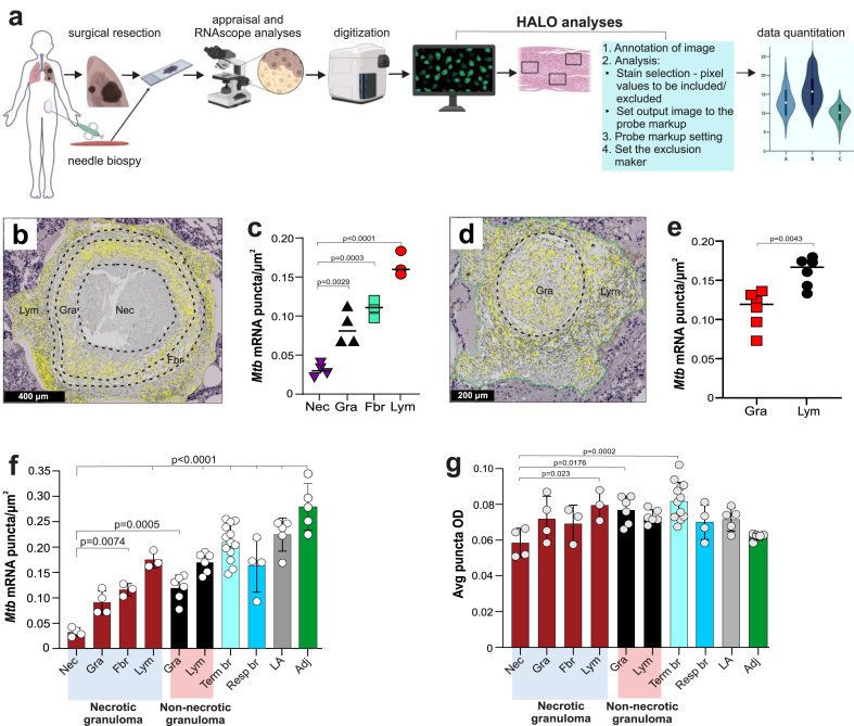
Findings: We identified Mtb mRNA within intact and disintegrating bacilli as well as extrabacillary mRNA. Mtb mRNA was distributed zonally within necrotic and non-necrotic granulomas. We also found Mtb mRNA within, and adjacent to, necrotic granulomas in ZN-negative lung tissue and in Ag85B-positive bronchiolar epithelium. Intriguingly, we observed accumulation of Mtb mRNA and Ag85B in the cytoplasm of host cells. Notably, many AFB were negative for Ag85B staining. Mtb mRNA was observed in ZN-negative antemortem lymph node biopsies.

Interpretation: RNAscope and dual ZN/immunohistochemistry staining are well-suited for identifying subsets of intact Mtb and/or bacillary remnants in human tissue. RNAscope can identify Mtb mRNA in ZN-negative tissues from patients with TB and may have diagnostic potential in complex TB cases.

Funding: Wellcome Leap Delta Tissue Program, Wellcome Strategic Core Award, the National Institutes of Health (NIH, USA), the Mary Heersink Institute for Global Health at UAB, the UAB Heersink School of Medicine.

Keywords: Antigen; Diagnosis; RNAscope; Tuberculosis; Ziehl-Neelsen.


Conflict of interest statement

Declaration of interests The authors have no competing interests or disclosures.

Figures

Fig. 1
Fig. 1
RNAscope and its application for detecting Mtb in human tissues. (a) Schematic of the RNAscope in situ hybridisation platform for mRNA detection. Following tissue fixation and permeabilisation, up to 20 “Z” oligonucleotide probe pairs hybridise with multiple target sequences (red lines) within a single mRNA across a ∼1000-bp region. For detection of Mtb in human tissue, 120 probe pairs (20 pairs per mRNA) bind complimentary sequences in six Mtb mRNAs. Subsequent steps include binding of a pre-amplifier to mRNA-bound “Z” only to probe pairs, addition of amplifiers molecules, and binding of chromogen label (fluorescent labels can also be used) for detection via microscopy where an individual mRNA molecule appears as a single dot or punctum. The binding of a single “Z” probe to mRNA does not result in pre-amplifier binding or a signal. (b) Diagram illustrating the potential of RNAscope to identify and track Mtb mRNA in resected, antemortem, or postmortem specimens in pulmonary or extrapulmonary tissue. Schematic in (a) adapted from Anderson, et al.
Fig. 2
Fig. 2
RNAscope detects intact and disintegrated Mtb bacilli and mRNA in human tissue. (a) low power image of a human testicular specimen containing ZN-positive AFB. Inset–note positive ZN staining within the seminiferous tubules. (b) low power image of a consecutive section of the testicular specimen in (a) revealing RNAscope Mtb mRNA signals, which are abundant within the seminiferous tubules. Medium power image showing ZN-positive AFB (c) or RNAscope Mtb signals (d) within seminiferous tubules. High power image revealing ZN-positive AFB (e) and RNAscope Mtb mRNA signals (f). (g) High power image showing a spectrum of RNAscope Mtb mRNA signals within a single field (hm). High power images of RNAscope signals from (h) intact bacilli, (i, j) partially intact bacilli, (k, l), disintegrated bacilli appearing as large, diffuse puncta (m) small puncta representing single extrabacillary Mtb mRNAs.
Fig. 3
Fig. 3
RNAscope detects Mtb mRNA in human TB lung tissue. Lung tissue from a patient with active TB. (a) ZN-positive tissue section revealing numerous intracellular (circled) and extracellular AFB. (b) ZN-negative lung tissue specimen from the patient in (a) containing a necrotic granuloma with RNAscope Mtb mRNA puncta; boxed regions correspond to high power images showing RNAscope puncta. (c) High power image revealing RNAscope Mtb mRNA puncta within a necrotic area (NE), granulomatous area (oval) and granulomatous inflammatory layer (red arrows). (d) High power image of numerous RNAscope Mtb mRNA puncta within the bronchiolar epithelial layer (BEL). (e) ZN staining showing the absence of AFB in the BEL. (f) High power image of Ag85B-positive staining within the cytoplasm (yellow asterisks) and nuclei (red arrows) of bronchiolar epithelial cells.
Fig. 4
Fig. 4
Accumulation of Mtb mRNA and secreted antigens in cells from pulmonary and extrapulmonary tissue. (a–e, g) The same ZN-negative lung tissue specimen analysed in Fig. 3b–f. (ad) High power images showing prominent RNAscope Mtb mRNA signals within alveolar epithelial cells (yellow arrows). (e) High power image of RNAscope signals within lymphoid aggregates of necrotic granulomas (area in yellow oval). (f) Medium power image of Ag85B-positive staining in the cytoplasm of macrophages in the ZN-positive extrapulmonary (testicular) human tissue specimen shown in Fig. 2. (g) Medium power image of Ag85B-positive staining in the cytoplasm of alveolar macrophages and epithelioid histiocytes within pulmonary human TB specimens. (f, g) Insets show higher power images of the cytoplasmic localisation of Ag85B staining.
Fig. 5
Fig. 5
Quantitation of RNAscope signals to inform the biology of TB lesions. (a) Flow diagram depicting sample acquisition, RNAscope analysis, digitisation, and quantitation of RNAscope Mtb mRNA puncta using HALO® analysis software. (bg) The same ZN-negative lung tissue specimen analysed in Fig. 3, Fig. 4. (b) Representative zonation of a single necrotic granuloma and (c) number of RNAscope Mtb mRNA puncta per square micron in each lesion zone. (d) Representative zonation of a single non-necrotic granuloma and (e) number of RNAscope Mtb mRNA puncta per square micron in each lesion zone. Yellow puncta (b and d) are false-color RNAscope Mtb mRNA signals. (f) Number of RNAscope Mtb mRNA puncta per square micron and (g) average puncta optical density (OD) in TB lesions and tissue. Zones include necrotic (Nec), granulomatous inflammatory (Gra), fibrotic (Fbr), lymphocytic (Lym), terminal bronchiole (Term br), respiratory bronchiole (Resp br), lymphocytic aggregates (LA) and tissue adjacent to diseased areas (Adj). In (c, e, f, and g), each data point represents the number of RNAscope signal puncta per square micron (c, e, and f) or average puncta OD (g) in an individual zone within a granuloma or other lung feature. In (c, f, and g), four necrotic granulomas were examined that exhibited Nec and Gra zones (n = 4) and Fbr and Lym zones (n = 3). In (e, f, and g), six non-necrotic granulomas were examined that contained Gra and Lym zones (n = 6). (f, g) Term br; n = 13, Resp br; n = 4, LA; n = 5, Adj; n = 5. Data in (c, e, f and g) represent the mean ± SD. Data were analysed using one-way ANOVA and Bonferroni's multiple comparison test (c, f, and g) or by unpaired Mann–Whitney test (e). (c, f, and g) all comparisons are with respect to the Nec zone.
Fig. 6
Fig. 6
Phenotypically distinct Mtb populations exist within human extrapulmonary TB tissue. (a) Medium power image of a seminiferous tubule with combined ZN staining (pink) and Ag85B IHC staining (brown). Inset; note the presence of Ag85B-positive and -negative bacilli within the seminiferous tubule. (b) High power image of ZN/Ag85B staining showing Ag85B-positive bacilli near ZN-positive/Ag85B-negative bacilli. (c, d) High power images of ZN/Ag85B dual staining showing Ag85B-positive (blue arrow) and -negative (red arrow) bacilli. (e) Ag85B-positive Mtb bacillus (blue arrow) and cytoplasm (yellow asterisk). (f) translucent, weakly acid-fast “ghost” bacillus (arrowhead) and an AFB with Ag85B positivity at the one pole (asterisk). (g) intracellular and extracellular AFB (red arrows) and Ag85B-positive bacilli (blue arrows), with some Ag85B-positive bacilli within Ag85B-positive host cell cytoplasm (yellow asterisks). (h) Ag85B-positive host cell cytoplasm (yellow asterisk) with ZN-positive/Ag85B-negative Mtb bacillus (yellow arrow). (i) Two Ag85-positive Mtb bacilli (blue arrows); one is inside an Ag85B-positive host cell (yellow asterisk).
Fig. 7
Fig. 7
Application of RNAscope may help guide therapeutic intervention. (a) Flowchart depicting CT-guided biopsies, hospitalisation, and postmortem analyses of tissues from a patient with undiagnosed TB. (b) Medium power image of a ZN-negative antemortem left inguinal lymph node needle biopsy specimen obtained 414 days prior to hospital admission. (c) High power images of the inguinal lymph node in (b) with RNAscope Mtb mRNA puncta within and around giant cells. (d) Number of Mtb mRNA puncta per square micron. Horizontal red line indicates the average number of puncta per square micron across the entire lymph node specimen. (e) Average puncta optical density (OD) in and around giant cells (Gc), lymphocytic aggregates (LA) and adjacent (Adj) lymphoid tissue. Horizontal red line indicates the average OD of puncta across the entire lymph node specimen. (f) Ag85B staining in and around giant cells in the ZN-negative inguinal lymph node specimen. The circled area shows weakly positive giant cells with several Ag85B-positive bacilli (yellow arrows). (g) High power image of a strongly Ag85B-positive giant cell in the inguinal lymph node specimen. (h) High power image of Ag85B accumulation within the cytoplasm of lymphocytes in the inguinal lymph node specimen. (i) Low power image of Ag85B-positive cells in the ZN-negative antemortem bone marrow biopsy. (j) Medium power image showing Ag85B positivity in the cytoplasm of lymphocytes within bone marrow shown in (i). (k) Ag85B positivity in the ZN-positive postmortem periaortic lymph node specimen. Data in (d) and (e) represent the mean ± SD and each data point represents the number of Mtb mRNA puncta per square micron (d) or average puncta OD (e) in each zone (Gc, LA, and Adj) of the inguinal lymph node (n = 5–6). Data in (d) were analyzed using one-way ANOVA and Bonferroni's multiple comparison test.

References

    1. Goletti D., Delogu G., Matteelli A., Migliori G.B. The role of IGRA in the diagnosis of tuberculosis infection, differentiating from active tuberculosis, and decision making for initiating treatment or preventive therapy of tuberculosis infection. Int J Infect Dis. 2022;124(Suppl 1):S12–S19. - PubMed
    1. Petruccioli E., Scriba T.J., Petrone L., et al. Correlates of tuberculosis risk: predictive biomarkers for progression to active tuberculosis. Eur Respir J. 2016;48(6):1751–1763. - PMC - PubMed
    1. Barcellini L., Borroni E., Brown J., et al. First evaluation of QuantiFERON-TB Gold Plus performance in contact screening. Eur Respir J. 2016;48(5):1411–1419. - PubMed
    1. Zaporojan N., Negrean R.A., Hodisan R., Zaporojan C., Csep A., Zaha D.C. Evolution of laboratory diagnosis of tuberculosis. Clin Pract. 2024;14(2):388–416. - PMC - PubMed
    1. Chakravorty S., Simmons A.M., Rowneki M., et al. The new Xpert MTB/RIF ultra: improving detection of Mycobacterium tuberculosis and resistance to rifampin in an assay suitable for point-of-care testing. mBio. 2017;8(4) - PMC - PubMed

LinkOut - more resources