. 2024 Jun 17;9(1):163.
doi: 10.1038/s41392-024-01881-6.

DNA of neutrophil extracellular traps promote NF-κB-dependent autoimmunity via cGAS/TLR9 in chronic obstructive pulmonary disease

Affiliations

DNA of neutrophil extracellular traps promote NF-κB-dependent autoimmunity via cGAS/TLR9 in chronic obstructive pulmonary disease

Jun Chen et al. Signal Transduct Target Ther. .

Abstract

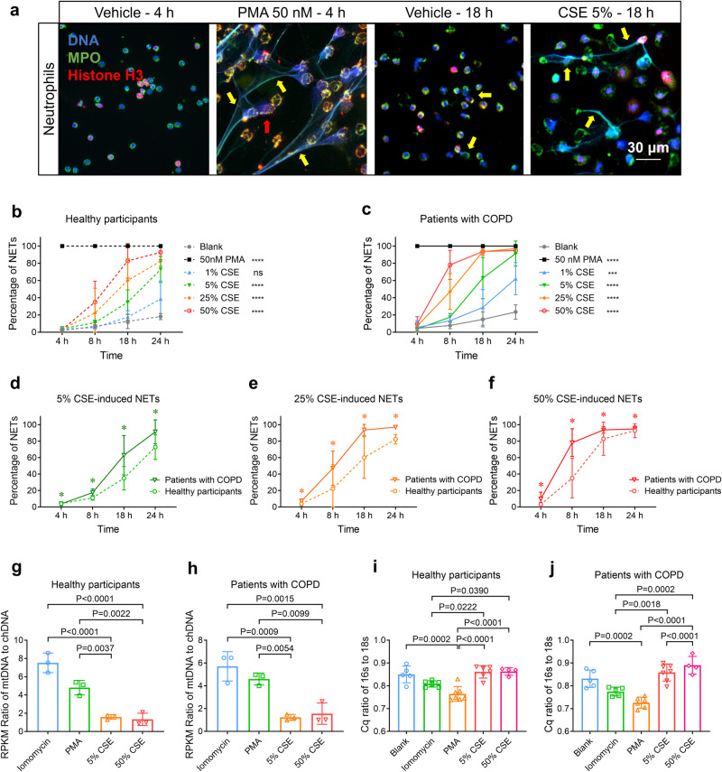
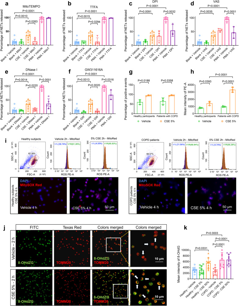
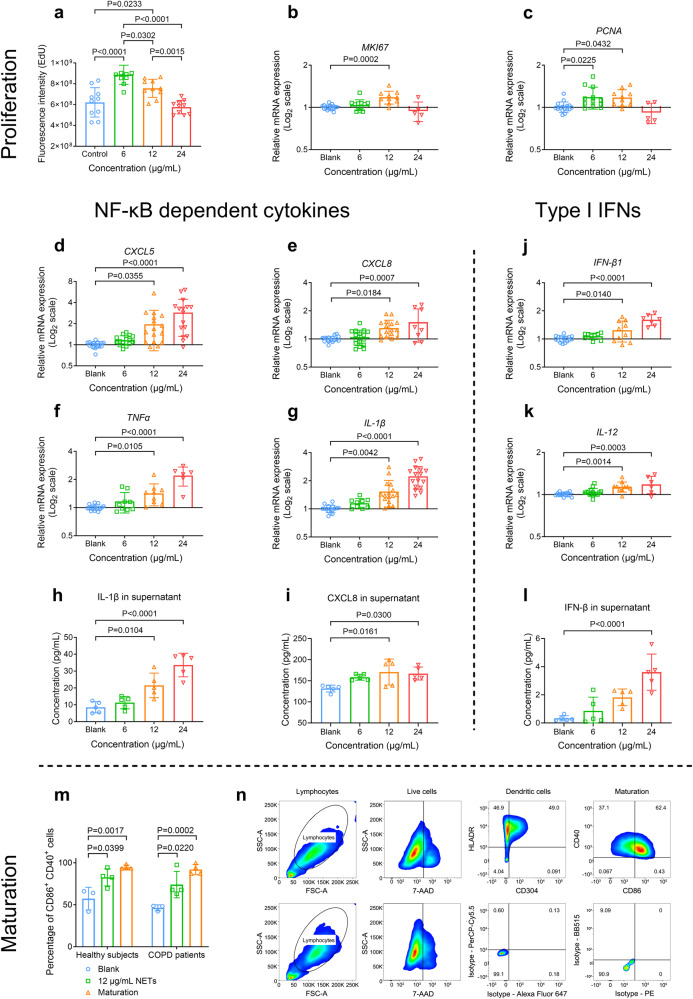
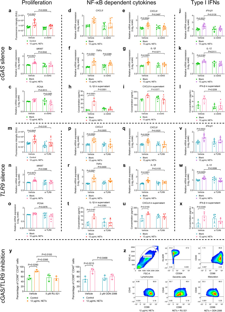
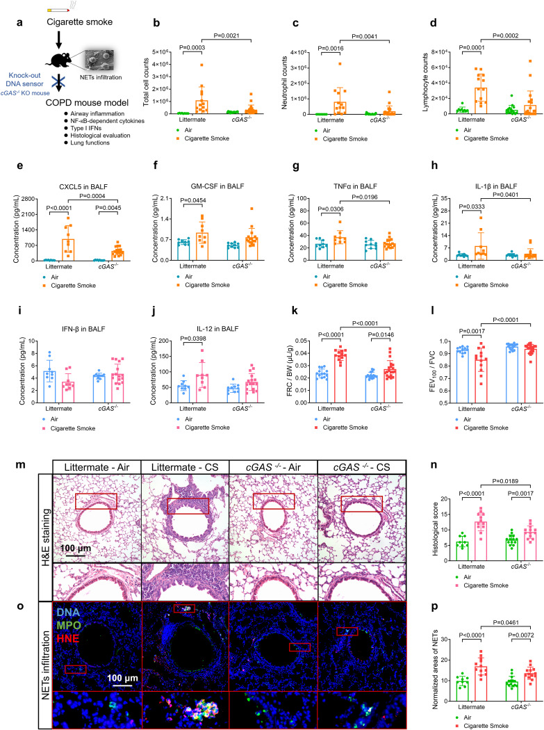
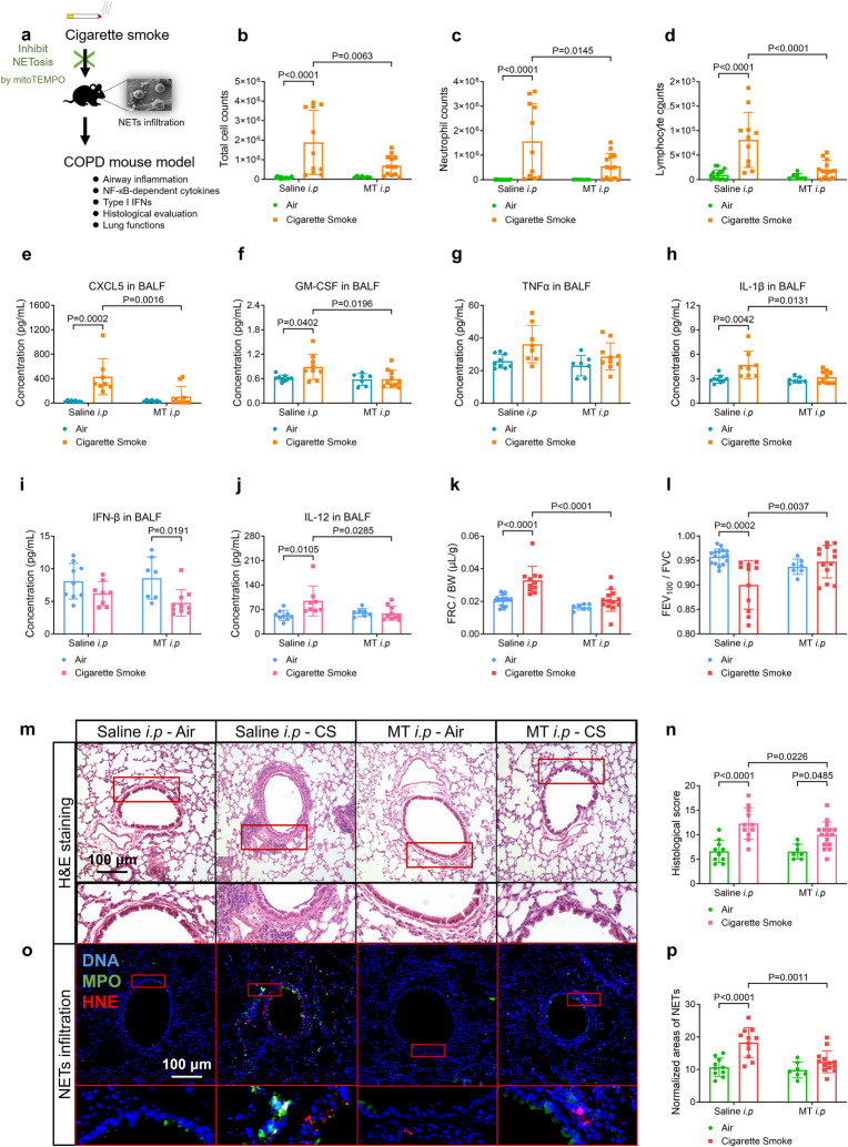
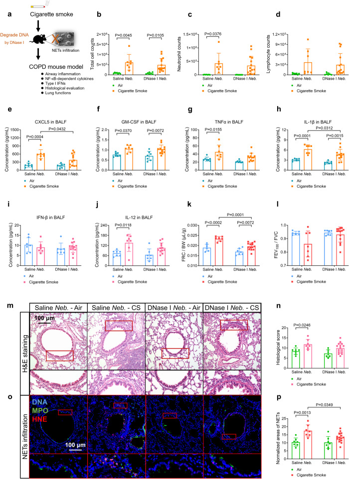
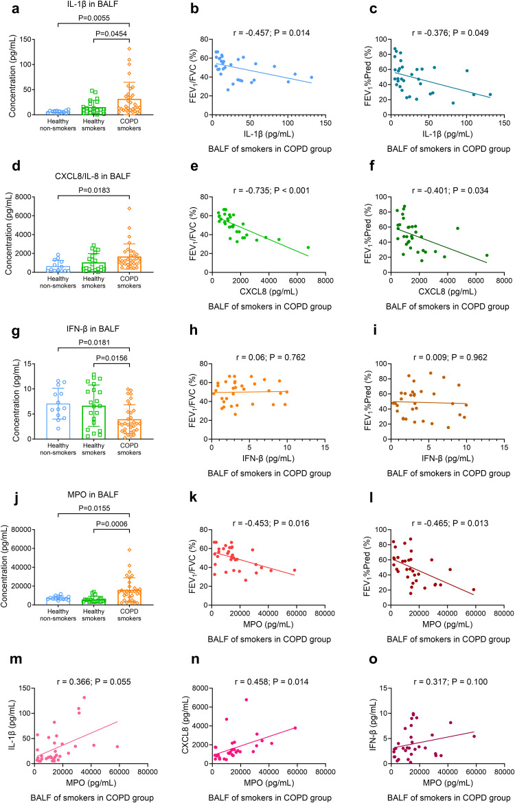
Chronic obstructive pulmonary disease (COPD) is characterised by persistent airway inflammation even after cigarette smoking cessation. Neutrophil extracellular traps (NETs) have been implicated in COPD severity and acute airway inflammation induced by short-term cigarette smoke (CS). However, whether and how NETs contribute to sustained airway inflammation in COPD remain unclear. This study aimed to elucidate the immunoregulatory mechanism of NETs in COPD, employing human neutrophils, airway epithelial cells (AECs), dendritic cells (DCs), and a long-term CS-induced COPD mouse model, alongside cyclic guanosine monophosphate-adenosine monophosphate synthase and toll-like receptor 9 knockout mice (cGAS--/-, TLR9-/-); Additionally, bronchoalveolar lavage fluid (BALF) of COPD patients was examined. Neutrophils from COPD patients released greater cigarette smoke extract (CSE)-induced NETs (CSE-NETs) due to mitochondrial respiratory chain dysfunction. These CSE-NETs, containing oxidatively-damaged DNA (NETs-DNA), promoted AECs proliferation, nuclear factor kappa B (NF-κB) activation, NF-κB-dependent cytokines and type-I interferons production, and DC maturation, which were ameliorated/reversed by silencing/inhibition of cGAS/TLR9. In the COPD mouse model, blocking NETs-DNA-sensing via cGAS-/- and TLR9-/- mice, inhibiting NETosis using mitoTEMPO, and degrading NETs-DNA with DNase-I, respectively, reduced NETs infiltrations, airway inflammation, NF-κB activation and NF-κB-dependent cytokines, but not type-I interferons due to IFN-α/β receptor degradation. Elevated NETs components (myeloperoxidase and neutrophil elastase activity) in BALF of COPD smokers correlated with disease severity and NF-κB-dependent cytokine levels, but not type-I interferon levels. In conclusion, NETs-DNA promotes NF-κB-dependent autoimmunity via cGAS/TLR9 in long-term CS exposure-induced COPD. Therefore, targeting NETs-DNA and cGAS/TLR9 emerges as a potential strategy to alleviate persistent airway inflammation in COPD.


Conflict of interest statement

The authors declare no competing interests.

Figures

Fig. 1
Fig. 1
Cigarette smoke extract (CSE) induces human peripheral blood neutrophils (hPBNs) to release neutrophil extracellular traps (NETs) in a dose- and time-dependent manner; CSE-induced NETs contain both mitochondrial DNA (mtDNA) and chromatin DNA (chDNA). Statistical analysis: n = 3–11 for each point or each bar in (bj) from 3–11 healthy participants or 3–10 patients with COPD, respectively, data were presented as the mean ± standard deviation; Differences are assessed by the (bf) two-way or (gj) one-way ANOVA analysis of variance, followed Tukey’s honest significant test; *P < 0.05, ***P < 0.001 and ****P < 0.0001 represent a significant difference from the group of blank or group of healthy participants, the scattered samples and the p values are displayed in (gj). a Representative immunofluorescence co-staining images display the NETs stimulated by 50 nM of phorbol-12-myristate-13-acetate (PMA) for 4 h and 5% CSE for 18 h; The NETs are costained with DNA (blue), MPO (myeloperoxidase, green) and histone H3 (red), and indicated by yellow arrows, scale bar: 30 μm. b, c The percentage of hPBNs, derived from b healthy participants and c patients with COPD, to release NETs upon stimulation with increasing dose of CSE and time of incubation, as charcterized by immunofluorescence staining of NETs components (supplementary Method 7); the stimulation of 50 nM of PMA as a positive control. df hPBNs derived from the patients with COPD release a higher percentage of NETs than those of healthy participants under the stimulation of d) 5% CSE, e 25% CSE, and f 50% CSE. g, h The ratio of the reads per kilobase per million mapped reads (RPKM) of mtDNA to the RPKM of chDNA in NETs induced by ionomycin (4 μM, incubated for 4 h), PMA (50 nM, for 4 h), 5% CSE (for 18 h) and 50% CSE (for 4 h) derived from g healthy participants and h patients with COPD, as assessed by next-generation sequencing (supplementary Method 10). i, j The ratio of the quantification cycle (Cq) value of 16 s to Cq of 18 s in spontaneous NETs (Blank) and those induced by ionomycin, PMA, 5% CSE and 50% CSE, derived from i healthy participants and j patients with COPD, as assessed by quantitative real-time polymerase chain reaction (qPCR) (supplementary Method 13)
Fig. 2
Fig. 2
Cigarette smoke extract (CSE)-induced NETosis requires mitochondrial reactive oxygen species (ROS) and mitochondrial respiratory chain, but not nicotinamide adenine dinucleotide phosphate (NADPH) oxidase. Statistical analysis: n = 3–10 for each bar in (af, h, i, k), from at least three healthy participants or three patients with COPD, data were presented as the mean ± standard deviation; Differences are assessed by the (af, k) one-way or (h, i) two-way ANOVA analysis of variance, followed Tukey’s honest significant test; P < 0.05 represents a significant difference, the scattered samples and the p values are displayed in figures. af The effects of several chemicals on the percentage of NETosis of human peripheral blood neutrophils (hPBNs, derived from healthy participants), stimulated by 5% CSE and 50 nM of PMA for 18 h: a 50 μM of mitoTEMPO (a mitochondrially targeted antioxidant), b 50 μM of thenoyltrifluoroacetone (TTFA, a mitochondrial respiration inhibitor), c 50 μM of diphenyleneiodonium chloride (DPI, a NADPH oxidase inhibitor), d 50 μM of VAS2870 (VAS, a NADPH oxidase inhibitor), e 200 IU/mL of deoxyribonuclease-I (DNase-I, an endonuclease for single- and double-stranded DNA) and f 50 μM of GW311616A (a selective human neutrophil elastase inhibitor), as characterized by immunofluorescence staining of NETs components (supplementary Method 7). gi The incubation of 5% CSE for 2 or 4 h induces the release of mitochondrial ROS (stained by mitoSOX Red, supplementary Method 8, 17) by hPBNs from both healthy participants and patients with COPD, as assessed by i flow cytometry and fluorescence staining (scale bar: 50 μm), and summarised in g the percentage of positive events and h the mean intensity of mitoSOX Red. j The immunofluorescence colocalization of 8-hydroxy-2’-deoxyguanosine (8-OHdG, a marker of oxidative stress to DNA, green) and translocase of the outer mitochondrial membrane complex subunit 20 (TOMM20, a mitochondrial outer membrane marker, red), with and without the treatment of 5% CSE (supplementary Method 8). Note: oxidative-damaged DNA are presented on the mitochondrial membrane following the treatment of 5% CSE for 2 h (scale bar: 10 μm). k The fluorescence intensity (mean) of 8-OHdZG on hPBNs treated with 5% CSE and 50% CSE from healthy participants and patients with COPD
Fig. 3
Fig. 3
Neutrophil extracellular traps (NETs) induced by cigarette smoke extract (CSE-NETs) dose-dependently promote the proliferation and the gene expressions of both nuclear factor kappa B (NF-κB)-dependent inflammatory cytokines and type-I interferons (IFNs) in the human airway epithelial cells (hAECs); CSE-NETs promote the maturation of human dendritic cells (DCs). Statistical analysis: n = 3–16 for each bar in (am) from at least three independent experiments, data were presented as the mean ± standard deviation; Differences are assessed by the (al) one-way or (m) two-way ANOVA analysis of variance, followed Tukey’s honest significant test; P < 0.05 represents a significant difference, the scattered samples and the p values are displayed in figures. ac Human peripheral neutrophils were stimulated by 5% CSE for 18 h to induce NETs, which were subsequently collected and quantified for DNA concentrations. Effects of 6, 12, and 24 μg/mL NETs (incubated for 48 h) on the proliferation of hAECs, as assessed by the a EdU proliferation assay (supplementary Method 11) and mRNA expressions of b MKI67 and c PCNA (both are markers of proliferation, supplementary Method 13). dl Effects of 6, 12, and 24 μg/mL NETs on the mRNA expressions and soluble levels of NF-κB-dependent inflammatory cytokines and type-I IFNs of hAECs (supplementary Method 13, 26): d mRNA expression of CXCL5 (C-X-C motif chemokine ligand 5), e mRNA expression of CXCL8, f mRNA expression of TNFα (tumour necrosis factor-alpha), g mRNA expression of IL-1β (interleukin 1β), h soluble levels of IL-1β in cell-culture supernatants, i soluble levels of CXCL8 in cell-culture supernatants, j mRNA expression of IFN-β1, k mRNA expression of IL-12 and l soluble levels of IFN-β in cell-culture supernatants. m, n 12 μg/mL of NETs promote the maturation of dendritic cells (differentiated from peripheral blood monocytes) from both healthy participants and patients with COPD (supplementary Method 15); the maturation is evaluated by m the percentage of CD86+ CD40+ cells, as assessed by flow cytometry with a gating strategy illustrated in (n) (Supplementary Method 17)
Fig. 4
Fig. 4
Cyclic guanosine monophosphate-adenosine monophosphate synthase (cGAS), and toll-like receptor 9 (TLR9) are required for the neutrophil extracellular traps (NETs)-stimulated proliferation, expressions of both nuclear factor kappa B (NF-κB)-dependent inflammatory cytokines and type-I interferons (IFNs) in human airway epithelial cells (hAECs), and maturation of human dendritic cells (hDCs). Statistical analysis: n = 6–10 for each bar in (ax), n = 3 for each bar in y from at least three independent experiments, data were presented as the mean ± standard deviation; Differences are assessed by the ay two-way ANOVA analysis of variance, followed Tukey’s honest significant test; P < 0.05 represents a significant difference, the scattered samples and the p values are displayed in figures. Effects of ac cGAS and mo TLR9 silencing on 12 μg/mL of NETs-stimulated proliferation of hAECs, as assessed by the a, m EdU proliferation assay (supplementary Method 11), and the mRNA expression of b, n MKI67 and c, o PCNA (both are markers of proliferation, supplementary Method 13). Effects of dl cGAS and p, x TLR9 silencing on 12 μg/mL of NETs-induced mRNA expression and soluble levels of NF-κB-dependent inflammatory cytokines and type-I interferons (IFNs) in hAECs (supplementary Method 13, 26): d, p mRNA expression of CXCL5 (C-X-C motif chemokine ligand 5), e, q mRNA expression of CXCL8, f, r mRNA expression of TNFα (tumour necrosis factor-alpha), g, s mRNA expression of IL-1β (interleukin 1β), h, t soluble levels of IL-1β in cell-culture supernatants, i, u soluble levels of CXCL8 in cell-culture supernatants, j, v mRNA expression of IFN-β1, k, w mRNA expression of IL-12 and l, x soluble levels of IFN-β in cell-culture supernatants. y The inhibition of cGAS and TLR9 by 5 μM of RU.521 and 2 μM of ODN 2088, respectively, reduces 12 μg/mL of NETs-mediated maturation of hDCs (supplementary Method 16). z Representative flow cytometry images display the gating strategy and maturation of DCs treated with either RU.521 or ODN 2088, as evaluated by the percentage of CD86+ CD40+ cells (supplementary Method 17)
Fig. 5
Fig. 5
Guanosine monophosphate-adenosine monophosphate synthase (cGAS) knock-out (cGAS−/−) mice treated with cigarette smoke (CS) exposure display decreased productions of nuclear factor kappa B (NF-κB)-dependent inflammatory cytokines, but not type-I interferons (IFNs), alleviated airway inflammation, infiltration of neutrophil extracellular traps (NETs) and improved lung functions, as compared with CS-treated littermate. Statistical analysis: n = 9–20 for each bar in (bl, n, p), data were presented as the mean ± standard deviation; Differences are assessed by the bl, n, p two-way ANOVA analysis of variance, followed by Tukey’s honest significant test; P < 0.05 represents a significant difference, the scattered samples and the p values are displayed in figures. a A brief outline for the experiments of the COPD mouse model (supplementary Method 18, 19). bd CS-treated cGAS−/− mice display alleviated airway inflammation as reflected by: b total cell counts, c neutrophil counts and d lymphocyte counts in the bronchoalveolar lavage fluid (BALF, supplementary Method 22). eh CS-treated cGAS/− mice reveal overall reduced productions of NF-κB-dependent inflammatory cytokines in the BALF (supplementary Method 22, 26): e C-X-C motif chemokine ligand 5 (CXCL5), f granulocyte-macrophage colony-stimulating factor (GM-CSF), g tumour necrosis factor-alpha (TNFα) and h interleukin 1β (IL-1β). i, j No significant changes of type-I IFNs are shown in BALF of either CS-treated littermate or cGAS/− mice: i IFN-β, j IL-12. k, l CS-treated cGAS/− mice reveal alleviated emphysema and airflow limitation in lung function tests (supplementary Method 21) as evaluated by: k functional residual capacity/body weight (FRC/BW), l forced expiratory volume at 100 ms/forced vital capacity (FEV100/FVC). m Representative images of hematoxylin-eosin (H&E)-stained lung slices display the decreased severity of airway inflammation in the CS-treated cGAS−/− mice, compared with CS-treated littermate (scale bar: 100 μm, Supplementary Method 23), as summarised in n histological score. o Representative immunofluorescence images display the decreased infiltration of NETs (co-stained with DNA, myeloperoxidase, and histone H3) in the lung slices of CS-treated cGAS−/− mice, compared with CS-treated littermate (scale bar: 100 μm, supplementary Method 24), as summarised in (p) normalised area of NETs
Fig. 6
Fig. 6
Cigarette smoke (CS)-exposed wild-type mice treated with an intraperitoneal injection (i.p) of mitoTEMPO (MT) reveal decreased productions of nuclear factor kappa B (NF-κB)-dependent inflammatory cytokines, but not type-I interferons (IFNs), alleviated airway inflammation, infiltration of neutrophil extracellular traps (NETs), and improved lung functions, as compared with CS-treated saline i.p mice. Statistical analysis: n = 7–16 for each bar in (bl, n, p), data were presented as the mean ± standard deviation; Differences are assessed by the (bl, n, p) two-way ANOVA analysis of variance, followed Tukey’s honest significant test; P < 0.05 represents a significant difference, the scattered samples and the p values are displayed in figures. a A brief outline for the experiments of the COPD mouse model (Supplementary Method 18, 19). bd CS-treated MT i.p mice reveal alleviated airway inflammation as reflected by the b total cell counts, c neutrophil counts and d lymphocyte counts in the bronchoalveolar lavage fluid (BALF, Supplementary Method 22). eh CS-treated MT i.p mice reveal overall reduced production of NF-κB-dependent inflammatory cytokines in the BALF (supplementary Method 22, 26): e C-X-C motif chemokine ligand 5 (CXCL5), f granulocyte-macrophage colony-stimulating factor (GM-CSF), g tumour necrosis factor-alpha (TNFα) and h interleukin 1β (IL-1β). i, j CS-treated MT i.p mice reveal reduced levels of j IL-12, but not i IFN-β, in the BALF, compared with CS-treated saline i.p mice. k, l CS-treated MT i.p mice reveal alleviated emphysema and airflow limitation in lung function tests (supplementary Method 21) as evaluated by k functional residual capacity/body weight (FRC/BW) and l forced expiratory volume at 100 ms/forced vital capacity (FEV100/FVC). m Representative images of hematoxylin-eosin (H&E)-stained lung slices display the decreased severity of airway inflammation in CS-treated MT i.p mice, compared with CS-treated saline i.p mice (scale bar: 100 μm, supplementary Method 23), as summarised in n histological score. o Representative immunofluorescence images reveal the decreased infiltration of NETs (co-stained with DNA, myeloperoxidase and histone H3) in lung slices of CS-treated MT i.p mice, compared with CS-treated saline i.p mice (scale bar: 100 μm, supplementary Method 24), as summarised in (p) normalised area of NETs
Fig. 7
Fig. 7
Cigarette smoke (CS)-exposed wild-type mice treated with the nebulization (Neb.) of deoxyribonuclease-I (DNase-I) reveal the decreased infiltration of neutrophil extracellular traps (NETs) and improved lung function, as compared with CS-treated saline Neb. mice. Statistical analysis: n = 7–15 for each bar in (bl, n, p), data were presented as the mean ± standard deviation; Differences are assessed by the (bl, n, p) two-way ANOVA analysis of variance, followed Tukey’s honest significant test; P < 0.05 represents a significant difference, the scattered samples and the p values are displayed in figures. a A brief outline for the experiments of the COPD mouse model (Supplementary Method 18, 19). bd No significant changes of cell counts in bronchoalveolar lavage fluid (BALF) of CS-treated DNase-I Neb. mice are observed (Supplementary Method 22): b total cell counts, c neutrophil counts and d lymphocyte counts. eh CS-treated DNase-I Neb. mice reveal the decreased levels of e C-X-C motif chemokine ligand 5 (CXCL5) and h interleukin 1β (IL-1β), but not that of f granulocyte-macrophage colony-stimulating factor (GM-CSF) or g TNFα (tumour necrosis factor-alpha) in the BALF (supplementary Method 22, 26). i, j No significant changes of type-I interferons (IFNs) level in the BALF of either CS-treated saline Neb. or DNase-I Neb. mice are observed: i IFN-β, j IL-12. k, l CS-treated DNase-I Neb. mice reveal alleviated emphysema, but not airflow limitation, in lung function tests (Supplementary Method 21) as evaluated by: k functional residual capacity/body weight (FRC/BW), l forced expiratory volume at 100 ms/forced vital capacity (FEV100/FVC). m Representative images of hematoxylin-eosin (H&E)-stained lung slices reveal that significant changes in the severity of airway inflammation are absent in the CS-treated DNase-I Neb. mice (scale bar: 100 μm, Supplementary Method 23), as summarised in n histological score. o Representative immunofluorescence images display the decreased infiltration of NETs (co-stained with DNA, MPO and Histone H3) in lung slices of the CS-treated DNase-I Neb. mice, compared with CS-treated saline Neb. mice (scale bar: 100 μm, Supplementary Method 24), as summarised in (p) normalised area of NETs
Fig. 8
Fig. 8
The level of myeloperoxidase (MPO) is correlated with the level of interleukin 1β (IL-1β) and level C-X-C motif chemokine ligand 8 (CXCL8), but not correlated with the level of interferons-β (IFN-β) in bronchoalveolar lavage fluid (BALF) of smokers in the group of patients with COPD, after controlling for their age, sex, body mass index (BMI) and smoking history. Statistical analysis: n = 13 non-smokers and 21 smokers in group of healthy participants, n = 32 smokers in group of patients with COPD in (a, d, g, j), n = 32 in (b, c, e, f, h, i, ko), data were presented as the mean ± standard deviation; Differences in (a, d, g, j) are assessed by one-way analysis of variance, followed Tukey’s honest significant test; In (b, c, e, f, h, i, k, l, mo), Pearson’s partial correlation test are performed by controlling for age, sex, BMI and smoking history of smokers in the group of patients with COPD, followed by the multiple linear regression analysis; P < 0.05 represents a significant difference, the scattered samples and the p values are displayed in figures. a IL-1β level is significantly increased in the BALF of smokers in the group of patients with COPD, compared with that of non-smokers and smokers in the healthy group, and negatively correlated with b ratio of FEV1 to forced vital capacity (FEV1/FVC), and c ratio of forced expiratory volume at 1 s (FEV1) to predicted FEV1 (FEV1%Pred, supplementary Method 1, 2, 3, 26), respectively. d CXCL8 level is significantly increased in the BALF of smokers in the group of patients with COPD, compared with that of non-smokers in the healthy group, and negatively correlated with e FEV1/FVC and f FEV1%Pred, respectively. g IFN-β level is significantly decreased in the BALF of smokers in the group of patients with COPD, compared with that of non-smokers and smokers in the healthy group; There is no correlation between IFN-β level and h FEV1/FVC, or IFN-β level and i FEV1%Pred. j MPO level is significantly increased in the BALF of smokers in the group of patients with COPD, compared with that of non-smokers and smokers in the healthy group, and negatively correlated with k FEV1/FVC and l FEV1%Pred, respectively. In the BALF of COPD smokers, MPO level is correlated with the level of n CXCL8, but not the level of m IL-1β or o IFN-β

References

    1. Global Initiative for Chronic Obstructive Lung Disease. Global strategy for the diagnosis, management, and prevention of COPD. http://goldcopd.org/ (2024).
    1. Brightling C, Greening N. Airway inflammation in COPD: progress to precision medicine. Eur. Respir. J. 2019;54:1900651. doi: 10.1183/13993003.00651-2019. - DOI - PubMed
    1. Yang, I. A., Clarke, M. S., Sim, E. H. A. & Fong, K. M. Inhaled corticosteroids for stable chronic obstructive pulmonary disease. Cochrane Database Syst. Rev. 2012, CD002991 (2012). - PMC - PubMed
    1. Hogg J, Paré P, Hackett T. The contribution of small airway obstruction to the pathogenesis of chronic obstructive pulmonary disease. Physiol. Rev. 2017;97:529–552. doi: 10.1152/physrev.00025.2015. - DOI - PMC - PubMed
    1. Oelsner E, et al. Lung function decline in former smokers and low-intensity current smokers: a secondary data analysis of the NHLBI Pooled Cohorts Study. Lancet Respir. Med. 2020;8:34–44. doi: 10.1016/S2213-2600(19)30276-0. - DOI - PMC - PubMed

Publication types

MeSH terms