

CT and MRI radiomic features of lung cancer (NSCLC): comparison and software consistency
- PMID: 38880866
- PMCID: PMC11180643
- DOI: 10.1186/s41747-024-00468-8
CT and MRI radiomic features of lung cancer (NSCLC): comparison and software consistency
Abstract
Background: Radiomics is a quantitative approach that allows the extraction of mineable data from medical images. Despite the growing clinical interest, radiomics studies are affected by variability stemming from analysis choices. We aimed to investigate the agreement between two open-source radiomics software for both contrast-enhanced computed tomography (CT) and contrast-enhanced magnetic resonance imaging (MRI) of lung cancers and to preliminarily evaluate the existence of radiomic features stable for both techniques.
Methods: Contrast-enhanced CT and MRI images of 35 patients affected with non-small cell lung cancer (NSCLC) were manually segmented and preprocessed using three different methods. Sixty-six Image Biomarker Standardisation Initiative-compliant features common to the considered platforms, PyRadiomics and LIFEx, were extracted. The correlation among features with the same mathematical definition was analyzed by comparing PyRadiomics and LIFEx (at fixed imaging technique), and MRI with CT results (for the same software).
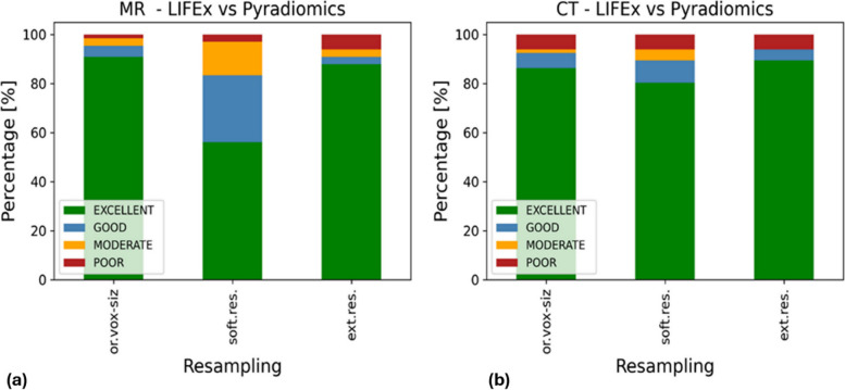
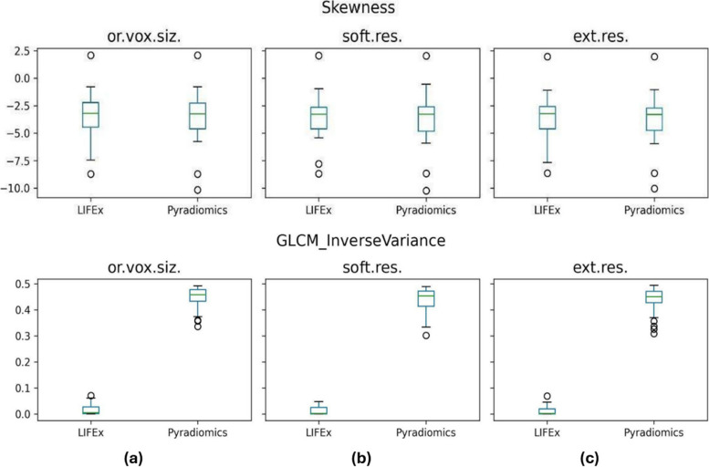
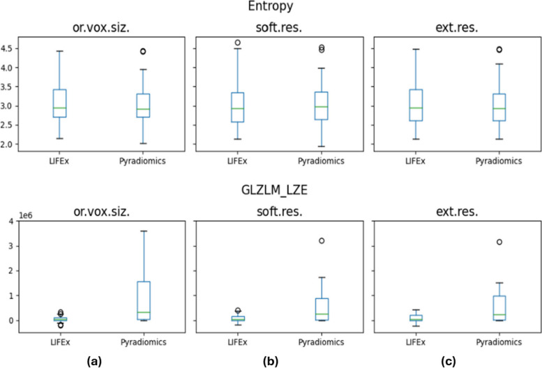
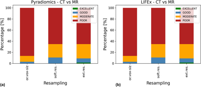
Results: When assessing the agreement between LIFEx and PyRadiomics across the considered resampling, the maximum statistically significant correlations were observed to be 94% for CT features and 95% for MRI ones. When examining the correlation between features extracted from contrast-enhanced CT and MRI using the same software, higher significant correspondences were identified in 11% of features for both software.
Conclusions: Considering NSCLC, (i) for both imaging techniques, LIFEx and PyRadiomics agreed on average for 90% of features, with MRI being more affected by resampling and (ii) CT and MRI contained mostly non-redundant information, but there are shape features and, more importantly, texture features that can be singled out by both techniques.
Relevance statement: Identifying and selecting features that are stable cross-modalities may be one of the strategies to pave the way for radiomics clinical translation.
Key points: • More than 90% of LIFEx and PyRadiomics features contain the same information. • Ten percent of features (shape, texture) are stable among contrast-enhanced CT and MRI. • Software compliance and cross-modalities stability features are impacted by the resampling method.
Keywords: Biomarkers; Lung neoplasms; Magnetic resonance imaging; Radiomics; Tomography (x-ray computed).
© 2024. The Author(s).
Conflict of interest statement
Andrea Riccardo Filippi discloses speakers’ bureau support from Astra Zeneca, MSD Italia (
Figures
References
Publication types
MeSH terms
Substances
Grants and funding
LinkOut - more resources
Full Text Sources
Medical