

Peripapillary RNFL cross-sectional area and its association with other parameters in a Chinese population
- PMID: 38880871
- PMCID: PMC11181622
- DOI: 10.1186/s12886-024-03481-y
Peripapillary RNFL cross-sectional area and its association with other parameters in a Chinese population
Abstract
Background: Quantitative analysis of retinal nerve fibers is important for the diagnosis and treatment of optic nerve diseases. Peripapillary retinal nerve fiber layer (RNFL) cross-sectional area may give a more accurate quantitative assessment of retinal nerve fibers than RNFL thickness but there have been no previous reports of the peripapillary RNFL cross-sectional area or other parameters. The purpose of the current study was to determine peripapillary RNFL cross-sectional area and its association with other factors in an adult Chinese population.
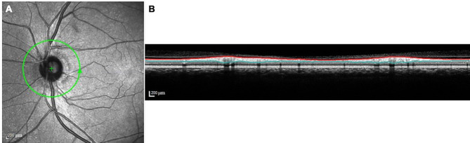
Methods: RNFL cross-sectional area was measured during peripapillary circular optical coherence tomography (OCT) scan with a diameter of 12° centered on the optic disc. Correlation between RNFL cross-sectional area and other parameters was evaluated by linear regression analysis in a cross-sectional study of an adult Chinese population.
Results: A total of 2404 eyes from 2404 subjects were examined. Multivariate linear regression analysis showed that larger RNFL cross-sectional area correlated with younger age (p < 0.001), female gender (p = 0.001), no history of diabetes (p = 0.012) and larger optic disc area (p < 0.001).
Conclusions: Peripapillary RNFL cross-sectional area is correlated positively with optic disc area, suggesting that eyes with larger optic discs have thicker RNFL. Further studies are needed to confirm whether this correlation is due to differences in the numbers of retinal nerve fibers or other factors.
Keywords: Ocular magnification; Optic disc area; Peripapillary RNFL cross-sectional area.
© 2024. The Author(s).
Conflict of interest statement
The authors declare that they have no competing interests.
Figures
Similar articles
-
Ganglion cell-inner plexiform layer and retinal nerve fiber layer thickness according to myopia and optic disc area: a quantitative and three-dimensional analysis.BMC Ophthalmol. 2017 Mar 11;17(1):22. doi: 10.1186/s12886-017-0419-1. BMC Ophthalmol. 2017. PMID: 28283025 Free PMC article.
-
Optical coherence tomography study of peripapillary retinal nerve fiber layer and choroidal thickness in eyes with tilted optic disc.J Glaucoma. 2015 Jan;24(1):45-50. doi: 10.1097/IJG.0b013e3182883c29. J Glaucoma. 2015. PMID: 23429636
-
Analysis of Optic Disc Morphology and the Peripapillary Retinal and Choroidal Thickness by the Swept Source Optical Coherence Tomography in Patients with Moyamoya Disease.Ophthalmic Res. 2025;68(1):61-70. doi: 10.1159/000542801. Epub 2024 Nov 25. Ophthalmic Res. 2025. PMID: 39586258 Free PMC article.
-
Optical Coherence Tomography Angiography and Structural Analyses of the Pale Optic Discs: Is It Possible to Differentiate the Cause?Curr Eye Res. 2021 Dec;46(12):1876-1885. doi: 10.1080/02713683.2021.1929331. Epub 2021 May 24. Curr Eye Res. 2021. PMID: 33980086
-
Determinants of normal retinal nerve fiber layer thickness measured by Stratus OCT.Ophthalmology. 2007 Jun;114(6):1046-52. doi: 10.1016/j.ophtha.2006.08.046. Epub 2007 Jan 8. Ophthalmology. 2007. PMID: 17210181 Free PMC article.
References
-
- Song Y, Li F, Chong RS, Wang W, Ran AR, Lin F, Wang P, Wang Z, Jiang J, Kong K, Jin L, Chen M, Sun J, Wang D, Tham CC, Lam DSC, Zangwill LM, Weinreb RN, Aung T, Jonas JB, Ohno-Matsui K, Cheng CY, Bressler NM, Sun X, Cheung CY, Chen S, Zhang X. Glaucoma Suspects with High Myopia Study Group. High Myopia Normative Database of Peripapillary Retinal Nerve Fiber Layer Thickness to Detect Myopic Glaucoma in a Chinese Population. Ophthalmology. 2023:S0161-6420(23)00518-3. - PubMed
MeSH terms
Grants and funding
LinkOut - more resources
Full Text Sources
