Case Reports
. 2024 Jun 16;24(1):522.
doi: 10.1186/s12877-024-05142-7.

Mikulicz's disease combined with IgG4-related hypophysitis: a case report

Affiliations
Case Reports

Mikulicz's disease combined with IgG4-related hypophysitis: a case report

Shu-Fan Zhang et al. BMC Geriatr. .

Abstract

Background: IgG4-related diseases are very uncommon, and its diagnosis and treatment are complicated as it encompasses multiple disciplines.

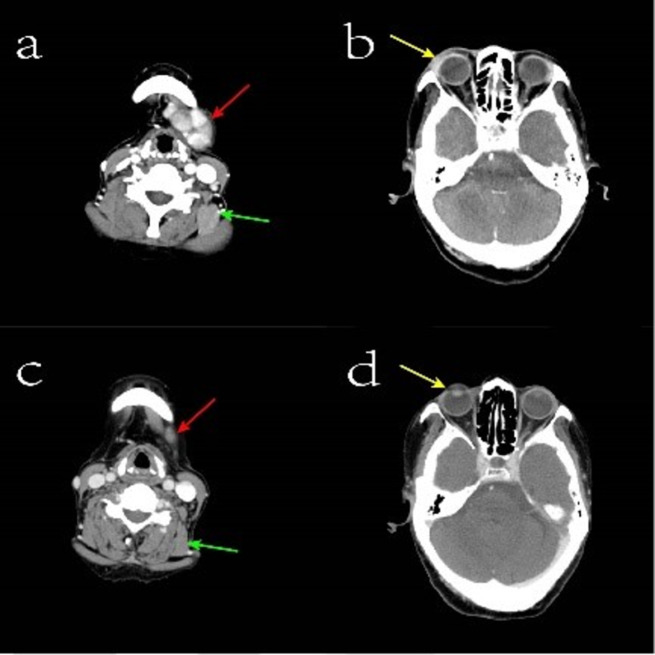
Case presentation: A 77-year-old woman was admitted with a jaw mass and nausea and vomiting. Laboratory tests showed elevated serum IgG4, pituitary MRI suggested thickening of the pituitary stalk, and head and neck CT suggested orbital and mandibular masses. Patients with mandibular mass were diagnosed with Mikulicz's disease with IgG4-related hypophysitis. We found no other evidence of causing thickening of the pituitary stalk. She was given oral prednisolone 30 mg daily, and her nausea and vomiting improved significantly, and the mandibular and ocular masses decreased in size.

Conclusion: Mikulicz's disease combined with IgG4-related hypophysitis is a rare case of IgG4-RD in elderly women. IgG4-RD is one of the causes of head and neck exocrine gland mass and pituitary stalk thickening in the elderly.

Keywords: Case report; IgG4-related disease; IgG4-related hypophysitis; Mikulicz’s disease; Pituitary stalk.


Conflict of interest statement

The Authors declare they have no competing interests.

Figures

Fig. 1
Fig. 1
Enhanced computerized tomography image of the head and neck. Figure a: The red arrow indicates the mandibular mass before hormonal treatment, and the green arrow indicates the involved supraspinatus muscle before treatment; Figure b: The yellow arrow refers to the orbital tissues enhanced and strengthened before treatment; Figure c: The red arrows show the reduced submandibular mass after hormonal treatment, and the green arrows refer to the normalized supraspinatus muscle after treatment. Figure d: The yellow arrows refer to the orbital tissues after treatment, with no significant enhancement shadow
Fig. 2
Fig. 2
Magnetic resonance image of the pituitary gland. The T1 sagittal fat suppression sequence shows a thickened pituitary stalk with the widest point being 5.6 mm
Fig. 3
Fig. 3
Biopsied tissue of the submandibular mass (HE staining, x200) proliferative fibrous tissue and lymphocytic infiltration are observed in the tissue
Fig. 4
Fig. 4
A sample viewed at 200x magnification using IgG4 immunohistochemistry. A large number of plasma cells and IgG4-positive plasma cells > 50 per high-power field

Similar articles

References

    1. Wallace ZS, Naden RP, Chari S, Choi H, Della-Torre E, Dicaire JF, et al. The 2019 American College of Rheumatology/European League against Rheumatism Classification Criteria for IgG4-Related disease. Arthritis Rheumatol. 2020;72(1):7–19. doi: 10.1002/art.41120. - DOI - PubMed
    1. Zen Y, Nakanuma Y. IgG4-related disease: a cross-sectional study of 114 cases. Am J Surg Pathol. 2010;34(12):1812–9. doi: 10.1097/PAS.0b013e3181f7266b. - DOI - PubMed
    1. Wang L, Zhang P, Zhang X, Lin W, Tang H, Li J, et al. Sex disparities in clinical characteristics and prognosis of immunoglobulin G4-related disease: a prospective study of 403 patients. Rheumatology (Oxford) 2019;58(5):820–30. doi: 10.1093/rheumatology/key397. - DOI - PubMed
    1. Wallace ZS, Zhang Y, Perugino CA, Naden R, Choi HK, Stone JH, et al. Clinical phenotypes of IgG4-related disease: an analysis of two international cross-sectional cohorts. Ann Rheum Dis. 2019;78(3):406–12. doi: 10.1136/annrheumdis-2018-214603. - DOI - PMC - PubMed
    1. Uccella S, Amaglio C, Brouland JP, Bianconi E, Ippolito S, Messerer M, et al. Disease heterogeneity in IgG4-related hypophysitis: report of two histopathologically proven cases and review of the literature. Virchows Arch. 2019;475(3):373–81. doi: 10.1007/s00428-019-02564-2. - DOI - PubMed

Publication types

MeSH terms

LinkOut - more resources