Review
. 2024 Aug 6;150(6):e109-e128.
doi: 10.1161/CIR.0000000000001254. Epub 2024 Jun 17.

Unraveling the Mechanisms of Valvular Heart Disease to Identify Medical Therapy Targets: A Scientific Statement From the American Heart Association

Review

Unraveling the Mechanisms of Valvular Heart Disease to Identify Medical Therapy Targets: A Scientific Statement From the American Heart Association

Aeron M Small et al. Circulation. .

Abstract

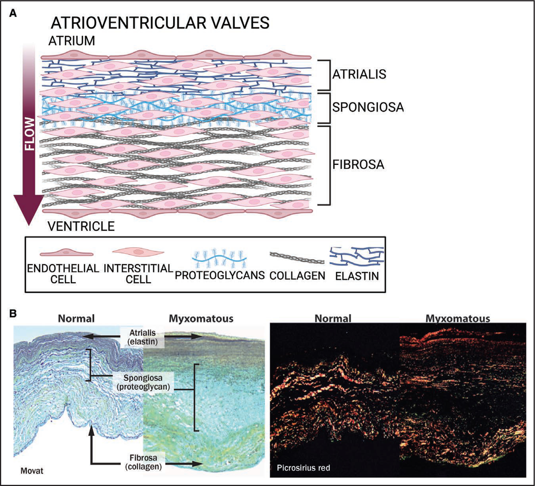
Valvular heart disease is a common cause of morbidity and mortality worldwide and has no effective medical therapy. Severe disease is managed with valve replacement procedures, which entail high health care-related costs and postprocedural morbidity and mortality. Robust ongoing research programs have elucidated many important molecular pathways contributing to primary valvular heart disease. However, there remain several key challenges inherent in translating research on valvular heart disease to viable molecular targets that can progress through the clinical trials pathway and effectively prevent or modify the course of these common conditions. In this scientific statement, we review the basic cellular structures of the human heart valves and discuss how these structures change in primary valvular heart disease. We focus on the most common primary valvular heart diseases, including calcific aortic stenosis, bicuspid aortic valves, mitral valve prolapse, and rheumatic heart disease, and outline the fundamental molecular discoveries contributing to each. We further outline potential therapeutic molecular targets for primary valvular heart disease and discuss key knowledge gaps that might serve as future research priorities.

Keywords: AHA Scientific Statements; aortic valve stenosis; bicuspid aortic valve disease; cellular structures; heart valve diseases; mitral valve prolapse; rheumatic heart disease.


Conflict of interest statement

The American Heart Association makes every effort to avoid any actual or potential conflicts of interest that may arise as a result of an outside relationship or a personal, professional, or business interest of a member of the writing panel. Specifically, all members of the writing group are required to complete and submit a Disclosure Questionnaire showing all such relationships that might be perceived as real or potential conflicts of interest.

Figures

Figure 1.
Figure 1.. Anatomical location of heart valves in diastole.
Netter illustration used with permission of Elsevier Inc. All rights reserved. www.netterimages.com
Figure 2.
Figure 2.. Semilunar valve leaflet structure.
A, Heart valves are coated in a layer of endothelial cells that encapsulate the valve interstitial cells in 3 layers defined by the extracellular matrix content. The ventricularis (V) is the elastin-rich layer of these structures connecting to the major arteries; the middle layer is the proteoglycan-rich spongiosa (S); and the outer layer is the collagen-rich fibrosa (F). Created with BioRender.com. B, Movat pentachrome staining of a calcified human aortic valve. Calcification is shown in purple. Bottom, Images of valves from a control and a fibrotic porcine valve. B, Reprinted with permission from Chen and Simmons.
Figure 3.
Figure 3.. Atrioventricular valve leaflet structure.
A, The tricuspid and mitral valves bridge the chambers of the heart and are thus referred to as atrioventricular valves. Blood flows from the atrium to the ventricle. The atrialis abutting the atrium contains elastin fibers; the middle layers, the proteoglycan-rich spongiosa; and the outer layer, the collagen-rich fibrosa. Created with BioRender.com. B, Movat pentachrome stain (left; collagen in yellow, proteoglycans in blue-green, elastin in black) of a normal mitral valve and mitral valve with myxomatous degeneration. Picrosirius red staining (right) of normal and myxomatous mitral valves viewed under polarized light illustrates disruption birefringence of collagen fibers in myxomatous leaflets. Magnification ×100. B, Reprinted with permission from Rabkin et al.
Figure 4.
Figure 4.. Mechanisms driving calcification of the aortic valve.
Although clinical trials to date have not shown that statin therapy is beneficial for aortic stenosis/sclerosis, lipids contribute to the osteogenic transition of valve interstitial cells. Low-density lipoprotein (LDL) and lipoprotein(A) [Lp(a)] enter the fibrosa, triggering an inflammatory cascade that leads to increased oxidative damage and cellular death. Secreted cytokines act in a paracrine manner to trigger the activation of osteogenic transcriptional programs in valvular interstitial cells. Apoptotic bodies and necrotic cells may also serve as loci for the nucleation of minerals. An intact extracellular matrix (ECM) is critical for leaflet function, and the breakdown of ECM proteins not only prevents proper leaflet movement but also activates mechano-sensing pathways that stimulate osteogenic transcriptional programs. Damaged ECM also captures secreted vesicles that, when released from activated cells, contain the enzymes necessary to produce the calcium and phosphate building blocks of minerals, as well as microRNAs that may further alter cell homeostasis. Amyloid plaques can also accumulate along these broken ECM components. αSMA indicates α-smooth muscle actin; IFN, interferon; IL, interleukin; LPA, lysophosphatidic acid; MMP, matrix metalloproteinase; NF-κB, nuclear factor-κB; oxLDL, oxidized low-density lipoprotein; RANKL, receptor activator of nuclear factor-κB ligand; TGFβ, transforming growth factor-β; TNF-α, tumor necrosis factor-α; and VIC, valve interstitial cell. Created with BioRender.com.
Figure 5.
Figure 5.. Pathogenesis of MVP.
A central role for transforming growth factor-β (TGF-β) signaling has emerged from genetic studies and is likely augmented by tissue damage from mechanical stress. TGF-β contributes to activation of valve interstitial cells (VICs). Other genes with possible roles in mechano-sensing/mechano-transduction include FLNA and TNS1. Serotonin signaling has been implicated in mitral valve prolapse (MVP) pathogenesis with an impact on TGF-β signaling and extracellular matrix (ECM) composition. DZIP1 and COL3A1 contribute to alteration of ECM integrity. Not pictured are genes involved in valvular development, including DCHS1. Created with BioRender.com.

References

    1. Coffey S, Roberts-Thomson R, Brown A, Carapetis J, Chen M, Enriquez-Sarano M, Zuhlke L, Prendergast BD. Global epidemiology of valvular heart disease. Nat Rev Cardiol. 2021;18:853–864. doi: 10.1038/s41569-021-00570-z - DOI - PubMed
    1. Tsao CW, Aday AW, Almarzooq ZI, Alonso A, Beaton AZ, Bittencourt MS, Boehme AK, Buxton AE, Carson AP, Commodore-Mensah Y, et al.; on behalf of the American Heart Association Council on Epidemiology and Prevention Statistics Committee and Stroke Statistics Subcommittee. Heart disease and stroke statistics–2022 update: a report from the American Heart Association [published correction appears in Circulation. 2022;146:e141]. Circulation. 2022;145:e153–e639. doi: 10.1161/CIR.0000000000001052 - DOI - PubMed
    1. Roth GA, Mensah GA, Johnson CO, Addolorato G, Ammirati E, Baddour LM, Barengo NC, Beaton AZ, Benjamin EJ, Benziger CP, et al.; GBD-NHLBI-JACC Global Burden of Cardiovascular Diseases Writing Group. Global burden of cardiovascular diseases and risk factors, 1990–2019: update from the GBD 2019 Study. J Am Coll Cardiol. 2020;76:2982–3021. doi: 10.1016/j.jacc.2020.11.010 - DOI - PMC - PubMed
    1. Messika-Zeitoun D, Baumgartner H, Burwash IG, Vahanian A, Bax J, Pibarot P, Chan V, Leon M, Enriquez-Sarano M, Mesana T, et al. Unmet needs in valvular heart disease. Eur Heart J. 2023;44:1862–1873. doi: 10.1093/eurheartj/ehad121 - DOI - PubMed
    1. Fedak PW, McCarthy PM, Bonow RO. Evolving concepts and technologies in mitral valve repair. Circulation. 2008;117:963–974. doi: 10.1161/CIRCULATIONAHA.107.702035 - DOI - PubMed