. 2024 Jun 12:17:1387-1404.
doi: 10.2147/CCID.S460845. eCollection 2024.

MALAT1 Knockdown Inhibits the Proliferation, Migration, and Collagen Deposition of Human Hypertrophic Scar Fibroblasts via Targeting miR-29a-3p/Smurf2 Axis

Affiliations

MALAT1 Knockdown Inhibits the Proliferation, Migration, and Collagen Deposition of Human Hypertrophic Scar Fibroblasts via Targeting miR-29a-3p/Smurf2 Axis

Chunyan Guo et al. Clin Cosmet Investig Dermatol. .

Abstract

Purpose: Hypertrophic scarring (HS) is commonly described as an abnormal post-traumatic tissue repair characterized by excessive hypercellularity and extracellular matrix (ECM) deposition. Mounting evidence suggests that MALAT1 is maladjusted in many fibrotic diseases, but its contribution to HS progression remains poorly understood. Hence, we sought to elucidate the fundamental role of MALAT1 in HS.

Methods: The expression of MALAT1, miR-29a-3p, and Smurf2 in skin tissues and fibroblasts was assessed by RT-qPCR and Western blotting. Furthermore, lentiviruses, RNAi, or plasmids were utilized to transfect hypertrophic scar fibroblasts (HSFs) for gene overexpression or downregulation. The biological behaviors of HSFs were quantified by the CCK-8 assay, wound healing assay, transwell assay, and flow cytometry. Mechanistically, bioinformatics analysis, dual-luciferase reporter assays, and rescue experiments were performed to verify the relationship between miR-29a-3p and MALAT1 or Smurf2.

Results: Our data indicate that MALAT1, Smurf2 were overexpressed while miR-29a-3p was suppressed in HS tissues and fibroblasts. Downregulation of MALAT1 may lead to decreased proliferation, migration, and invasion of fibroblasts, accompanied by enhanced apoptosis, reduced TGF-β signal transduction, and ECM accumulation in HSFs, by enhancing miR-29a-3p and suppressing Smurf2 expression. Mechanistically, MALAT1 acted as a sponge for miR-29a-3p, while miR-29a-3p directly targeted Smurf2. More importantly, rescue experiments suggested that MALAT1 downregulation induced impact on the proliferation, migration, and invasion of HSFs could be partially overturned through miR-29a-3p knockdown or Smurf2 overexpression.

Conclusion: MALAT1 knockdown inhibits the proliferation, migration, invasion, and collagen deposition of HSFs via targeting the miR-29a-3p/Smurf2 axis, which may reveal a promising therapeutic exploitable vulnerability to HS.

Keywords: MALAT1; Smurf2; TGF-β signaling pathway; fibroblasts; hypertrophic scar; miR-29a-3p.


Conflict of interest statement

The authors declare that they have no known competing financial interests or personal relationships that could have appeared to influence the work reported in this paper.

Figures

None
Graphical abstract
Figure 1
Figure 1
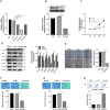
MALAT1 was up-regulated and TGF-β signaling pathway was over activated in HS and HSFs. (A and B) HE and Masson staining were used to analyze the histopathological features of HS and the matched NS tissues (×100, scale bar = 100 µm). (C) The process of primary skin fibroblasts culture was described (×100, scale bar = 100 µm). (D) Immunofluorescence staining was applied for fibroblasts identification (×200, scale bar = 50 µm). (EJ) RT-qPCR and Western Blot were employed to determine the abundance of MALAT1, the genes associated with TGF-β signaling pathway, such as TβRI, Smad7, Smad2, Smad3, and fibrosis indicators such as α-SMA and COL1A1 in HS and NS, as well as in HSFs and NSFs. ns P≥0.05,**P<0.01, *** P<0.001.
Figure 2
Figure 2
MALAT1 downregulation impeded the development of HS. HSFs were transfected with sh-NC, sh-MALAT1-1, sh-MALAT1-2 or sh-MALAT1-3. (A) The interference efficiency of MALAT1 downregulation lentiviruses was evaluated by RT-qPCR. (B) Cell proliferation ability was manifested by CCK-8 assay. (C and D) Wound healing assay and Transwell migration assay were utilized both to determine cell migration. (E) Transwell invasion assay was performed to measure cell invasion abilities (×50, scale bar = 100 µm). (F) FCM was applied to analyze cell apoptosis. (G) The protein expression of TβRI, Smad2, Smad3, Smad7, α-SMA, COL1A1 were estimate by Western blot assay. *P<0.05, **P<0.01, *** P<0.001.
Figure 3
Figure 3
MALAT1 directly targeted miR-29a-3p in HSFs. (A) MALAT1 had several possible interaction regions with miR-29a-3p. (B) The relative luciferase activity was evaluated by dual-luciferase reporter assay in HSFs. (C and D) RT-qPCR was utilized to analyze the expression of miR-29a-3p in HS and NS, as well as in HSFs and NSFs. (E) The expression of miR-29a-3p was calculated by RT-qPCR in HSFs transfected with MALAT1 knockdown lentiviruses. (F) Pearson correlation analysis was carried out to analyze the expression correlation between MALAT1 and miR-29a-3p in HS tissues. **P<0.01, *** P<0.001.
Figure 4
Figure 4
MALAT1 knockdown suppressed the progression of HS by impacting miR-29a-3p. (AE) HSFs were transfected with mimic-NC, miR-29a-3p mimic, Inhibitor-NC, or miR-29a-3p inhibitor. (FI) HSFs were transfected with sh-NC, sh-MALAT1-2, sh-MALAT1-2+inhibitor-NC, or sh-MALAT1-2 + miR-29a-3p inhibitor. (A) Transfection efficiency of miR-29a-3p was quantified by RT-qPCR. (Band F) CCK-8 assay was performed to evaluate cell proliferation ability. (Cand H) Transwell migration assay was utilized to determine cell migration abilities (×50, scale bar = 100 µm). (Dand G) Wound healing assay was performed to exam cell migration abilities (×100, scale bar = 100 µm). (Eand I) Transwell invasion assay was applied to measure cell invasion abilities (×50, scale bar = 100 µm). *P<0.05, **P<0.01, *** P<0.001.
Figure 5
Figure 5
MiR-29a-3p negatively regulated Smurf2 expression in HSFs. (A) The predicted potential interaction sites between miR-29a-3p and Smurf2, along with the mutated nucleotides of Smurf2 were displayed. (B) Dual-luciferase reporter assay was applied to analyze the luciferase activities in HSFs. (CF) The mRNA and protein expression of Smurf2 were assessed using RT-qPCR and Western blot assays in HS and NS, as well as in HSFs and NSFs. (G and H) RT-qPCR and Western blot assays were performed to quantify the expression of Smurf2 in HSFs after silencing or overexpressing of miR-29a-3p. (I) The relationship between miR-29a-3p and Smurf2 was conducted by Pearson correlation analysis in HS tissues. *P<0.05, **P<0.01, *** P<0.001.
Figure 6
Figure 6
Smurf2 downregulation suppressed the development of HS. Transfection of HSFs was performed using si-NC or si-Smurf2. (A and B) RT−qPCR and Western blot assay were performed both to measure the interference efficiency of si-Smurf2. (C) CCK-8 assay was utilized to analyze cell proliferation. (D) The expression of TGF-β signaling-related and ECM-related proteins were estimated by Western blot assay. (E and F) Scratch Assays and Transwell migration assay were used both to determine cell migration abilities (×50, scale bar = 100 µm). (G) Transwell invasion assay was utilized to assess migratory capabilities of cells (×50, scale bar = 100 µm). (H) Flow cytometry was applied to test cell apoptosis. **P<0.01, ***P<0.001.
Figure 7
Figure 7
MALAT1 knockdown hindered the progression of HS via downregulating Smurf2 expression. (A and B) RT-qPCR and Western blot assays were performed to detect the mRNA and protein expression of Smurf2 in HSFs after downregulation of MALAT1. (C–F) HSFs were transfected with sh-NC, sh-MALAT1-2, sh-MALAT1-2 + oe-NC, or sh-MALAT1-2 +oe-Smurf2. (C) Cell proliferation ability was evaluated by CCK-8 assay. (D and E) Cell migration capacity was determined by Scratch Assays (×100, scale bar = 100 µm) and Transwell migration assay (×50, scale bar = 100 µm). (F) Cell invasion ability was tested by Transwell invasion assay (×50, scale bar = 100 µm). *** P<0.001.

References

    1. Finnerty CC, Jeschke MG, Branski LK, Barret JP, Dziewulski P, Herndon DN. Hypertrophic scarring: the greatest unmet challenge after burn injury. Lancet. 2016;388(10052):1427–1436. doi:10.1016/S0140-6736(16)31406-4 - DOI - PMC - PubMed
    1. Zhang J, Li Y, Bai X, Li Y, Shi J, Hu D. Recent advances in hypertrophic scar. Histol Histopathol. 2018;33(1):27–39. doi:10.14670/HH-11-908 - DOI - PubMed
    1. Ogawa R. The most current algorithms for the treatment and prevention of hypertrophic scars and keloids: a 2020 update of the algorithms published 10 years ago. Plast Reconstr Surg. 2022;149(1):79–94. doi:10.1097/PRS.0000000000008667 - DOI - PMC - PubMed
    1. Frech FS, Hernandez L, Urbonas R, Zaken GA, Dreyfuss I, Nouri K. Hypertrophic scars and keloids: advances in treatment and review of established therapies. Am J Clin Dermatol. 2023;24(2):225–245. doi:10.1007/s40257-022-00744-6 - DOI - PubMed
    1. Shirakami E, Yamakawa S, Hayashida K. Strategies to prevent hypertrophic scar formation: a review of therapeutic interventions based on molecular evidence. Burns Trauma. 2020;8:tkz003. doi:10.1093/burnst/tkz003 - DOI - PMC - PubMed