

Polydopamine-Modified Liposomes: Preparation and Recent Applications in the Biomedical Field
- PMID: 38882106
- PMCID: PMC11170693
- DOI: 10.1021/acsomega.4c02555
Polydopamine-Modified Liposomes: Preparation and Recent Applications in the Biomedical Field
Abstract
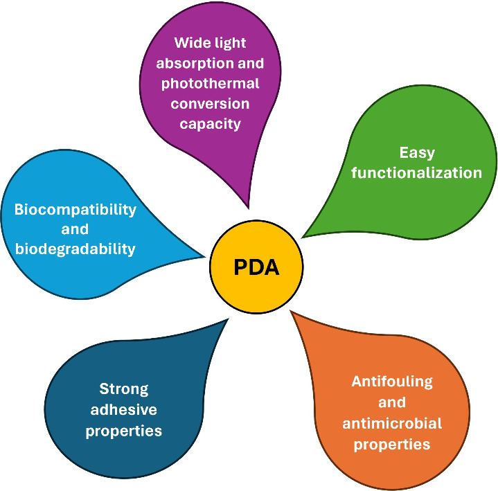
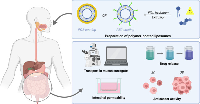
Polydopamine (PDA) is a bioinspired polymer that has unique and desirable properties for emerging applications in the biomedical field, such as extraordinary adhesiveness, extreme ease of functionalization, great biocompatibility, large drug loading capacity, good mucopenetrability, strong photothermal capacity, and pH-responsive behavior. Liposomes are consolidated and attractive biomimetic nanocarriers widely used in the field of drug delivery for their biocompatibility and biodegradability, as well as for their ability to encapsulate hydrophobic, hydrophilic, and amphiphilic compounds, even simultaneously. In addition, liposomes can be decorated with appropriate functionalities for targeted delivery purposes. Thus, combining the interesting properties of PDA with those of liposomes allows us to obtain multifunctional nanocarriers with enhanced stability, biocompatibility, and functionality. In this review, a focus on the most recent developments of liposomes modified with PDA, either in the form of polymer layers trapping multiple vesicles or in the form of PDA-coated nanovesicles, is proposed. These innovative PDA coatings extend the application range of liposomes into the field of biomedical applications, thereby allowing for easier functionalization with targeting ligands, which endows them with active release capabilities and photothermal activity and generally improves their interaction with biological fluids. Therefore, hybrid liposome/PDA systems are proposed for surface-mediated drug delivery and for the development of nanocarriers intended for systemic and oral drug delivery, as well as for multifunctional nanocarriers for cancer therapy. The main synthetic strategies for the preparation of PDA-modified liposomes are also illustrated. Finally, future prospects for PDA-coated liposomes are discussed, including the suggestion of potential new applications, deeper evaluation of side effects, and better personalization of medical treatments.
© 2024 The Authors. Published by American Chemical Society.
Conflict of interest statement
The authors declare no competing financial interest.
Figures


Similar articles
-
[Advances in polydopamine surface modification for capillary electrochromatography].Se Pu. 2020 Sep 8;38(9):1057-1068. doi: 10.3724/SP.J.1123.2020.03004. Se Pu. 2020. PMID: 34213272 Chinese.
-
Nanoparticles modified by polydopamine: Working as "drug" carriers.Bioact Mater. 2020 Apr 18;5(3):522-541. doi: 10.1016/j.bioactmat.2020.04.003. eCollection 2020 Sep. Bioact Mater. 2020. PMID: 32322763 Free PMC article. Review.
-
Combinatorial Polydopamine-Liposome Nanoformulation as an Effective Anti-Breast Cancer Therapy.Int J Nanomedicine. 2023 Feb 17;18:861-879. doi: 10.2147/IJN.S382109. eCollection 2023. Int J Nanomedicine. 2023. PMID: 36844433 Free PMC article.
-
A comparative study of polydopamine modified and conventional chemical synthesis method in doxorubicin liposomes form the aspect of tumor targeted therapy.Int J Pharm. 2019 Mar 25;559:76-85. doi: 10.1016/j.ijpharm.2019.01.033. Epub 2019 Jan 21. Int J Pharm. 2019. PMID: 30677481
-
Liposomes for Tumor Targeted Therapy: A Review.Int J Mol Sci. 2023 Jan 31;24(3):2643. doi: 10.3390/ijms24032643. Int J Mol Sci. 2023. PMID: 36768966 Free PMC article. Review.
Cited by
-
Bioinspired Nanoplatforms: Polydopamine and Exosomes for Targeted Antimicrobial Therapy.Polymers (Basel). 2025 Jun 16;17(12):1670. doi: 10.3390/polym17121670. Polymers (Basel). 2025. PMID: 40574198 Free PMC article. Review.
-
Innovative Approaches to Enhancing the Biomedical Properties of Liposomes.Pharmaceutics. 2024 Nov 27;16(12):1525. doi: 10.3390/pharmaceutics16121525. Pharmaceutics. 2024. PMID: 39771504 Free PMC article. Review.
-
Innovative Strategies in Oncology: Bacterial Membrane Vesicle-Based Drug Delivery Systems for Cancer Diagnosis and Therapy.Pharmaceutics. 2025 Jan 3;17(1):58. doi: 10.3390/pharmaceutics17010058. Pharmaceutics. 2025. PMID: 39861706 Free PMC article. Review.
References
-
- Liebscher J. Chemistry of Polydopamine – Scope, Variation, and Limitation. Eur. J. Org. Chem. 2019, 2019 (31–32), 4976–4994. 10.1002/ejoc.201900445. - DOI
-
- Gao S.; Zhang D.; Pedrero M.; Guo Z.; Pingarrón J. M.; Campuzano S.; Zou X. Advances and opportunities of polydopamine coating in biosensing: Preparation, functionality, and applications. Coord. Chem. Rev. 2024, 501, 21556410.1016/j.ccr.2023.215564. - DOI
Publication types
LinkOut - more resources
Full Text Sources