. 2024 Mar 26;6(6):101073.
doi: 10.1016/j.jhepr.2024.101073. eCollection 2024 Jun.

Glycogen synthase kinase 3 activity enhances liver inflammation in MASH

Affiliations

Glycogen synthase kinase 3 activity enhances liver inflammation in MASH

Mireille Khoury et al. JHEP Rep. .

Abstract

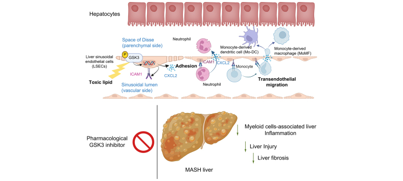
Background & aims: Metabolic dysfunction-associated steatohepatitis (MASH) is characterized by excessive circulating toxic lipids, hepatic steatosis, and liver inflammation. Monocyte adhesion to liver sinusoidal endothelial cells (LSECs) and transendothelial migration (TEM) are crucial in the inflammatory process. Under lipotoxic stress, LSECs develop a proinflammatory phenotype known as endotheliopathy. However, mediators of endotheliopathy remain unclear.

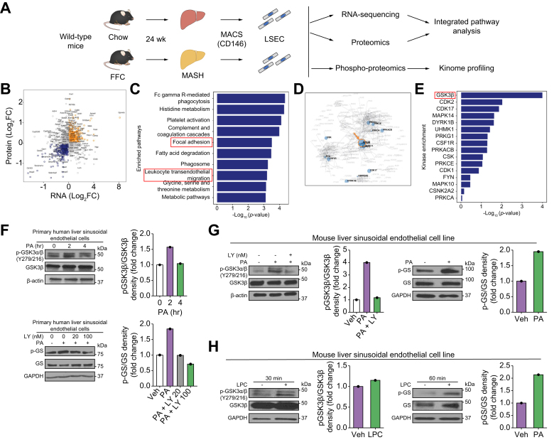
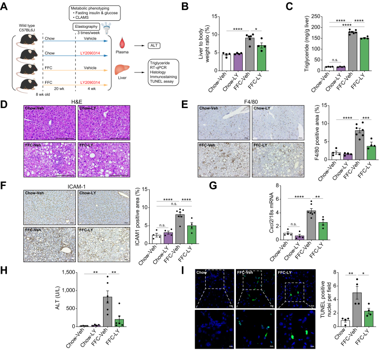
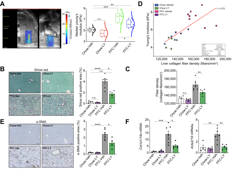
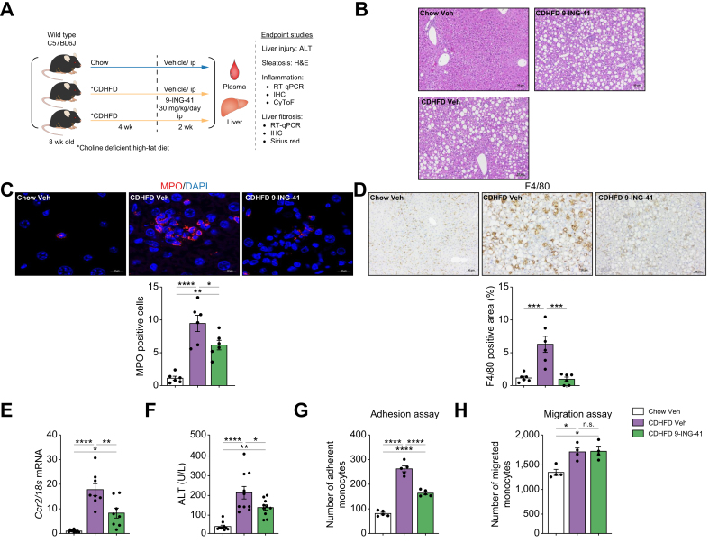
Methods: Primary mouse LSECs isolated from C57BL/6J mice fed chow or MASH-inducing diets rich in fat, fructose, and cholesterol (FFC) were subjected to multi-omics profiling. Mice with established MASH resulting from a choline-deficient high-fat diet (CDHFD) or FFC diet were also treated with two structurally distinct GSK3 inhibitors (LY2090314 and elraglusib [9-ING-41]).

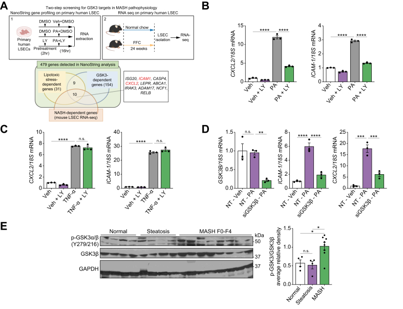
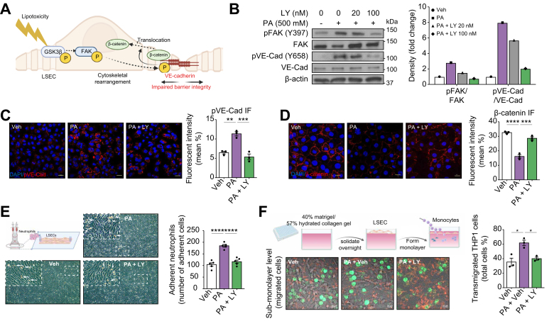
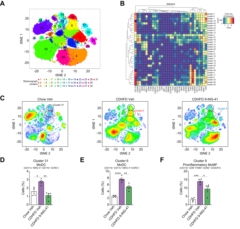
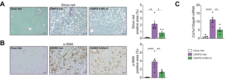
Results: Integrated pathway analysis of the mouse LSEC proteome and transcriptome indicated that leukocyte TEM and focal adhesion were the major pathways altered in MASH. Kinome profiling of the LSEC phosphoproteome identified glycogen synthase kinase (GSK)-3β as the major kinase hub in MASH. GSK3β-activating phosphorylation was increased in primary human LSECs treated with the toxic lipid palmitate and in human MASH. Palmitate upregulated the expression of C-X-C motif chemokine ligand 2, intracellular adhesion molecule 1, and phosphorylated focal adhesion kinase, via a GSK3-dependent mechanism. Congruently, the adhesive and transendothelial migratory capacities of primary human neutrophils and THP-1 monocytes through the LSEC monolayer under lipotoxic stress were reduced by GSK3 inhibition. Treatment with the GSK3 inhibitors LY2090314 and elraglusib ameliorated liver inflammation, injury, and fibrosis in FFC- and CDHFD-fed mice, respectively. Immunophenotyping using cytometry by mass cytometry by time of flight of intrahepatic leukocytes from CDHFD-fed mice treated with elraglusib showed reduced infiltration of proinflammatory monocyte-derived macrophages and monocyte-derived dendritic cells.

Conclusion: GSK3 inhibition attenuates lipotoxicity-induced LSEC endotheliopathy and could serve as a potential therapeutic strategy for treating human MASH.

Impact and implications: LSECs under lipotoxic stress in MASH develop a proinflammatory phenotype known as endotheliopathy, with obscure mediators and functional outcomes. The current study identified GSK3 as the major driver of LSEC endotheliopathy, examined its pathogenic role in myeloid cell-associated liver inflammation, and defined the therapeutic efficacy of pharmacological GSK3 inhibitors in murine MASH. This study provides preclinical data for the future investigation of GSK3 pharmacological inhibitors in human MASH. The results of this study are important to hepatologists, vascular biologists, and investigators studying the mechanisms of inflammatory liver disease and MASH, as well as those interested in drug development.

Keywords: Adhesion; Chemokines; Glycogen synthase kinase 3 (GSK3); Inflammation; Liver fibrosis; Liver sinusoidal endothelial cells (LSEC); Metabolic dysfunction associated steatohepatitis (MASH); Migration; Myeloid cells; Non-alcoholic steatohepatitis (NASH).


Conflict of interest statement

The authors declare no conflicts of interest. Please refer to the accompanying ICMJE disclosure forms for further details.

Figures

Image 1
Graphical abstract
Fig. 1
Fig. 1
Glycogen synthase kinase (GSK)-3β is a central mediator of liver sinusoidal endothelial cell (LSEC) endotheliopathy in metabolic dysfunction-associated steatohepatitis (MASH). (A) Schematic of the in vivo multi-omics study. (B) Scatter plot showing differentially expressed genes (p <0.05) in chow vs. fat, fructose, and cholesterol (FFC)-fed mice based on either LSEC total proteomic or RNA-sequencing (RNA-seq) analysis. The x- and y-axes represent the log 2-converted fold change (log2FC) of the signals in FFC- vs. chow-fed mice in the proteomic and RNA-seq studies, respectively. (C) Top-10 enriched pathways representative of the differentially regulated genes in FFC vs. chow-fed mice based on both proteomics and RNA-seq analyses. (D) Kinome map generated from the phosphoproteomic study shown in A. (E) Top-16 ranked putative altered kinases in FFC vs. chow-fed mice. (F) Primary human LSECs were treated with the vehicle (Veh), 500 μM palmitate (PA), or 500 μM PA ± the GSK3 inhibitor LY2090314 (LY). Protein levels of phosphorylated (p)-GSK3α/β (Y279/216) and GSK3β (top) and phosphorylated glycogen synthase (p-GS) and glycogen synthase (GS) (bottom) were assessed using Western blotting. (G) A mouse LSEC line was treated with Veh or 500 μM PA ± 20 nM LY for 4 h, and the protein levels of p-GSK3α/β (Y279/216) and GSK3β were assessed (left). Mouse LSEC lines were also treated with Veh or 500 μM PA for 2 h and p-GS and GS were assessed. (H) A mouse LSCE line was treated with Veh or lysophosphatidylcholine (LPC), and the protein levels of p-GSK3α/β (Y279/216), GSK3β, p-GS, and GS were assessed. β-Actin or glyceraldehyde-3-phosphate dehydrogenase (GAPDH) was used as loading control. F–H were repeated for at least three times; representative results are shown with quantification.
Fig. 2
Fig. 2
Glycogen synthase kinase (GSK)-3 mediates toxic lipid-induced proinflammatory responses in liver sinusoidal endothelial cells (LSECs). (A) Primary human LSECs were treated with vehicle (Veh) or 500 μM palmitate (PA) ± 20 nM LY2090314 (LY) for 16 h, and gene expression profiling was performed using the NanoString nCounter system (1). LSECs were isolated from chow- and fat, fructose, and cholesterol (FFC)-fed mice and subjected to RNA-sequencing (RNA-seq) (2). Venn diagram (bottom) of genes classified as ‘lipotoxic stress-dependent genes’ and ‘GSK3-dependent genes’ in NanoString analysis and ‘Metabolic dysfunction-associated steatohepatitis (MASH)-dependent genes’ in mouse LSEC RNA-seq. Primary human LSECs were treated with Veh or 500 μM of PA (B) or 10 ng/ml tumor necrosis factor (TNF)-α (C) ± 20 nM LY for 16 h, and mRNA expression of C-X-C motif chemokine ligand 2 (CXCL2) and intracellular adhesion molecule 1 (ICAM1) was examined. (D) Primary human LSECs were transfected with siGSK3β or non-target small interfering (si)RNA for 72 h, then treated with Veh or 500 μM PA for 24 h, and the mRNA expression of GSK3B, CXCL2, and ICAM1 was examined (n = 3 per group). (E) Phospho-GSK3α/β (Y279/216) and GSK3β protein levels were assessed in normal controls and patients with steatosis, and MASH by Western blotting with quantification. Glyceraldehyde-3-phosphate dehydrogenase (GAPDH) was used as the loading control (n = 4–8 per group). Bar graphs represent mean ± SEM; ∗p <0.05, ∗∗p <0.01, ∗∗∗p <0.001, ∗∗∗∗p <0.0001, ns, non-significant (one-way ANOVA with Bonferroni’s multiple comparison).
Fig. 3
Fig. 3
Glycogen synthase kinase (GSK)-3 mediates liver sinusoidal endothelial cell (LSEC) lipotoxicity-induced monocyte transendothelial migration (TEM). (A) Schematic showing that lipotoxicity-induced GSK3β activation triggers a multistep phosphorelay signal transduction mechanism (GSK3β–focal adhesion kinase [FAK]–vascular endothelial [VE]-cadherin), leading to LSEC cytoskeletal rearrangement, adherence junction disassembly, and β-catenin dissociation from VE-cadherin and internalization, resulting in endothelial barrier dysfunction. (B) Primary human LSECs were treated with vehicle (Veh) or 500 μM palmitate (PA) ± LY2090314 (LY) at the indicated concentrations for 4 h, and protein levels of phosphorylated FAK (p-FAK), total FAK, phosphorylated (p)-VE-cadherin, and total VE-cadherin were assessed by Western blotting. β-Actin was used as a loading control. B was repeated at least three times, and representative results are shown, along with quantification. (C) Localization of p-VE-cadherin was assessed using immunofluorescence and quantified (right). (D) β-Catenin localization was assessed by immunofluorescence and quantified (right). DAPI was used for the nuclear staining. (E) Schematic of the flow-based neutrophil adhesion assay. Representative microscopic images of neutrophils adherent to primary human LSECs under the three experimental conditions. White arrows indicate adherent neutrophils. Five random fields in a 20 × microscopic field for each condition were captured and quantified (right). (F) Schematic of the collagen gel-based TEM assay. Representative images from confocal microscopy for the TEM assay at 1 h (left). THP-1 cells (green fluorescence) in focus on the apical surface of the endothelial monolayer (red fluorescence) were defined as non-migrated, those in focus below the plane of the endothelial monolayer were defined as transmigrated, and three random fields in a 63 × microscopic field from each condition were captured and quantified. Bar graphs represent mean ± SEM; ∗p <0.05, ∗∗p <0.01, ∗∗∗p <0.001, ∗∗∗∗p <0.0001 (one-way ANOVA with Bonferroni’s multiple comparison; n = 3–5 per group). Scale bars: 20 μm (C,D,F).
Fig. 4
Fig. 4
Glycogen synthase kinase (GSK)-3 inhibition ameliorates liver injury and inflammation in fat, fructose, and cholesterol (FFC) diet-induced murine metabolic dysfunction-associated steatohepatitis (MASH). Wild-type (WT) C57BL/6J mice were fed either a chow or FFC diet for 24 weeks and treated with either vehicle (Veh) or the GSK3 inhibitor LY2090314 (LY) intraperitoneally 10 mg/kg three times per week for 4 weeks. (A) Schematic of the feeding experiment. (B) Liver-to-body weight ratio. (C) Liver triglyceride content. (D) Representative images of H&E staining of the liver sections. (E) Representative images of F4/80 immunostaining of the liver sections (left). F4/80-positive areas were quantified in five random 10 × microscopic fields and averaged for each animal (right). (F) Representative images of intracellular adhesion molecule 1 (ICAM-1) immunostaining in liver sections (left). ICAM-1-positive area is quantified (right). (G) Hepatic mRNA expression of C-X-C motif chemokine ligand 2 (Cxcl2). (H) Plasma alanine aminotransferase (ALT) level. (I) Representative images of terminal deoxynucleotidyl transferase dUTP nick-end labeling (TUNEL) staining of the liver sections (left). Quantification of TUNEL-positive cells (right). The marked boxes in the top images indicate the magnified images at the bottom. Bar graphs represent the mean ± SEM; ∗p <0.05, ∗∗p <0.01, ∗∗∗p <0.001, ∗∗∗∗p <0.0001, ns, non-significant (one-way ANOVA with Bonferroni’s multiple comparison; n = 4–7). Scale bars: 10 μm (I, insets), 20 μm (I), 50 μm (E,F), 100 μm (D).
Fig. 5
Fig. 5
Glycogen synthase kinase (GSK)-3 inhibition by LY2090314 (LY)-ameliorated liver fibrosis in diet-induced murine metabolic dysfunction-associated steatohepatitis (MASH). (A) Ultrasound images of the liver using shear wave elastography segmentation overlay. Median Young’s modulus (kPa) of the vehicle (Veh) and LY-treated mice fed chow or a fat, fructose, and cholesterol (FFC) diet. (B) Representative images of Sirius Red staining of liver sections (left). Sirius Red-positive areas were quantified in five random 10 × microscopic fields and averaged for each animal (right). (C) Ex vivo validation of collagen fiber density after the final time point using CT-FIRE (left). (D) Correlation between in vivo ultrasound and ex vivo CT-FIRE measurements of liver fibrosis pooled across all groups. Pearson’s r correlation = 0.7 (right). (E) Representative images of alpha-smooth muscle actin (α-SMA) immunostaining (left) and quantification (right) of liver sections. (F) α-SMA and Collagen1α1 mRNA expression levels. Bar graphs represent mean ± SEM; ∗p <0.05, ∗∗p <0.01, ∗∗∗p <0.001, ∗∗∗∗p <0.0001, ns, non-significant (one-way ANOVA with Bonferroni’s multiple comparison; n = 4–7). Scale bars: 50 μm (B,E).
Fig. 6
Fig. 6
Glycogen synthase kinase (GSK)-3 inhibition with elraglusib improves liver injury and inflammation in choline-deficient high-fat diet (CDHFD)-induced murine metabolic dysfunction-associated steatohepatitis (MASH). Eight-week-old wild-type (WT) C57BL/6J mice were fed either a chow diet or CDHFD for 6 weeks and treated with either vehicle (Veh) or the GSK3 inhibitor 9-ING-41 (intraperitoneally 30 mg/kg daily for 2 weeks). (A) Schematic of the feeding experiment. (B) Representative images of H&E staining of the liver sections. (C) Myeloperoxidase (MPO) immunofluorescence staining (red) Nuclei were stained with DAPI (blue). Images are shown at a 63 × magnification. (D) F4/80 immunostaining of liver sections (left). F4/80-positive areas were quantified in five random 10 × microscopic fields and averaged for each animal (right). (E) Hepatic mRNA expression levels of Ccr2. (F) Plasma alanine aminotransferase (ALT) levels (n = 5–10). Quantification of (G) monocytes adherent to liver sinusoidal endothelial cells (LSECs) (n = 5), (H) and migrating monocytes (n = 3) in the different experimental groups. Bar graphs represent mean ± SEM; ∗p <0.05, ∗∗p <0.01, ∗∗∗p <0.001, ∗∗∗∗p <0.0001, ns, non-significant (one-way ANOVA with Bonferroni’s multiple comparison). Scale bars: 10 μm (C), 50 μm (B,D).
Fig. 7
Fig. 7
Glycogen synthase kinase (GSK)-3 inhibition with elraglusib attenuates the recruitment of proinflammatory myeloid cells in the liver of choline-deficient high-fat diet (CDHFD)-fed mice. Cytometry by time of flight (CyTOF) was performed on intrahepatic leukocytes (IHLs) of chow-fed mice and CDHFD-fed mice treated with the GSK3 inhibitor elraglusib (9-ING-41). (A) Thirty-one unique clusters were defined by a panel of 32 cell-surface markers, including CD45 and two intracellular markers (myeloperoxidase [MPO] and S100A8) using the Rphenograph clustering algorithm and visualized on a tSNE plot. (B) Heatmap demonstrating the distribution and relative intensity of the markers used in clustering analysis. (C) Representative tSNE plots of each experimental group. Red indicates high-frequency categorization of cells into a cluster, and blue indicates low frequency. Clusters 31 (D) and 6 (E) are consistent with monocyte-derived dendritic cells (MoDCs), whereas cluster 9 (F) is consistent with proinflammatory monocyte-derived macrophages (MoMFs). Bar graphs represent the mean ± SEM; ∗p <0.05, ∗∗p <0.01, ∗∗∗p <0.001, ∗∗∗∗p <0.0001 (one-way ANOVA with Bonferroni’s multiple comparison; n = 4). tSNE, t-distributed stochastic neighbor embedding.
Fig. 8
Fig. 8
Elraglusib (9-ING-41) treatment in choline-deficient high-fat diet (CDHFD)-fed mice attenuates liver fibrosis. (A) Representative images of Sirius Red staining of liver sections (left). Sirius Red-positive areas were quantified (right). (B) Representative images of α-SMA immunostaining of the liver sections (left). α-SMA-positive areas were quantified (right). (C) Hepatic mRNA expression levels of collagen 1α1 were assessed. Bar graphs represent the mean ± SEM; ∗p <0.05, ∗∗p <0.01, ∗∗∗∗p <0.0001 (one-way ANOVA with Bonferroni’s multiple comparison) (n = 5 or 6). Scale bars: 50 μm (A,B).

Similar articles

Cited by

References

    1. Younossi Z.M. Non-alcoholic fatty liver disease – a global public health perspective. J Hepatol. 2019;70:531–544. - PubMed
    1. Neuschwander-Tetri B.A. Hepatic lipotoxicity and the pathogenesis of nonalcoholic steatohepatitis: the central role of nontriglyceride fatty acid metabolites. Hepatology. 2010;52:774–788. - PubMed
    1. Friedman S.L., Neuschwander-Tetri B.A., Rinella M., et al. Mechanisms of NAFLD development and therapeutic strategies. Nat Med. 2018;24:908–922. - PMC - PubMed
    1. McConnell M.J., Kostallari E., Ibrahim S.H., et al. The evolving role of liver sinusoidal endothelial cells in liver health and disease. Hepatology. 2023;78:649–669. - PMC - PubMed
    1. Ibrahim S.H. Sinusoidal endotheliopathy in nonalcoholic steatohepatitis: therapeutic implications. Am J Physiol Gastrointest Liver Physiol. 2021;321:G67–G74. - PMC - PubMed

LinkOut - more resources