. 2023 Oct 27;32(2):121-133.
doi: 10.4103/jmu.jmu_68_23. eCollection 2024 Apr-Jun.

High-quality Agar and Polyacrylamide Tumor-mimicking Phantom Models for Magnetic Resonance-guided Focused Ultrasound Applications

Affiliations

High-quality Agar and Polyacrylamide Tumor-mimicking Phantom Models for Magnetic Resonance-guided Focused Ultrasound Applications

Panagiotis Sofokleous et al. J Med Ultrasound. .

Abstract

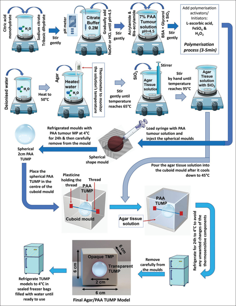
Background: Tissue-mimicking phantoms (TMPs) have been used extensively in clinical and nonclinical settings to simulate the thermal effects of focus ultrasound (FUS) technology in real tissue or organs. With recent technological developments in the FUS technology and its monitoring/guided techniques such as ultrasound-guided FUS and magnetic resonance-guided FUS (MRgFUS) the need for TMPs are more important than ever to ensure the safety of the patients before being treated with FUS for a variety of diseases (e.g., cancer or neurological). The purpose of this study was to prepare a tumor-mimicking phantom (TUMP) model that can simulate competently a tumor that is surrounded by healthy tissue.

Methods: The TUMP models were prepared using polyacrylamide (PAA) and agar solutions enriched with MR contrast agents (silicon dioxide and glycerol), and the thermosensitive component bovine serum albumin (BSA) that can alter its physical properties once thermal change is detected, therefore offering real-time visualization of the applied FUS ablation in the TUMPs models. To establish if these TUMPs are good candidates to be used in thermoablation, their thermal properties were characterized with a custom-made FUS system in the laboratory and a magnetic resonance imaging (MRI) setup with MR-thermometry. The BSA protein's coagulation temperature was adjusted at 55°C by setting the pH of the PAA solution to 4.5, therefore simulating the necrosis temperature of the tissue.

Results: The experiments carried out showed that the TUMP models prepared by PAA can change color from transparent to cream-white due to the BSA protein coagulation caused by the thermal stress applied. The TUMP models offered a good MRI contrast between the TMPs and the TUMPs including real-time visualization of the ablation area due to the BSA protein coagulation. Furthermore, the T2-weighted MR images obtained showed a significant change in T2 when the BSA protein is thermally coagulated. MR thermometry maps demonstrated that the suggested TUMP models may successfully imitate a tumor that is present in soft tissue.

Conclusion: The TUMP models developed in this study have numerous uses in the testing and calibration of FUS equipment including the simulation and validation of thermal therapy treatment plans with FUS or MRgFUS in oncology applications.

Keywords: Agar; focus ultrasound; magnetic resonance imaging; phantom; polyacrylamide; tumor.


Conflict of interest statement

The authors declare that they have no known competing financial interests or personal relationships that could have appeared to influence the work reported in this paper.

Figures

Figure 1
Figure 1
Methodology followed in Experiment 1 showing the preparation of the transparent polyacrylamide tumor-mimicking phantom (PAA TUMP) and the opaque agar TMP, including the fabrication of the final Agar/PAA TUMP model with bovine serum albumin protein
Figure 2
Figure 2
Shows (a) the placement of the spherical polyacrylamide tumor-mimicking phantom (PAA TUMP) in the acrylic mould before adding the TMP solution, (b) a rendered cross-section image of the opaque Agar/PAA TUMP model and (c) a rendered image of the transparent PAA/PAA TUMP model (rendered in OPENAI DALL-E online software)
Figure 3
Figure 3
Shows (a) a schematic of the focus ultrasound (FUS) ablation to the polyacrylamide (PAA)/PAA tumor-mimicking phantom model and (b) the realistic custom-made FUS setup used for the thermal ablation
Figure 4
Figure 4
Experimental setup inside the 3T magnetic resonance imaging (MRI) scanner with the focus ultrasound custom-made setup, in which the tumor-mimicking phantom models were placed on the MRI table and a GPFLEX coil placed on top of it to take the MR images
Figure 5
Figure 5
Shows photos of (a) the opaque Agar/polyacrylamide tumor-mimicking phantom (PAA TUMP) model, (b) the transparent PAA/PAA TUMP model and (c) a cross section of Agar/PAA TUMP model and (d) a cross section of PAA/PAA TUMP model
Figure 6
Figure 6
Shows photos of (a) the transparent polyacrylamide tumor-mimicking phantom (PAA TUMP) before heating it in a water bath, (b) the coagulated PAA TUMP after immersing it in a water bath with temperature >55°C and (c) the coagulated region in the centre of the PAA/PAA TUMP model after focus ultrasound ablation
Figure 7
Figure 7
Shows magnetic resonance imaging images of the opaque Agar/polyacrylamide tumor-mimicking phantom model acquired by using. (a and b) The T1W FSE sequence and (c and d) the T2W FSE sequence
Figure 8
Figure 8
Shows magnetic resonance (MR)-Thermometry images acquired for the opaque Agar/polyacrylamide tumor-mimicking phantom model and the temperature evolution observed in a region of interest set within the focal spot with (a) a coronal thermal map with the sonication power set to 200W for 60 s and (b) an axial thermal map with the sonication power set to 200W for 60 s. MR parameters used: Sequence = FLASH 2D, Coil type: Body_12_BM, TR = 25 ms, TE = 10 ms, FA = 30°, acquisition matrix: 96 × 96, slice thickness: 3 mm, acquisition time/slice: 2.4 s, Echo train length: 1, Pixel BW: 501 Hz/pixel, FOV: 280 mm × 280 mm × 3 mm
Figure 9
Figure 9
Shows photos of (a) the coagulation of the bovine serum albumin (BSA) protein from transparent to cream white in the (PAA)/PAA tumor-mimicking phantom model (TUMP) model formed by the thermal stress applied with FUS ablation and (b) the coagulation of the BSA protein in the FUS focal spot located in the transparent TUMP fused in the centre of the also transparent PAA TMP material
Figure 10
Figure 10
Shows magnetic resonance (MR)-Thermometry images acquired for the transparent (PAA)/PAA tumor-mimicking phantom model model and the temperature evolution observed in a region of interest set within the focal spot with (a) a coronal thermal map with the sonication power set to 200W for 30 s and (b) an axial thermal map with the sonication power set to 250W for 120 s. MR parameters used: Sequence = FLASH 2D, Coil type: Body_12_BM, TR = 25 ms, TE = 10 ms, FA = 30°, acquisition matrix: 96 × 96, slice thickness: 3 mm, acquisition time/slice: 2.4 s, Echo train length: 1, Pixel BW: 501 Hz/pixel, FOV: 280 mm × 280 mm × 3 mm

Similar articles

Cited by

References

    1. Baek JH, Lee JH, Valcavi R, Pacella CM, Rhim H, Na DG. Thermal ablation for benign thyroid nodules: Radiofrequency and laser. Korean J Radiol. 2011;12:525–40. - PMC - PubMed
    1. Rhim H, Dodd GD., 3rd Radiofrequency thermal ablation of liver tumors. J Clin Ultrasound. 1999;27:221–9. - PubMed
    1. Zhao Z, Wu F. Minimally-invasive thermal ablation of early-stage breast cancer: A systemic review. Eur J Surg Oncol. 2010;36:1149–55. - PubMed
    1. Johnson SL, Dillon C, Odéen H, Parker D, Christensen D, Payne A. Development and validation of a MRgHIFU non-invasive tissue acoustic property estimation technique. Int J Hyperthermia. 2016;32:723–34. - PMC - PubMed
    1. Furusawa H, Namba K, Thomsen S, Akiyama F, Bendet A, Tanaka C, et al. Magnetic resonance-guided focused ultrasound surgery of breast cancer: Reliability and effectiveness. J Am Coll Surg. 2006;203:54–63. - PubMed