. 2024 May 30:39:562-581.
doi: 10.1016/j.bioactmat.2024.04.022. eCollection 2024 Sep.

A cuttlefish ink nanoparticle-reinforced biopolymer hydrogel with robust adhesive and immunomodulatory features for treating oral ulcers in diabetes

Affiliations

A cuttlefish ink nanoparticle-reinforced biopolymer hydrogel with robust adhesive and immunomodulatory features for treating oral ulcers in diabetes

Yajing Xiang et al. Bioact Mater. .

Abstract

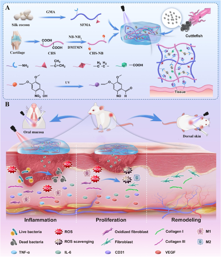
Oral ulcers can be managed using a variety of biomaterials that deliver drugs or cytokines. However, many patients experience minimal benefits from certain medical treatments because of poor compliance, short retention times in the oral cavity, and inadequate drug efficacy. Herein, we present a novel hydrogel patch (SCE2) composed of a biopolymer matrix (featuring ultraviolet-triggered adhesion properties) loaded with cuttlefish ink nanoparticles (possessing pro-healing functions). Applying a straightforward local method initiates the formation of a hydrogel barrier that adheres to mucosal injuries under the influence of ultraviolet light. SCE2 then demonstrates exceptional capabilities for near-infrared photothermal sterilization and neutralization of reactive oxygen species. These properties contribute to the elimination of bacteria and the management of the oxidation process, thus accelerating the healing phase's progression from inflammation to proliferation. In studies involving diabetic rats with oral ulcers, the SCE2 adhesive patch significantly quickens recovery by altering the inflamed state of the injured area, facilitating rapid re-epithelialization, and fostering angiogenesis. In conclusion, this light-sensitive hydrogel patch offers a promising path to expedited wound healing, potentially transforming treatment strategies for clinical oral ulcers.

Keywords: Cuttlefish ink nanoparticles; Hydrogel patches; Oral ulcers; Tissue adhesives; Wound healing.


Conflict of interest statement

Jianliang Shen is an editorial board member for Bioactive Materials and was not involved in the editorial review or the decision to publish this article. All authors declare that there are no competing interests.

Figures

Image 1
Graphical abstract
Scheme 1
Scheme 1
Fabrication and usage of SCE2 hydrogel for accelerated healing in oral ulcers among bacteria-infected diabetic rats. (A) Synthesis process of the SCE2 hydrogel. (B) Mechanisms of SCE2 hydrogel in enhancing wound recovery.
Fig. 1
Fig. 1
Characterization of CFI nanoparticles. (A) SEM image of CFI. (B) TEM image of CFI. (C) Thermographs of different CFI concentrations over time. (D) Temperature increase profiles of CFI nanoparticles. (E) Photothermal cycling curve of 1 mg/mL CFI. (F–H) The effectiveness of CFI nanoparticles in neutralizing O2•− (F), ABTS (G), and DPPH radicals (H). (I and J) Evaluation of cell survival in RAW 264.7 (I) and RS1 (J) cells exposed to different concentrations of CFI nanoparticles in a solution with 670 μM H2O2, using the CCK-8 assay. (K and L) Flow cytometric assessment of RAW 264.7 macrophages (K) and RS1 cells (L). Error bars indicate the average ± standard deviation (n = 3). Levels of significance are denoted as *** for P < 0.001 and * for P < 0.05.
Fig. 2
Fig. 2
Analyzing the physiochemical characteristics of SCE2 hydrogel. (A and B) Displaying 1H NMR spectra for SFMA (A) and CHS-NB (B). (C) TGA profiles of CFI and SCE hydrogels. (D and E) Diverse magnifications of SEM images featuring SCE0 (D) and SCE2 (E). (F and G) Performance in swelling (F) and deswelling (G) of SCE hydrogels. (H–J) Examining the radical scavenging abilities of SCE2 hydrogel against O2•− (H), ABTS (I), and DPPH (J). Error bars indicate the average ± standard deviation (n = 3). Significance levels: *** for P < 0.001.
Fig. 3
Fig. 3
Evaluating the mechanical attributes and adhesive strength of SCE2 hydrogel. (A) Illustration of the hydrogel's adhesion process with tissue and the corresponding lap-shear examination. (B) The hydrogels' stress−strain profile obtained through lap-shear testing. (C) Illustrative depiction of conducting a tensile strength assessment. (D) Capturing the stress−strain relationship during the tensile evaluation of the hydrogels. (E–H) Analysis of SCE2 hydrogel's characteristics includes strain-responsive rheological behavior (E), frequency-responsive rheological properties (F), evaluations of step-strain (G), and shear-thinning attributes (H). Displaying SCE2 hydrogel's ease of injection through representative images. (I) Illustration of SCE2 hydrogel's injectability within a PBS setting. (J) A sequence of images showcasing the self-healing capabilities of the SCE2 hydrogel.
Fig. 4
Fig. 4
In vitro cellular and hemolytic assays with SCE2 hydrogel. (A and B) Viability of RAW 264.7 macrophages (A) and RS1 cells (B) following exposure to PBS and SCE groups for durations of 1, 3, and 5 days. (C) Hemolysis assessment and images of SCE hydrogels post 2-h exposure to erythrocytes at 37 °C. (D and E) Fluorescence imaging of calcein-AM/PI staining in RAW 264.7 (D) and RS1 cells (E) after 3 days of varied treatments (scale bar: 100 μm). (F) Assessment of intracellular ROS neutralization by SCE hydrogels in RAW 264.7 cells employing DCFH-DA (scale bar: 20 μm). (G) Evaluation of ROS neutralization in RS1 cells by SCE hydrogels, indicated by DCFH-DA (scale bar: 50 μm). (H and I) Flow cytometric analysis of ROS in RAW 264.7 (H) and RS1 cells (I) using DCFH-DA as the detecting agent. (J and K) Survival rates of RAW 264.7 (J) and RS1 (K) cells post-incubation in SCE and H2O2 solution. (L) Images showing the migration of RS1 cells at intervals of 0, 12, and 24 h (scale bar: 300 μm). (M) Visuals captured through the fluorescence of HUVECs forming vessels (scale bar: 100 μm). Error bars represent the average ± standard deviation (n = 3). Levels of significance are denoted as *** for P < 0.001, ** for P < 0.01, * for P < 0.05, and NS for P > 0.05.
Fig. 5
Fig. 5
Examining the SCE2 hydrogel's in vitro photothermal antibacterial properties (A) Analyzing the temperature rise patterns among various hydrogels. (B) Studying the thermal increase in SCE2 hydrogel under different NIR light exposures. (C) Investigating the photothermal resilience of SCE2 hydrogel through four alternating heating and cooling cycles. (D) Analyzing the heating and cooling phases of SCE2 hydrogel's photothermal process. (E) Assessing SCE2 hydrogel's antibacterial effectiveness against MRSA and MRPA through methods like agar plate counting, SEM, and bacterial live-dead staining. (F) Presenting TEM imagery of bacteria pre and post exposure to SCE2 hydrogel combined with NIR treatment.
Fig. 6
Fig. 6
RNA sequencing evaluation of SCE2 hydrogel's therapeutic efficacy. (A) Differential gene count comparison in a Venn diagram for M1, SCE0, and SCE2 categories. (B) Three-group PCA plot illustrating proteomic data with three biological replicates per group. (C) Volcano chart presenting the downregulated and upregulated gene expressions in the SCE2 versus M1 gene expressions. (D) Analysis of GO pathways enriched among detected DEGs. (E) Dot chart depicting selected DEGs' KEGG enrichment analysis outcomes between M1 and SCE2 groups. (F) Heat map representing downregulated genes in both M1 and SCE2 groups. (G) Chord diagram illustrating GO enrichment terms linked to specific downregulated genes associated with oxidative stress. (H) GSEA results indicating cytosolic TNF and NF-kB signaling pathways' gene set enrichment in cells treated with SCE2 hydrogel.
Fig. 7
Fig. 7
Assessment of SCE2 hydrogel's therapeutic efficacy on back skin wounds in diabetic rats. (A) Depictions of skin injury progression in PBS, 3 M, SCE0, SCE2, and SCE2 + NIR. (B) Visual representation of healing stages in wounds. (C) Comparative measurement of wound sizes in all groups. (D–F) Staining for histology in rat skin sections from the wound region: H&E (D), Masson's trichrome (E), and markers TNF-α, MPO, CD31, VEGF, CD86, and CD206 (F). Error bars represent the average ± standard deviation (n = 3).
Fig. 8
Fig. 8
Assessment of SCE2 hydrogel's therapeutic efficacy on oral ulcer wounds in type 1 diabetic rats. (A) Diagram illustrating the creation and treatment approach for oral ulcers. (B) Digital images showing oral ulcer healing in PBS, DEX, SCE2, and SCE2 + NIR sets throughout 1–5 days. (C–E) Stained sections of oral ulcer wounds: H&E (C), Masson's trichrome (D), and markers TNF-α, MPO, CD31, VEGF, CD86, and CD206 (E).

Similar articles

Cited by

References

    1. Choi S., Jeon J., Bae Y., Hwang Y., Cho S.W. Mucoadhesive phenolic pectin hydrogels for saliva substitute and oral patch. Adv. Funct. Mater. 2023;33(44)
    1. Cui C., Mei L., Wang D., Jia P., Zhou Q., Liu W. A self-stabilized and water-responsive deliverable coenzyme-based polymer binary elastomer adhesive patch for treating oral ulcer. Nat. Commun. 2023;14(1):7707. - PMC - PubMed
    1. Edmans J.G., Ollington B., Colley H.E., Santocildes-Romero M.E., Siim Madsen L., Hatton P.V., Spain S.G., Murdoch C. Electrospun patch delivery of anti-TNFalpha F(ab) for the treatment of inflammatory oral mucosal disease. J. Contr. Release. 2022;350:146–157. - PubMed
    1. Mohammed E., Aboulkhair A.G., Tawifq M.M. Effect of nano-chitosan and nano-doxycycline gel on healing of induced oral ulcer in rat model: histological and immunohistochemical study. Clin. Oral Invest. 2021;26(3):3109–3118. - PubMed
    1. Wu J., Pan Z., Zhao Z.Y., Wang M.H., Dong L., Gao H.L., Liu C.Y., Zhou P., Chen L., Shi C.J., Zhang Z.Y., Yang C., Yu S.H., Zou D.H. Anti-swelling, robust, and adhesive extracellular matrix-mimicking hydrogel used as intraoral dressing. Adv. Mater. 2022;34(20) - PubMed

LinkOut - more resources