. 2024 May 30:39:582-594.
doi: 10.1016/j.bioactmat.2024.04.025. eCollection 2024 Sep.

Orthogonally woven 3D nanofiber scaffolds promote rapid soft tissue regeneration by enhancing bidirectional cell migration

Affiliations

Orthogonally woven 3D nanofiber scaffolds promote rapid soft tissue regeneration by enhancing bidirectional cell migration

Jiayi Yuan et al. Bioact Mater. .

Abstract

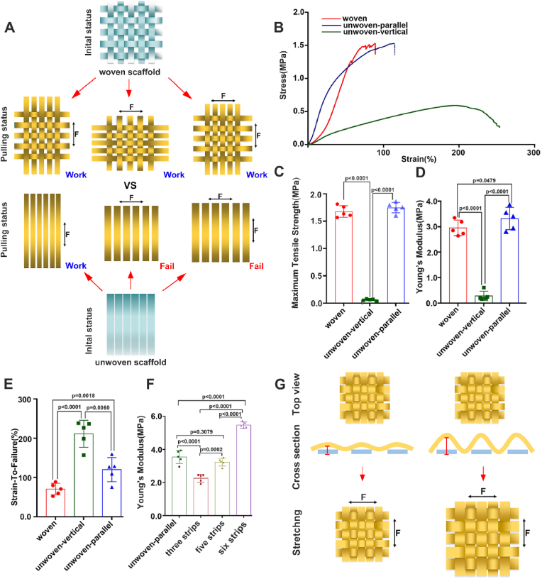
Repairing large-area soft tissue defects caused by traumas is a major surgical challenge. Developing multifunctional scaffolds with suitable scalability and favorable cellular response is crucial for soft tissue regeneration. In this study, we developed an orthogonally woven three-dimensional (3D) nanofiber scaffold combining electrospinning, weaving, and modified gas-foaming technology. The developed orthogonally woven 3D nanofiber scaffold had a modular design and controlled fiber alignment. In vitro, the orthogonally woven 3D nanofiber scaffold exhibited adjustable mechanical properties, good cell compatibility, and easy drug loading. In vivo, for one thing, the implantation of an orthogonally woven 3D nanofiber scaffold in a full abdominal wall defect model demonstrated that extensive granulation tissue formation with enough mechanical strength could promote recovery of abdominal wall defects while reducing intestinal adhesion. Another result of diabetic wound repair experiments suggested that orthogonally woven 3D nanofiber scaffolds had a higher wound healing ratio, granulation tissue formation, collagen deposition, and re-epithelialization. Taken together, this novel orthogonally woven 3D nanofiber scaffold may provide a promising and effective approach for optimal soft tissue regeneration.

Keywords: 3D nanofiber scaffold; Cell migration; Electrospinning; Orthogonal weaving; Tissue regeneration.


Conflict of interest statement

All authors declare no conflicts of interest in this work, and all authors have read and agreed to this submission.

Figures

Image 1
Graphical abstract
Fig. 1
Fig. 1
Schematic illustrates the applications of orthogonally woven 3D nanofiber scaffold in soft tissue regeneration. For one thing, the orthogonally woven 3D nanofiber scaffold could mimic the abdominal muscle tissue consisting of biaxially-aligned muscle fibers. It can potentially be used for hernia repair. For another, the orthogonally woven 3D nanofiber scaffold is able to recruit cells (e.g., fibroblasts, endothelial cells, and macrophages) from four directions simultaneously, and it is expected to be used to repair skin wounds.
Fig. 2
Fig. 2
The fabrication of an orthogonally woven 3D nanofiber scaffold. (A) The schematic illustrates the fabrication processes of an orthogonally woven 3D nanofiber scaffold containing electrospinning, cryo-cutting, weaving, and modified gas-foaming expansion. (B) The woven PCL nanofiber strips before expansion and the expanded orthogonally woven 3D nanofiber scaffold, the number of knitted strips could be adjustable. (C) The cross-section and top view of the orthogonally woven 3D nanofiber scaffold. (D) The nanofiber diameter quantification of the orthogonally woven 3D nanofiber scaffold. (E) The inner pore size of the orthogonally woven 3D nanofiber scaffold. (F) The thickness of one layer and the whole scaffold. (G) The proliferation and interaction between BMSCs and HUVECs after 7 days of co-culture. (H) The proliferation and interaction between BMSCs and HUVECs after 14 days of co-culture.
Fig. 3
Fig. 3
The mechanical properties of orthogonally woven 3D nanofiber scaffold. (A) The schematic illustrates the situation of the orthogonally woven 3D nanofiber scaffold under stretching compared to the uniaxial aligned 3D nanofiber scaffold. (B) The stress-strain curve of the orthogonally woven 3D nanofiber scaffold and uniaxial aligned 3D nanofiber scaffold in two different directions. (C, D) The maximum tensile strength and Young's modulus of the orthogonally woven 3D nanofiber scaffold and uniaxial aligned 3D nanofiber scaffold in two different directions. (E) The breaking strain of the orthogonally woven 3D nanofiber scaffold and uniaxial aligned 3D nanofiber scaffold in two different directions. (F) The Young's modulus of uniaxial aligned 3D nanofiber scaffold and the orthogonally woven 3D nanofiber scaffold with 3 × 3, 5 × 5, and 6 × 6 strips. (G) The schematic illustrates the stain of the orthogonally woven 3D nanofiber scaffold could be adjusted by increasing the wavelength of expanded strips.
Fig. 4
Fig. 4
The drug loading of the orthogonally woven 3D nanofiber scaffold. (A) The demonstration of one drug loading in the whole orthogonally woven 3D nanofiber scaffold, and two drug loading in two different directions of the scaffold. (B) The schematic illustrates a single bFGF or VEGF loading and both bFGF and VEGF loading in the orthogonally woven 3D nanofiber scaffold. (C) The transection trichrome staining of the scaffold only, bFGF loaded scaffold, VEGF loaded scaffold, and bFGF/VEGF co-loaded scaffold with surrounding tissues after subcutaneous implantation for 1 week. (D) The quantification of newly formed blood vessels among the four groups after subcutaneous implantation for 1 week. (E) The quantification of collagen deposition among the four groups after subcutaneous implantation for 1 week. (F) The cross-section trichrome staining shows the scaffold only, bFGF loaded scaffold, VEGF loaded scaffold, and bFGF/VEGF co-loaded scaffold with surrounding tissues after subcutaneous implantation for 1 week. The green arrows indicate the newly formed blood vessels.
Fig. 5
Fig. 5
The application of orthogonally woven 3D nanofiber scaffold in hernia repair. (A) The schematic illustrates the application of an orthogonally woven 3D nanofiber scaffold in hernia repair. Two methods were used in this study. One method was directly used (DU), and another was subcutaneously implanted for 2 weeks, then cut three sides of the scaffold and flipped horizontally to the hernia area. (B) The photographs of the repaired hernia were treated with scaffold directly for 2 months, or subcutaneously implantation for 2 weeks first and then applied in the hernia area for 2 months. (C) The trichrome staining of regenerated hernia tissue was treated with scaffolds directly for 1, 2 months, or subcutaneously implantation of scaffolds for 2 weeks first and then applied in the hernia area for 1, 2 months. (D) The strain-stress curve of regenerated hernia tissue was treated with scaffolds directly for 1, 2 months, or subcutaneously implantation of scaffolds for 2 weeks first and then applied in the hernia area for 1, 2 months. (E, F) The maximum tensile strength and Young's modulus of the regenerated hernia tissue that treated with scaffolds directly for 1, 2 months, or subcutaneously implanted scaffolds for 2 weeks first and then applied in the hernia area for 1, 2 months.
Fig. 6
Fig. 6
The effects of orthogonally woven 3D nanofiber scaffold on skin wound healing. (A) Photographs of chronic skin wounds treated with Lando, PELNAC, PCL orthogonally woven 3D nanofiber scaffold (PCL), the wounds without any treatment as the Control group after 3, 7, 14, and 21 days, respectively. (B) The changes in wound size of control, Lando, PELNAC, and PCL groups from day 0 to day 21. (C) The wound closure rate of the Control, Lando, PELNAC, and PCL groups at each indicated time point. (D) The histological observations of wound area in the Control, Lando, PELNAC, and PCL groups on day 3 and 7 days, respectively. (E) The wound re-epithelialization rate of the Control, Lando, PELNAC, and PCL groups on day 3, 7, 14, and 21 days, respectively. (F) The trichrome staining of the wound area in the Control, Lando, PELNAC, and PCL groups after 3 and 7 days of treatment.
Fig. 7
Fig. 7
The potential pro-healing mechanism of orthogonally woven 3D nanofiber scaffold. (A) The heatmap exhibits the differentially expressed genes in the PCL nanofiber scaffold-treated wounds compared to PELNAC-treated wounds. (B) The GO functional enrichment of these differentially expressed genes. (C) Immunohistochemical staining of CCR7 in wound center area of the Control, Lando, PELNAC, and PCL groups after 3 and 7 days of treatment. (D, F) The quantification of CCR7 (M1 macrophage) and CD206 (M2 macrophage) positive cells in the wound center area of the Control, Lando, PELNAC, and PCL groups after 3 and 7 days of treatment. (E) Immunohistochemical staining of CD206 in wound center area of the Control, Lando, PELNAC, and PCL groups after 3 and 7 days of treatment. (G) The ratio between the numbers of M2 type macrophage and M1 type macrophage of the Control, Lando, PELNAC, and PCL groups on day 3 and day 7. (H) Immunohistochemical staining of LY6G in wound center area of the Control, Lando, PELNAC, and PCL groups after 3 and 7 days of treatment. (I) The quantification of LY6G (a pan marker of monocytes, granulocytes, and neutrophils) positive cells in the wound center area of the Control, Lando, PELNAC, and PCL groups after 3 and 7 days of treatment.

Similar articles

References

    1. Wong R., Geyer S., Weninger W., Guimberteau J.-C., Wong J.K. The dynamic anatomy and patterning of skin. Exp. Dermatol. 2016;25(2):92–98. - PubMed
    1. Smith D.R., Caban-Rivera D.A., McGarry M.D.J., Williams L.T., McIlvain G., Okamoto R.J., Van Houten E.E.W., Bayly P.V., Paulsen K.D., Johnson C.L. Anisotropic mechanical properties in the healthy human brain estimated with multi-excitation transversely isotropic MR elastography. Brain Multiphysics. 2022;3 - PMC - PubMed
    1. Liu Z., Zhang Z., Ritchie R.O. Structural orientation and anisotropy in biological materials: functional designs and mechanics. Adv. Funct. Mater. 2020;30(10)
    1. Chanda A., Callaway C. Tissue anisotropy modeling using soft composite materials. Appl. Bionics Biomech. 2018 2018. - PMC - PubMed
    1. Astruc L., De Meulaere M., Witz J.F., Nováček V., Turquier F., Hoc T., Brieu M. Characterization of the anisotropic mechanical behavior of human abdominal wall connective tissues. J. Mech. Behav. Biomed. Mater. 2018;82:45–50. - PubMed

LinkOut - more resources