

Pigs lacking TMPRSS2 displayed fewer lung lesions and reduced inflammatory response when infected with influenza A virus
- PMID: 38883409
- PMCID: PMC11176495
- DOI: 10.3389/fgeed.2023.1320180
Pigs lacking TMPRSS2 displayed fewer lung lesions and reduced inflammatory response when infected with influenza A virus
Abstract
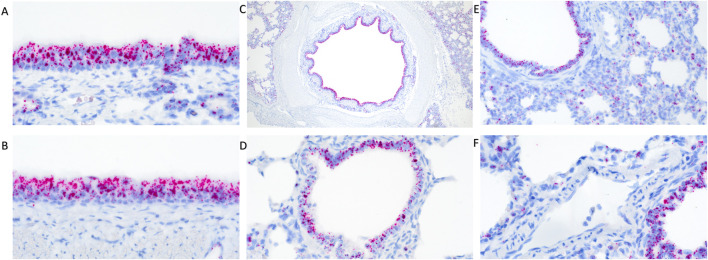
Influenza A virus (IAV) infection is initiated by hemagglutinin (HA), a glycoprotein exposed on the virion's lipid envelope that undergoes cleavage by host cell proteases to ensure membrane fusion, entry into the host cells, and completion of the viral cycle. Transmembrane protease serine S1 member 2 (TMPRSS2) is a host transmembrane protease expressed throughout the porcine airway epithelium and is purported to play a major role in the HA cleavage process, thereby influencing viral pathogenicity and tissue tropism. Pigs are natural hosts of IAV and IAV disease causes substantial economic impact on the pork industry worldwide. Previous studies in mice demonstrated that knocking out expression of TMPRSS2 gene was safe and inhibited the spread of IAV after experimental challenge. Therefore, we hypothesized that knockout of TMPRSS2 will prevent IAV infectivity in the swine model. We investigated this hypothesis by comparing pathogenesis of an H1N1pdm09 virus challenge in wildtype (WT) control and in TMPRSS2 knockout (TMPRSS2 -/-) pigs. We demonstrated that TMPRSS2 was expressed in the respiratory tract in WT pigs with and without IAV infection. No differences in nasal viral shedding and lung lavage viral titers were observed between WT and TMPRSS2 -/- pigs. However, the TMPRSS2 -/- pig group had significantly less lung lesions and significant reductions in antiviral and proinflammatory cytokines in the lung. The virus titer results in our direct challenge model contradict prior studies in the murine animal model, but the reduced lung lesions and cytokine profile suggest a possible role for TMPRSS2 in the proinflammatory antiviral response. Further research is warranted to investigate the role of TMPRSS2 in swine IAV infection and disease.
Keywords: RNAscope; TMPRSS2 gene; influenza A; knockout; proinflammatory response; somatic cell nuclear transfer; swine.
Copyright © 2024 Ciacci Zanella, Snyder, Arruda, Whitworth, Green, Poonooru, Telugu and Baker.
Conflict of interest statement
Author BT is a founding member and serves as a consultant for RenOVAte Biosciences Inc., (RBI). The remaining authors declare that the research was conducted in the absence of any commercial or financial relationships that could be construed as a potential conflict of interest.
Figures


Similar articles
-
TMPRSS2 Is the Major Activating Protease of Influenza A Virus in Primary Human Airway Cells and Influenza B Virus in Human Type II Pneumocytes.J Virol. 2019 Oct 15;93(21):e00649-19. doi: 10.1128/JVI.00649-19. Print 2019 Nov 1. J Virol. 2019. PMID: 31391268 Free PMC article.
-
Hemagglutinins of Avian Influenza Viruses Are Proteolytically Activated by TMPRSS2 in Human and Murine Airway Cells.J Virol. 2021 Sep 27;95(20):e0090621. doi: 10.1128/JVI.00906-21. Epub 2021 Jul 28. J Virol. 2021. PMID: 34319155 Free PMC article.
-
The host protease TMPRSS2 plays a major role in in vivo replication of emerging H7N9 and seasonal influenza viruses.J Virol. 2014 May;88(10):5608-16. doi: 10.1128/JVI.03677-13. Epub 2014 Mar 5. J Virol. 2014. PMID: 24600012 Free PMC article.
-
Role of host trypsin-type serine proteases and influenza virus-cytokine-trypsin cycle in influenza viral pathogenesis. Pathogenesis-based therapeutic options.Biochimie. 2019 Nov;166:203-213. doi: 10.1016/j.biochi.2019.09.006. Epub 2019 Sep 10. Biochimie. 2019. PMID: 31518617 Review.
-
Role of host cellular proteases in the pathogenesis of influenza and influenza-induced multiple organ failure.Biochim Biophys Acta. 2012 Jan;1824(1):186-94. doi: 10.1016/j.bbapap.2011.07.001. Epub 2011 Jul 23. Biochim Biophys Acta. 2012. PMID: 21801859 Review.
Cited by
-
Progress and persistence of diseases of high consequence to livestock in the United States.One Health. 2024 Jul 29;19:100865. doi: 10.1016/j.onehlt.2024.100865. eCollection 2024 Dec. One Health. 2024. PMID: 39185352 Free PMC article. Review.
-
Effects of Alkaline Solutions on the Structure and Function of Influenza A Virus.Viruses. 2024 Oct 19;16(10):1636. doi: 10.3390/v16101636. Viruses. 2024. PMID: 39459968 Free PMC article.
-
Exploring TMPRSS2 Drug Target to Combat Influenza and Coronavirus Infection.Scientifica (Cairo). 2025 Apr 21;2025:3687892. doi: 10.1155/sci5/3687892. eCollection 2025. Scientifica (Cairo). 2025. PMID: 40297833 Free PMC article. Review.
-
Genetic resilience or resistance in poultry against avian influenza virus: mirage or reality?J Virol. 2025 Jul 22;99(7):e0082025. doi: 10.1128/jvi.00820-25. Epub 2025 Jun 30. J Virol. 2025. PMID: 40586559 Free PMC article. Review.
References
-
- Arendsee Z. W., Chang J., Hufnagel D. E., Markin A., Janas-Martindale A., Vincent A. L., et al. (2021). Octoflushow: an interactive tool describing spatial and temporal trends in the genetic diversity of influenza A virus in U.S. Swine. Microbiol. Resour. Announc 10 (50), E0108121. 10.1128/MRA.01081-21 - DOI - PMC - PubMed
LinkOut - more resources
Full Text Sources