

This is a preprint.
The Neural Bases of Language Processing During Social and Non-Social Contexts: A fNIRS Study of Autistic and Neurotypical Preschool-Aged Children
- PMID: 38883761
- PMCID: PMC11177967
- DOI: 10.21203/rs.3.rs-4450882/v1
The Neural Bases of Language Processing During Social and Non-Social Contexts: A fNIRS Study of Autistic and Neurotypical Preschool-Aged Children
Update in
-
The neural bases of language processing during social and non-social contexts: a fNIRS study of autistic and neurotypical preschool-aged children.Mol Autism. 2025 Aug 6;16(1):40. doi: 10.1186/s13229-025-00655-3. Mol Autism. 2025. PMID: 40770353 Free PMC article.
Abstract
Background: Little is known about how the brains of autistic children process language during real-world "social contexts," despite the fact that challenges with language, communication, and social interaction are core features of Autism Spectrum Disorder (ASD).
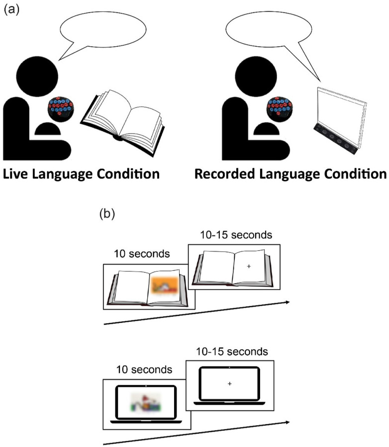
Methods: We investigated the neural bases of language processing during social and non-social contexts in a sample of N=20 autistic and N=20 neurotypical (NT) preschool-aged children, 3 to 6 years old. Functional near-infrared spectroscopy (fNIRS) was used to measure children's brain response to "live language" spoken by a live experimenter during an in-person social context (i.e., book reading), and "recorded language" played via an audio recording during a non-social context (i.e., screen time). We examined within-group and between-group differences in the strength and localization of brain response to live language and recorded language, as well as correlations between children's brain response and language skills measured by the Preschool Language Scales.
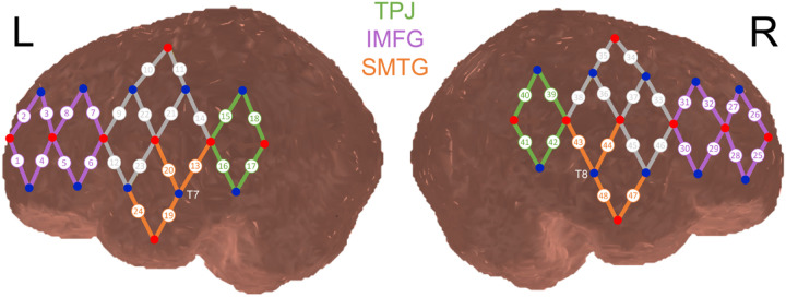
Results: In the NT group, brain response to live language was greater than brain response to recorded language in the right temporal parietal junction (TPJ). In the ASD group, the strength of brain response did not differ between conditions. The ASD group showed greater brain response to recorded language than the NT group in the right inferior and middle frontal gyrus (IMFG). Across groups, children's language skills were negatively associated with brain response to recorded language in the right IMFG, suggesting that processing recorded language required more cognitive effort for children with lower language skills. Children's language skills were also positively associated with the difference in brain response between conditions in the right TPJ, demonstrating that children who showed a greater difference in brain response to live language versus recorded language had higher language skills.
Limitations: Findings should be considered preliminary until they are replicated in a larger sample.
Conclusions: Findings suggest that the brains of NT children, but not autistic children, process language differently during social and non-social contexts. Individual differences in how the brain processes language during social and non-social contexts may help to explain why language skills are so variable across children with and without autism.
Keywords: autism; fNIRS; language; live; preschool; social context.
Conflict of interest statement
Competing interests: The authors declare that they have no competing interests.
Figures


References
-
- Ambarchi Z, Boulton KA, Thapa R, Arciuli J, DeMayo MM, Hickie IB, Thomas EE, Guastella AJ. Social and joint attention during shared book reading in young autistic children: a potential marker for social development. J Child Psychol Psychiatry. 2024. Apr 24. - PubMed
-
- Bennett TA, Szatmari P, Georgiades K, Hanna S, Janus M, Georgiades S, Duku E, Bryson S, Fombonne E, Smith IM, Mirenda P. Language impairment and early social competence in preschoolers with autism spectrum disorders: a comparison of DSM-5 profiles. J Autism Dev Disord. 2014;44:2797–808. - PubMed
Publication types
Grants and funding
LinkOut - more resources
Full Text Sources
Research Materials
