[Preprint]. 2024 Jun 3:rs.3.rs-4433306.
doi: 10.21203/rs.3.rs-4433306/v1.

CCR5 Decorated Rilpivirine Lipid Nanoparticles Build Myeloid Drug Depots Which Sustains Antiretroviral Activities

Affiliations

CCR5 Decorated Rilpivirine Lipid Nanoparticles Build Myeloid Drug Depots Which Sustains Antiretroviral Activities

Howard E Gendelman et al. Res Sq. .

Update in

Abstract

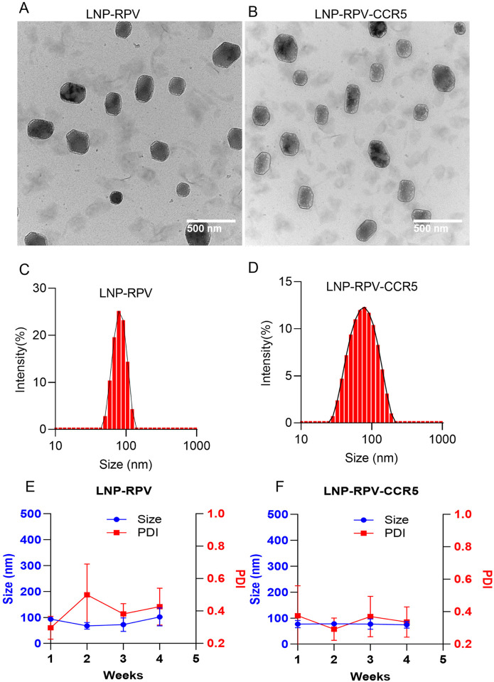
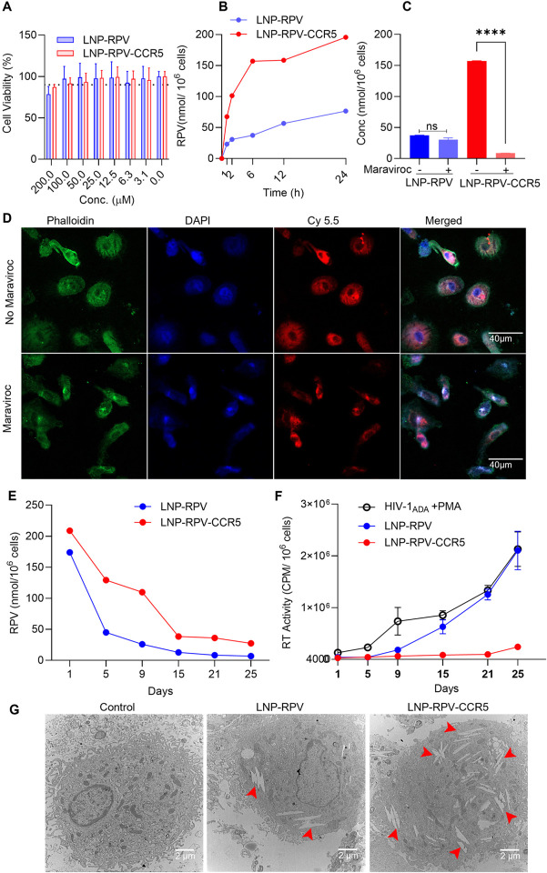
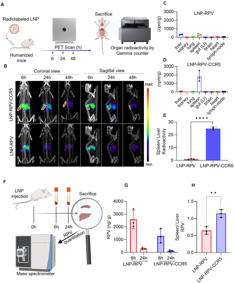
Antiretroviral therapy (ART) improves the quality of life for those living with the human immunodeficiency virus type one (HIV-1). However, poor compliance reduces ART effectiveness and leads to immune compromise, viral mutations, and disease co-morbidities. A novel drug formulation is made whereby a lipid nanoparticle (LNP) carrying rilpivirine (RPV) is decorated with the C-C chemokine receptor type 5 (CCR5). This facilitates myeloid drug depot deposition. Particle delivery to viral reservoirs is tracked by positron emission tomography. The CCR5-mediated RPV LNP cell uptake and retention reduce HIV-1 replication in human monocyte-derived macrophages and infected humanized mice. Focused ultrasound allows the decorated LNP to penetrate the blood-brain barrier and reach brain myeloid cells. These findings offer a role for CCR5-targeted therapeutics in antiretroviral delivery to optimize HIV suppression.

Keywords: CCR5-targeting; HIV; antiretroviral therapy; lipid nanoparticles; non-invasive imaging.


Conflict of interest statement

Additional Declarations: Yes there is potential Competing Interest. Howard Gendelman is the co-founder of Exavir Therapeutics, Inc, a company formed to develop LA ART.

Figures

Figure 1
Figure 1. Morphological characterization of the multimodal CuInEuS2 nanoprobe.
(A-B) Transmission electron microscope images of the multimodal rode shape nanoprobes. CuInEuS2 and its crystal lattice (red lines) are illustrated. (C) A scanning transmission electron microscopy map shows elemental localization within the nanoprobe from a corresponding high-angle annular dark-field electron microscopy image. The map shows the presence of copper (red), indium (blue), europium (cyan), and sulfur (green) in the nanoprobe. (D) XRD pattern of CuInEuS2 nanoprobe demonstrates a crystal structure.
Figure 2
Figure 2. Physicochemical LNP characterization.
(A-B) The TEM image shows a spherical LNP-RPV and LNP-RPV-CCR5 morphology. (C-D) The DLS size profile demonstrates the unimodular distribution of the LNPs. (E-F) The changes in size and polydispersity of the LNPs were recorded for one month at 4 °C. LNPs were stable without changes in size and polydispersity.
Figure 3
Figure 3. CCR5-receptor-modified LNPs facilitate the particle’s cell uptake, depot formation, and HIV-1 suppression.
(A) Dose-associated macrophage viability measurements following LNP exposures by the CTB assay after a 24 h incubation. The LNP- RPV and RPV-CCR5 treatments showed that cell viability was maintained at 100 μM of RPV doses. (B) LNP-RPV and LNP-RPV-CCR5 macrophage uptake was analyzed by measuring RPV concentration for 24 h at 20 μM RPV. Comparisons between LNP-RPV and RPV-CCR5 showed a 3-fold increase in drug uptake. (C) The CCR5 inhibitor (maraviroc, 1 nM) attenuated LNP uptake for the LNP-RPV CCR5 LNP. (D) Confocal microscopy was performed with Cy 5.5 dye-labeled LNP-RPV-CCR5 treated macrophages in the presence (lower panel) and absence (upper panel) of maraviroc. (E) RPV retention and (F) viral suppression in LNP- RPV and RPV-CCR5 macrophage uptake were evaluated by measuring RPV and virus levels in the cell supernatant fluids. The data were collected over 25 days after a 100 μM administered dose. (G) TEM image of LNP engulfed macrophage. Macrophages depots for LNP-RPV-CCR5 are shown. Statistical analysis was performed by an unpaired t-test. ****, P < 0.0001; and ns=not significant.
Figure 4
Figure 4. LNP and RPV tissue biodistribution in mice by PET imaging and mass spectrometry.
(A) Schematic presentation of LNP biodistribution by PET. (B) Humanized mice were injected with LNP-RPV or RPV-CCR5, and particle biodistribution was monitored by PET at 6, 24, and 48 h. Both coronal (left panel) and sagittal (right panel) views for LNP-RPV-CCR5 are illustrated. (C-D) Quantitative measurements of radiolabeled LNPs in tissue were recorded at 48 h by gamma counter measurements. LNP-RPV-CCR5 was concentrated in the spleen. (E) Spleen/liver RPV ratios for LNP-RPV-CCR5 and LNP-RPV are shown. (F) A schematic presentation of LNP injection measurements in humanized mice is shown. (G) Plasma RPV levels following NP- RPV and RPV-CCR5 treated mice are illustrated at 6 and 24 h after injection. (H) The spleen/liver RPV ratio at 24 h shows the highest RPV levels in the spleen after LNP-RPV injection.
Figure 5
Figure 5. Brain delivery of LNP-RPV-CCR5 nanoparticles.
(A) Schematic presentation of FUS-treated humanized microglial (MG) mice received LNP treatment. (B) IVIS images show bright yellow Cy5.5 signals in the brain of FUS-treated mice that received the LNP-RPV-CCR5 compared to LNP-RPV. Both mice showed good BBB disruption, as shown by the gadolinium enhancements (bright signals, blue arrows) on the coronal sections of the T1-weighted images (T1WI) on MRI. (C) Approximately 50% of microglia (IBA-1, red) showed human marker staining (HuNu, green), and only those showed the engulfment of LNPs. The Cy5.5 signals appear to be more intense in the animals that received the LNP-RPV-CCR5. Scale bar = 200 μm.
Figure 6
Figure 6. Viral suppression and toxicity profiles of LNP-carried RPV.
(A) Schematic representations of experimental timelines are shown for LNP treatments. (B) Viral suppression efficiency of LNP-RPV (individual humanized mice) and LNP-RPV-CCR5 (averages of all animals) on days 7 and 14 are illustrated. LNP-RPV-CCR5 showed complete viral suppression up to day 14. (C) The change in mice body weight in the untreated, LNP-RPV, and LNP-RPV-CCR5 treated group. (D) The histological images of hematoxylin and eosin-stained heart tissues, lung, kidney, spleen, and liver sections from the treatment groups. The images were captured at 20X magnification.

References

    1. Holmes EC (2001) On the origin and evolution of the human immunodeficiency virus (HIV). Biol Rev Camb Philos Soc 76(2):239–254. 10.1017/s1464793101005668From NLM - DOI - PubMed
    1. Senapathi J, Bommakanti A, Mallepalli S, Mukhopadhyay S, Kondapi AK (2020) Sulfonate modified Lactoferrin nanoparticles as drug carriers with dual activity against HIV-1. Colloids Surf B Biointerfaces 191:110979. 10.1016/j.colsurfb.2020.110979From NLM - DOI - PubMed
    1. Singh A, Kutscher HL, Bulmahn JC, Mahajan SD, He GS, Prasad P (2020) N. Laser ablation for pharmaceutical nanoformulations: Multi-drug nanoencapsulation and theranostics for HIV. Nanomedicine 25:102172. 10.1016/j.nano.2020.102172From NLM - DOI - PMC - PubMed
    1. Fotooh Abadi L, Kumar P, Paknikar K, Gajbhiye V, Kulkarni S (2023) Tenofovir-tethered gold nanoparticles as a novel multifunctional long-acting anti-HIV therapy to overcome deficient drug delivery-: an in vivo proof of concept. J Nanobiotechnol 21(1):19. 10.1186/s12951-022-01750-wFrom NLM - DOI - PMC - PubMed
    1. Chun TW, Moir S, Fauci AS (2015) HIV reservoirs as obstacles and opportunities for an HIV cure. Nat Immunol 16(6):584–589. 10.1038/ni.3152From NLM - DOI - PubMed

Publication types

LinkOut - more resources